|
Superior Vesical Artery
The superior vesical artery supplies numerous branches to the upper part of the bladder. This artery often also gives branches to the vas deferens and can provide minor collateral circulation for the testicles. Anatomy The superior vesical artery is a branch of the umbilical artery. The vesiculo-prostatic artery usually arises from the superior vesical artery in men. Distribution Other branches supply the ureter. Variation The middle vesical artery, usually a branch of the superior vesical artery, is distributed to the fundus of the bladder and the seminal vesicles. This artery is not usually described in modern anatomy textbooks. Instead, it is described that the superior vesical artery may exist as multiple vessels that arise from a common origin. Development The first part of the superior vesical artery represents the terminal section of the previous portion of the umbilical artery (fetal hypogastric artery The umbilical artery is a paired artery (with one for each ha ... [...More Info...] [...Related Items...] OR: [Wikipedia] [Google] [Baidu] |
Internal Iliac Artery
The internal iliac artery (formerly known as the hypogastric artery) is the main artery of the pelvis. Structure The internal iliac artery supplies the walls and viscera of the pelvis, the buttock, the reproductive organs, and the medial compartment of the thigh. The vesicular branches of the internal iliac arteries supply the bladder. It is a short, thick vessel, smaller than the external iliac artery, and about 3 to 4 cm in length. Course The internal iliac artery arises at the bifurcation of the common iliac artery, opposite the lumbosacral articulation, and, passing downward to the upper margin of the greater sciatic foramen, divides into two large trunks, an anterior and a posterior. It is posterior to the ureter, anterior to the internal iliac vein, anterior to the lumbosacral trunk, and anterior to the piriformis muscle. Near its origin, it is medial to the external iliac vein, which lies between it and the psoas major muscle. It is above the obturator nerve. ... [...More Info...] [...Related Items...] OR: [Wikipedia] [Google] [Baidu] |
Umbilical Artery
The umbilical artery is a paired artery (with one for each half of the body) that is found in the abdominal and pelvic regions. In the fetus, it extends into the umbilical cord. Structure Development The umbilical arteries supply deoxygenated blood from the fetus to the placenta. Although this blood is typically referred to as deoxygenated, this blood is fetal systemic arterial blood and will have the same amount of oxygen and nutrients as blood distributed to the other fetal tissues. There are usually two umbilical arteries present together with one umbilical vein in the umbilical cord. The umbilical arteries surround the urinary bladder and then carry all the deoxygenated blood out of the fetus through the umbilical cord. Inside the placenta, the umbilical arteries connect with each other at a distance of approximately 5 mm from the cord insertion in what is called the ''Hyrtl anastomosis''. Subsequently, they branch into chorionic arteries or ''intraplacental fetal arteri ... [...More Info...] [...Related Items...] OR: [Wikipedia] [Google] [Baidu] |
Vesical Venous Plexus
The vesical plexus envelops the lower part of the bladder and the base of the prostate The prostate is both an accessory gland of the male reproductive system and a muscle-driven mechanical switch between urination and ejaculation. It is found only in some mammals. It differs between species anatomically, chemically, and phys ... and communicates with the pudendal and prostatic plexuses. It is drained, by means of several vesical veins, into the internal iliac veins. References External links Anatomy at umich.edu Veins of the torso {{circulatory-stub ... [...More Info...] [...Related Items...] OR: [Wikipedia] [Google] [Baidu] |
Urinary Bladder
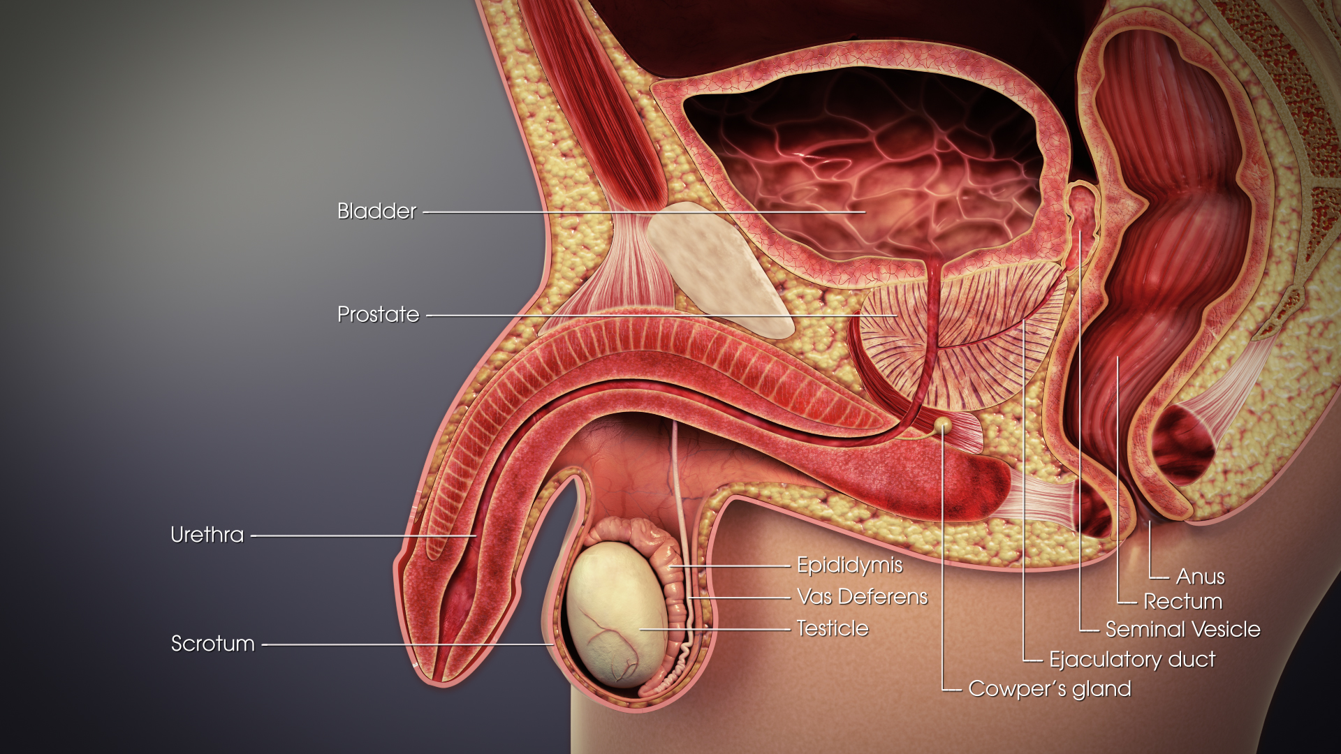
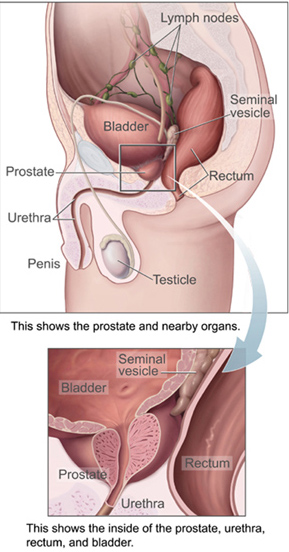
The urinary bladder, or simply bladder, is a hollow organ in humans and other vertebrates that stores urine from the kidneys before disposal by urination. In humans the bladder is a distensible organ that sits on the pelvic floor. Urine enters the bladder via the ureters and exits via the urethra. The typical adult human bladder will hold between 300 and (10.14 and ) before the urge to empty occurs, but can hold considerably more. The Latin phrase for "urinary bladder" is ''vesica urinaria'', and the term ''vesical'' or prefix ''vesico -'' appear in connection with associated structures such as vesical veins. The modern Latin word for "bladder" – ''cystis'' – appears in associated terms such as cystitis (inflammation of the bladder). Structure In humans, the bladder is a hollow muscular organ situated at the base of the pelvis. In gross anatomy, the bladder can be divided into a broad , a body, an apex, and a neck. The apex (also called the vertex) is directed fo ... [...More Info...] [...Related Items...] OR: [Wikipedia] [Google] [Baidu] |
Ureter
The ureters are tubes made of smooth muscle that propel urine from the kidneys to the urinary bladder. In a human adult, the ureters are usually long and around in diameter. The ureter is lined by urothelial cells, a type of transitional epithelium, and has an additional smooth muscle layer that assists with peristalsis in its lowest third. The ureters can be affected by a number of diseases, including urinary tract infections and kidney stone. is when a ureter is narrowed, due to for example chronic inflammation. Congenital abnormalities that affect the ureters can include the development of two ureters on the same side or abnormally placed ureters. Additionally, reflux of urine from the bladder back up the ureters is a condition commonly seen in children. The ureters have been identified for at least two thousand years, with the word "ureter" stemming from the stem relating to urinating and seen in written records since at least the time of Hippocrates. It is, however, ... [...More Info...] [...Related Items...] OR: [Wikipedia] [Google] [Baidu] |
Vas Deferens
The vas deferens or ductus deferens is part of the male reproductive system of many vertebrates. The ducts transport sperm from the epididymis to the ejaculatory ducts in anticipation of ejaculation. The vas deferens is a partially coiled tube which exits the abdominal cavity through the inguinal canal. Etymology ''Vas deferens'' is Latin, meaning "carrying-away vessel"; the plural version is ''vasa deferentia''. ''Ductus deferens'' is also Latin, meaning "carrying-away duct"; the plural version is ''ducti deferentes''. Structure There are two vasa deferentia, connecting the left and right epididymis with the seminal vesicles to form the ejaculatory duct in order to move sperm. The (human) vas deferens measures 30–35 cm in length, and 2–3 mm in diameter. The vas deferens is continuous proximally with the tail of the epididymis. The vas deferens exhibits a tortuous, convoluted initial/proximal section (which measures 2–3 cm in length). Distally, it form ... [...More Info...] [...Related Items...] OR: [Wikipedia] [Google] [Baidu] |
Testicles
A testicle or testis (plural testes) is the male reproductive gland or gonad in all bilaterians, including humans. It is homologous to the female ovary. The functions of the testes are to produce both sperm and androgens, primarily testosterone. Testosterone release is controlled by the anterior pituitary luteinizing hormone, whereas sperm production is controlled both by the anterior pituitary follicle-stimulating hormone and gonadal testosterone. Structure Appearance Males have two testicles of similar size contained within the scrotum, which is an extension of the abdominal wall. Scrotal asymmetry, in which one testicle extends farther down into the scrotum than the other, is common. This is because of the differences in the vasculature's anatomy. For 85% of men, the right testis hangs lower than the left one. Measurement and volume The volume of the testicle can be estimated by palpating it and comparing it to ellipsoids of known sizes. Another method is to use caliper ... [...More Info...] [...Related Items...] OR: [Wikipedia] [Google] [Baidu] |
Fundus Of The Urinary Bladder
The urinary bladder, or simply bladder, is a hollow organ in humans and other vertebrates that stores urine from the kidneys before disposal by urination. In humans the bladder is a distensible organ that sits on the pelvic floor. Urine enters the bladder via the ureters and exits via the urethra. The typical adult human bladder will hold between 300 and (10.14 and ) before the urge to empty occurs, but can hold considerably more. The Latin phrase for "urinary bladder" is ''vesica urinaria'', and the term ''vesical'' or prefix ''vesico -'' appear in connection with associated structures such as vesical veins. The modern Latin word for "bladder" – ''cystis'' – appears in associated terms such as cystitis (inflammation of the bladder). Structure In humans, the bladder is a hollow muscular organ situated at the base of the pelvis. In gross anatomy, the bladder can be divided into a broad , a body, an apex, and a neck. The apex (also called the vertex) is directed forwa ... [...More Info...] [...Related Items...] OR: [Wikipedia] [Google] [Baidu] |
Seminal Vesicle
The seminal vesicles (also called vesicular glands, or seminal glands) are a pair of two convoluted tubular glands that lie behind the urinary bladder of some male mammals. They secrete fluid that partly composes the semen. The vesicles are 5–10 cm in size, 3–5 cm in diameter, and are located between the bladder and the rectum. They have multiple outpouchings which contain secretory glands, which join together with the vas deferens at the ejaculatory duct. They receive blood from the vesiculodeferential artery, and drain into the vesiculodeferential veins. The glands are lined with column-shaped and cuboidal cells. The vesicles are present in many groups of mammals, but not marsupials, monotremes or carnivores. Inflammation of the seminal vesicles is called seminal vesiculitis, most often is due to bacterial infection as a result of a sexually transmitted disease or following a surgical procedure. Seminal vesiculitis can cause pain in the lower abdomen, scrot ... [...More Info...] [...Related Items...] OR: [Wikipedia] [Google] [Baidu] |
Fetal Hypogastric Artery
The umbilical artery is a paired artery (with one for each half of the body) that is found in the abdominal and pelvic regions. In the fetus, it extends into the umbilical cord. Structure Development The umbilical arteries supply deoxygenated blood from the fetus to the placenta. Although this blood is typically referred to as deoxygenated, this blood is fetal systemic arterial blood and will have the same amount of oxygen and nutrients as blood distributed to the other fetal tissues. There are usually two umbilical arteries present together with one umbilical vein in the umbilical cord. The umbilical arteries surround the urinary bladder and then carry all the deoxygenated blood out of the fetus through the umbilical cord. Inside the placenta, the umbilical arteries connect with each other at a distance of approximately 5 mm from the cord insertion in what is called the ''Hyrtl anastomosis''. Subsequently, they branch into chorionic arteries or ''intraplacental fetal arteri ... [...More Info...] [...Related Items...] OR: [Wikipedia] [Google] [Baidu] |


