|
Superficial Vein
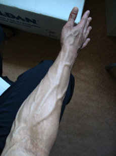
Superficial veins are veins that are close to the surface of the body, as opposed to deep veins, which are far from the surface. Superficial veins are not paired with an artery, unlike the deep veins, which are typically associated with an artery of the same name. Superficial veins are important physiologically for cooling of the body. When the body is too hot, the body shunts blood from the deep veins to the superficial veins to facilitate heat transfer to the body's surroundings. Superficial veins are often visible underneath the skin. Those below the level of the heart tend to bulge out, which can be readily witnessed in the hand, where the veins bulge significantly less after the arm has been raised above the head for a short time. Veins become more visually prominent when lifting heavy weight, especially after a period of proper strength training. Physiologically, the superficial veins are not as important as the deep veins (as they carry less blood) and are sometimes r ... [...More Info...] [...Related Items...] OR: [Wikipedia] [Google] [Baidu] |
Vein
Veins are blood vessels in humans and most other animals that carry blood towards the heart. Most veins carry deoxygenated blood from the tissues back to the heart; exceptions are the pulmonary and umbilical veins, both of which carry oxygenated blood to the heart. In contrast to veins, arteries carry blood away from the heart. Veins are less muscular than arteries and are often closer to the skin. There are valves (called ''pocket valves'') in most veins to prevent backflow. Structure Veins are present throughout the body as tubes that carry blood back to the heart. Veins are classified in a number of ways, including superficial vs. deep, pulmonary vs. systemic, and large vs. small. * Superficial veins are those closer to the surface of the body, and have no corresponding arteries. *Deep veins are deeper in the body and have corresponding arteries. *Perforator veins drain from the superficial to the deep veins. These are usually referred to in the lower limbs and feet. *Communic ... [...More Info...] [...Related Items...] OR: [Wikipedia] [Google] [Baidu] |
Venipuncture
In medicine, venipuncture or venepuncture is the process of obtaining intravenous access for the purpose of venous blood sampling (also called ''phlebotomy'') or intravenous therapy. In healthcare, this procedure is performed by medical laboratory scientists, medical practitioners, some EMTs, paramedics, phlebotomists, dialysis technicians, and other nursing staff. In veterinary medicine, the procedure is performed by veterinarians and veterinary technicians. It is essential to follow a standard procedure for the collection of blood specimens to get accurate laboratory results. Any error in collecting the blood or filling the test tubes may lead to erroneous laboratory results. Venipuncture is one of the most routinely performed invasive procedures and is carried out for any of five reasons: # to obtain blood for diagnostic purposes; # to monitor levels of blood components; # to administer therapeutic treatments including medications, nutrition, or chemotherapy; # to remov ... [...More Info...] [...Related Items...] OR: [Wikipedia] [Google] [Baidu] |
Vascularity
Vascularity, in bodybuilding, is the condition of having many highly visible, prominent, and often extensively-ramified superficial veins. The skin appears "thin"—sometimes virtually transparent—due to an extreme reduction of subcutaneous fat, allowing for maximum muscle definition. Vascularity is enhanced by extremely low body fat (usually below 10%) and low retained water, as well as the muscle engorgement ("pump") and venous distension accentuated by the vigorous flexing and potentially hazardous Valsalva effect which characterize competitive posing. Genetics and androgenic hormones will affect vascularity, as will ambient temperature. Additionally, although some bodybuilders develop arterial hypertension from performance-enhancing substances and practices, "high" venous pressure—being an order of magnitude lower than that of arteries— neither causes nor is caused by vascularity. Some bodybuilders use topical vasodilators to increase blood flow to the skin as well. Al ... [...More Info...] [...Related Items...] OR: [Wikipedia] [Google] [Baidu] |
Varicose Vein
Varicose veins, also known as varicoses, are a medical condition in which superficial veins become enlarged and twisted. These veins typically develop in the legs, just under the skin. Varicose veins usually cause few symptoms. However, some individuals may experience fatigue or pain in the area. Complications can include bleeding or superficial thrombophlebitis. Varices in the scrotum are known as a varicocele, while those around the anus are known as hemorrhoids. Due to the various physical, social, and psychological effects of varicose veins, they can negatively affect one's quality of life. Varicose veins have no specific cause. Risk factors include obesity, lack of exercise, leg trauma, and family history of the condition. They also develop more commonly during pregnancy. Occasionally they result from chronic venous insufficiency. Underlying causes include weak or damaged valves in the veins. They are typically diagnosed by examination, including observation by ultrasound. ... [...More Info...] [...Related Items...] OR: [Wikipedia] [Google] [Baidu] |
Deep Vein
A deep vein is a vein that is deep in the body. This contrasts with superficial veins that are close to the body's surface. Deep veins are almost always beside an artery with the same name (e.g. the femoral vein is beside the femoral artery). Collectively, they carry the vast majority of the blood. Occlusion of a deep vein can be life-threatening and is most often caused by thrombosis. Occlusion of a deep vein by thrombosis is called ''deep vein thrombosis''. Because of their location deep within the body, operation on these veins can be difficult. List *Internal jugular vein Upper limb *Brachial vein *Axillary vein *Subclavian vein Lower limb *Common femoral vein *Femoral vein *Profunda femoris vein *Popliteal vein *Peroneal vein *Anterior tibial vein *Posterior tibial vein The posterior tibial veins are veins of the leg in humans. They drain the posterior compartment of the leg and the plantar surface of the foot to the popliteal vein. Structure The posterior tibi ... [...More Info...] [...Related Items...] OR: [Wikipedia] [Google] [Baidu] |
Coronary Artery Bypass Surgery
Coronary artery bypass surgery, also known as coronary artery bypass graft (CABG, pronounced "cabbage") is a surgical procedure to treat coronary artery disease (CAD), the buildup of plaques in the arteries of the heart. It can relieve chest pain caused by CAD, slow the progression of CAD, and increase life expectancy. It aims to bypass narrowings in heart arteries by using arteries or veins harvested from other parts of the body, thus restoring adequate blood supply to the previously ischemic (deprived of blood) heart. There are two main approaches. The first uses a cardiopulmonary bypass machine, a machine which takes over the functions of the heart and lungs during surgery by circulating blood and oxygen. With the heart in arrest, harvested arteries and veins are used to connect across problematic regions—a construction known as surgical anastomosis. In the second approach, called the off-pump coronary artery bypass graft (OPCABG), these anastomoses are constructed while t ... [...More Info...] [...Related Items...] OR: [Wikipedia] [Google] [Baidu] |
Great Saphenous Vein
The great saphenous vein (GSV, alternately "long saphenous vein"; ) is a large, subcutaneous, superficial vein of the leg. It is the longest vein in the body, running along the length of the lower limb, returning blood from the foot, leg and thigh to the deep femoral vein at the femoral triangle. Structure The great saphenous vein originates from where the dorsal vein of the big toe (the hallux) merges with the dorsal venous arch of the foot. After passing in front of the medial malleolus (where it often can be visualized and palpated), it runs up the medial side of the leg. At the knee, it runs over the posterior border of the medial epicondyle of the femur bone. In the proximal anterior thigh inferolateral to the pubic tubercle, the great saphenous vein dives down deep through the cribriform fascia of the saphenous opening to join the femoral vein. It forms an arch, the saphenous arch, to join the common femoral vein in the region of the femoral triangle at the sapheno-femoral ... [...More Info...] [...Related Items...] OR: [Wikipedia] [Google] [Baidu] |
Small Saphenous Vein
The small saphenous vein (also short saphenous vein or lesser saphenous vein) is a relatively large superficial vein of the posterior leg. Structure The origin of the small saphenous vein, (SSV) is where the dorsal vein from the fifth digit (smallest toe) merges with the dorsal venous arch of the foot, which attaches to the great saphenous vein (GSV). It is a superficial vein, being Subcutaneous tissue, subcutaneous (just under the skin). From its origin, it courses around the lateral aspect of the foot (inferior and posterior to the lateral malleolus) and runs along the posterior aspect of the leg (with the sural nerve), where it passes between the heads of the gastrocnemius muscle. This vein presents a number of different draining points. Usually, it drains into the popliteal vein, at or above the level of the knee joint. Variation Sometimes, the SSV joins the common gastrocnemius vein before draining in the popliteal vein. Sometimes, it does not make contact with the popliteal ... [...More Info...] [...Related Items...] OR: [Wikipedia] [Google] [Baidu] |
Kidney Dialysis
Kidney dialysis (from Greek , , 'dissolution'; from , , 'through', and , , 'loosening or splitting') is the process of removing excess water, solutes, and toxins from the blood in people whose kidneys can no longer perform these functions naturally. This is referred to as renal replacement therapy. The first successful dialysis was performed in 1943. Dialysis may need to be initiated when there is a sudden rapid loss of kidney function, known as acute kidney injury (previously called acute renal failure), or when a gradual decline in kidney function, chronic kidney disease, reaches stage 5. Stage 5 chronic renal failure is reached when the glomerular filtration rate is 10–15% of normal, creatinine clearance is less than 10 mL per minute and uremia is present. Dialysis is used as a temporary measure in either acute kidney injury or in those awaiting kidney transplant and as a permanent measure in those for whom a transplant is not indicated or not possible.Pendse S, Singh A, ... [...More Info...] [...Related Items...] OR: [Wikipedia] [Google] [Baidu] |
Basilic Vein
The basilic vein is a large superficial vein of the upper limb that helps drain parts of the hand and forearm. It originates on the medial (ulnar) side of the dorsal venous network of the hand and travels up the base of the forearm, where its course is generally visible through the skin as it travels in the subcutaneous fat and fascia lying superficial to the muscles. The basilic vein terminates by uniting with the brachial veins to form the axillary vein. Anatomy Course As it ascends the medial side of the biceps in the arm proper (between the elbow and shoulder), the basilic vein normally perforates the brachial fascia (deep fascia) superior to the medial epicondyle, or even as high as mid-arm. Tributaries and anastomoses Near the region anterior to the cubital fossa (in the bend of the elbow joint), the basilic vein usually communicates with the cephalic vein (the other large superficial vein of the upper extremity) via the median cubital vein. The layout of superfic ... [...More Info...] [...Related Items...] OR: [Wikipedia] [Google] [Baidu] |
Median Cubital Vein
In human anatomy, the median cubital vein (or median basilic vein) is a superficial vein of the upper limb. It lies in the cubital fossa superficial to the bicipital aponeurosis. It connects the cephalic vein and the basilic vein. It becomes prominent when pressure is applied. It is routinely used for venipuncture (taking blood) and as a site for an intravenous cannula. This is due to its particularly wide lumen, and its tendency to remain stationary upon needle insertion. Structure The median cubital vein is a superficial vein of the upper limb. It lies in the cubital fossa superficial to the bicipital aponeurosis. It connects the cephalic vein and the basilic vein. It becomes prominent when pressure is applied to upstream veins, as venous blood builds up. Variations The median cubital vein shows a wide range of variations. More commonly, the vein forms an H-pattern with the cephalic and basilic veins making up the sides. Other forms include an M-pattern, where the vein bran ... [...More Info...] [...Related Items...] OR: [Wikipedia] [Google] [Baidu] |
Deep Vein
A deep vein is a vein that is deep in the body. This contrasts with superficial veins that are close to the body's surface. Deep veins are almost always beside an artery with the same name (e.g. the femoral vein is beside the femoral artery). Collectively, they carry the vast majority of the blood. Occlusion of a deep vein can be life-threatening and is most often caused by thrombosis. Occlusion of a deep vein by thrombosis is called ''deep vein thrombosis''. Because of their location deep within the body, operation on these veins can be difficult. List *Internal jugular vein Upper limb *Brachial vein *Axillary vein *Subclavian vein Lower limb *Common femoral vein *Femoral vein *Profunda femoris vein *Popliteal vein *Peroneal vein *Anterior tibial vein *Posterior tibial vein The posterior tibial veins are veins of the leg in humans. They drain the posterior compartment of the leg and the plantar surface of the foot to the popliteal vein. Structure The posterior tibi ... [...More Info...] [...Related Items...] OR: [Wikipedia] [Google] [Baidu] |


