|
Submandibular Triangle
The submandibular triangle (or submaxillary or digastric triangle) corresponds to the region of the neck immediately beneath the body of the mandible. Boundaries and coverings It is bounded: * ''above'', by the lower border of the body of the mandible, and a line drawn from its angle to the mastoid process; * ''below'', by the posterior belly of the Digastricus; in front, by the anterior belly of the Digastricus. It is covered by the integument, superficial fascia, Platysma, and deep fascia, ramifying in which are branches of the facial nerve and ascending filaments of the cutaneous cervical nerve. Its floor is formed by the Mylohyoideus anteriorly, and by the hyoglossus posteriorly. Triangles * Beclard Triangle * Lesser Triangle * Pirogoff Triangle Divisions It is divided into an anterior and a posterior part by the stylomandibular ligament. Anterior part The anterior part contains the submandibular gland, superficial to which is the anterior facial vein, while imbedded ... [...More Info...] [...Related Items...] OR: [Wikipedia] [Google] [Baidu] |
Human Mandible
In anatomy, the mandible, lower jaw or jawbone is the largest, strongest and lowest bone in the human facial skeleton. It forms the lower jaw and holds the lower tooth, teeth in place. The mandible sits beneath the maxilla. It is the only movable bone of the skull (discounting the ossicles of the middle ear). It is connected to the temporal bones by the temporomandibular joints. The bone is formed prenatal development, in the fetus from a fusion of the left and right mandibular prominences, and the point where these sides join, the mandibular symphysis, is still visible as a faint ridge in the midline. Like other symphyses in the body, this is a midline articulation where the bones are joined by fibrocartilage, but this articulation fuses together in early childhood.Illustrated Anatomy of the Head and Neck, Fehrenbach and Herring, Elsevier, 2012, p. 59 The word "mandible" derives from the Latin word ''mandibula'', "jawbone" (literally "one used for chewing"), from ''wikt:mandere ... [...More Info...] [...Related Items...] OR: [Wikipedia] [Google] [Baidu] |
Submental Artery
The submental artery is a branch of the facial artery that runs on the underside of the chin. Course The submental artery is the largest of the cervical branches of the facial artery, given off just as that vessel leaves the submandibular gland: it runs forward upon the mylohyoid, just below the body of the mandible, and beneath the digastric muscle. It supplies the surrounding muscles, and anastomoses with the sublingual artery and with the mylohyoid branch of the inferior alveolar artery; at the symphysis menti it turns upward over the border of the mandible. The submental vessels also supply a territory of skin in the submental area. Surgeons can use the skin and vessels in reconstruction of the face or the oral cavity. Branching When the submental artery turns upward over the border of the mandible it divides into a superficial and a deep branch. * The ''superficial branch'' passes between the integument and depressor labii inferioris, and anastomoses with the inferior ... [...More Info...] [...Related Items...] OR: [Wikipedia] [Google] [Baidu] |
Submandibular Space
The submandibular space is a fascial space of the head and neck (sometimes also termed fascial spaces or tissue spaces). It is a potential space, and is paired on either side, located on the superficial surface of the mylohyoid muscle between the anterior and posterior bellies of the digastric muscle. The space corresponds to the anatomic region termed the submandibular triangle, part of the anterior triangle of the neck. Location and structure Anatomic boundaries The anatomic boundaries of each submandibular space are: * the mylohyoid muscle superiorly, * the skin, superficial fascia, platysma muscle and superficial layer of the deep cervical fascia inferiorly and laterally, * the medial surface of the mandible anteriorly and laterally, * the hyoid bone posteriorly, * the anterior belly of the digastric muscle medially. Communications The communications of the submandibular space are: * medially and anteriorly to the submental space (located medial to the paired submandibu ... [...More Info...] [...Related Items...] OR: [Wikipedia] [Google] [Baidu] |
Anterior Triangle Of The Neck
The anterior triangle is a region of the neck. Structure The triangle is inverted with its apex inferior to its base which is under the chin. Investing fascia covers the roof of the triangle while visceral fascia covers the floor. Anatomy Muscles: * Suprahyoid muscles - Digastric (Ant and Post Belly), mylohyoid, geniohyoid and Stylohyoid. * Infrahyoid muscles - Omohyoid, Sternohyoid, Sternothyroid, and Thyrohyoid. Nerve supply 2 Bellies of Digastric * Anterior: Mylohyoid nerve * Posterior: Facial nerve Stylohyoid: by the facial nerve, by a branch from that to the posterior belly of digastric. Mylohyoid: by its own nerve, a branch of the inferior alveolar ( from the mandibular division of trigeminal nerve), which arises just before the parent nerve enters the mandibular foramen, pierces the sphenomandibular ligament, and runs forward on the inferior surface of the mylohyoid, supplying it and the anterior belly of the digastric. Geniohyoid: by a branch from the hypoglossal ... [...More Info...] [...Related Items...] OR: [Wikipedia] [Google] [Baidu] |
Hypoglossal Nerve
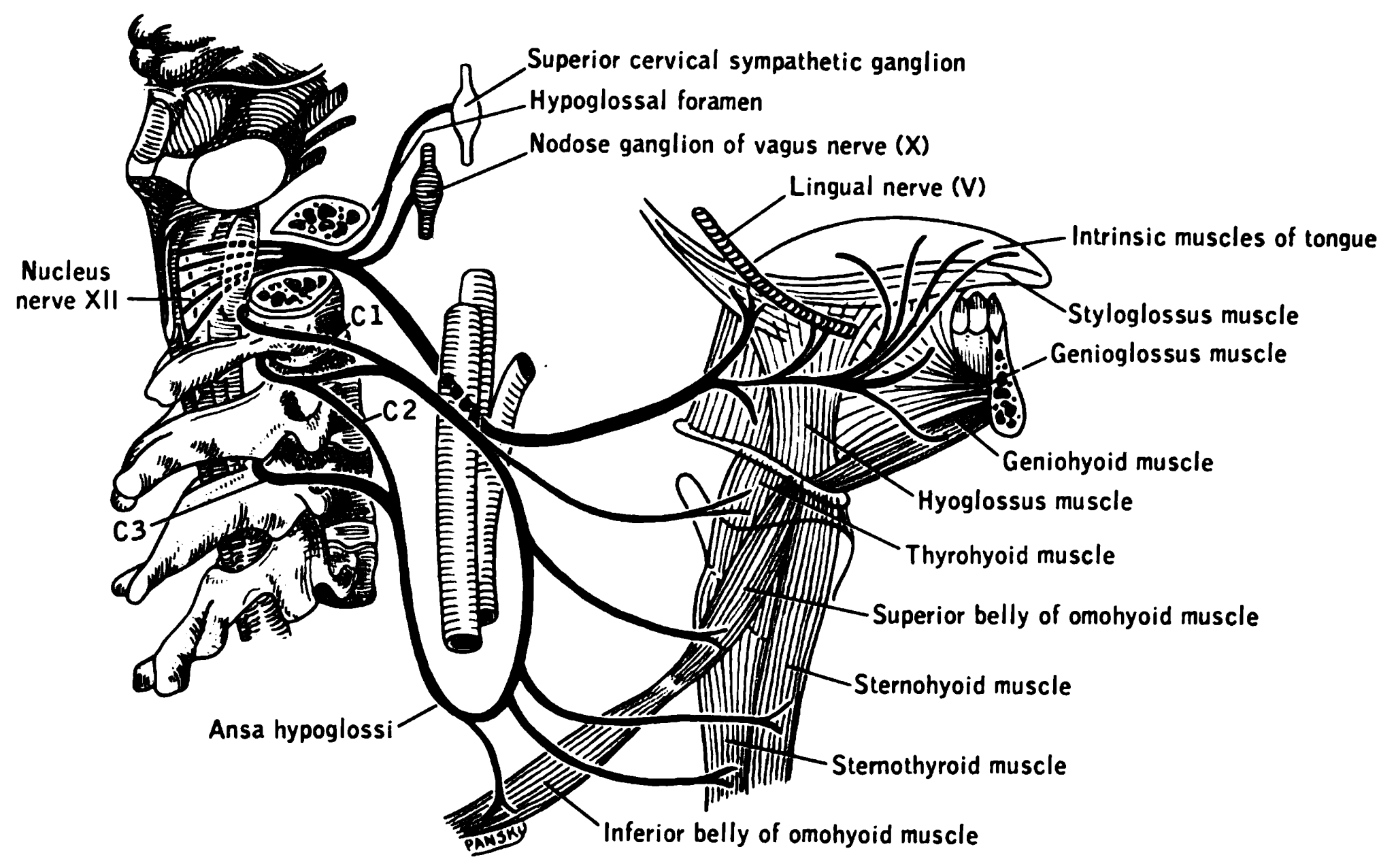
The hypoglossal nerve, also known as the twelfth cranial nerve, cranial nerve XII, or simply CN XII, is a cranial nerve that innervates all the extrinsic and intrinsic muscles of the tongue except for the palatoglossus, which is innervated by the vagus nerve. CN XII is a nerve with a solely motor function. The nerve arises from the hypoglossal nucleus in the medulla as a number of small rootlets, passes through the hypoglossal canal and down through the neck, and eventually passes up again over the tongue muscles it supplies into the tongue. The nerve is involved in controlling tongue movements required for speech and swallowing, including sticking out the tongue and moving it from side to side. Damage to the nerve or the neural pathways which control it can affect the ability of the tongue to move and its appearance, with the most common sources of damage being injury from trauma or surgery, and motor neuron disease. The first recorded description of the nerve is by Herophil ... [...More Info...] [...Related Items...] OR: [Wikipedia] [Google] [Baidu] |
Stylopharyngeus
The stylopharyngeus is a muscle in the head that stretches between the temporal styloid process and the pharynx. Structure The stylopharyngeus is a long, slender muscle, cylindrical above, flattened below. It arises from the medial side of the base of the temporal styloid process, passes downward along the side of the pharynx between the superior pharyngeal constrictor and the middle pharyngeal constrictor, and spreads out beneath the mucous membrane. Some of its fibers are lost in the constrictor muscles while others, joining the palatopharyngeus muscle, are inserted into the posterior border of the thyroid cartilage. The glossopharyngeal nerve runs on the lateral side of this muscle, and crosses over it to reach the tongue. Nerve supply The stylopharyngeus is the only muscle in the pharynx innervated by the glossopharyngeal nerve (CN IX) via branchial motor neurons with their cell bodies in the rostral part of the nucleus ambiguus. Development Embryological origin is the ... [...More Info...] [...Related Items...] OR: [Wikipedia] [Google] [Baidu] |
Styloglossus
The styloglossus, the shortest and smallest of the three styloid muscles, arises from the anterior and lateral surfaces of the styloid process near its apex, and from the stylomandibular ligament. Passing inferiorly and anteriorly between the internal and external carotid arteries, it divides upon the side of the tongue near its dorsal surface, blending with the fibers of the longitudinalis inferior in front of the hyoglossus; the other, oblique, overlaps the Hyoglossus and decussates with its fibers. Innervation The styloglossus is innervated by the hypoglossal nerve (CN XII) like all muscles of the tongue except palatoglossus which is innervated by the pharyngeal plexus of vagus nerve The pharyngeal plexus is a network of nerve fibers innervating most of the palate and pharynx. (The larynx, which is innervated by the superior and recurrent laryngeal nerves from vagus nerve (CN X), is not included.) It is located on the surfa ... (CN X). Action The styloglossus draws up the ... [...More Info...] [...Related Items...] OR: [Wikipedia] [Google] [Baidu] |
Vagus Nerve
The vagus nerve, also known as the tenth cranial nerve, cranial nerve X, or simply CN X, is a cranial nerve that interfaces with the parasympathetic control of the heart, lungs, and digestive tract. It comprises two nerves—the left and right vagus nerves—but they are typically referred to collectively as a single subsystem. The vagus is the longest nerve of the autonomic nervous system in the human body and comprises both sensory and motor fibers. The sensory fibers originate from neurons of the nodose ganglion, whereas the motor fibers come from neurons of the dorsal motor nucleus of the vagus and the nucleus ambiguus. The vagus was also historically called the pneumogastric nerve. Structure Upon leaving the medulla oblongata between the olive and the inferior cerebellar peduncle, the vagus nerve extends through the jugular foramen, then passes into the carotid sheath between the internal carotid artery and the internal jugular vein down to the neck, chest, and abdom ... [...More Info...] [...Related Items...] OR: [Wikipedia] [Google] [Baidu] |
Internal Jugular Vein
The internal jugular vein is a paired jugular vein that collects blood from the brain and the superficial parts of the face and neck. This vein runs in the carotid sheath with the common carotid artery and vagus nerve. It begins in the posterior compartment of the jugular foramen, at the base of the skull. It is somewhat dilated at its origin, which is called the ''superior bulb''. This vein also has a common trunk into which drains the anterior branch of the retromandibular vein, the facial vein, and the lingual vein. It runs down the side of the neck in a vertical direction, being at one end lateral to the internal carotid artery, and then lateral to the common carotid artery, and at the root of the neck, it unites with the subclavian vein to form the brachiocephalic vein (innominate vein); a little above its termination is a second dilation, the ''inferior bulb''. Above, it lies upon the rectus capitis lateralis, behind the internal carotid artery and the nerves passing ... [...More Info...] [...Related Items...] OR: [Wikipedia] [Google] [Baidu] |
Internal Carotid
The internal carotid artery (Latin: arteria carotis interna) is an artery in the neck which supplies the anterior circulation of the brain. In human anatomy, the internal and external carotids arise from the common carotid arteries, where these bifurcate at cervical vertebrae C3 or C4. The internal carotid artery supplies the brain, including the eyes, while the external carotid nourishes other portions of the head, such as the face, scalp, skull, and meninges. Classification Terminologia Anatomica in 1998 subdivided the artery into four parts: "cervical", "petrous", "cavernous", and "cerebral". However, in clinical settings, the classification system of the internal carotid artery usually follows the 1996 recommendations by Bouthillier, describing seven anatomical segments of the internal carotid artery, each with a corresponding alphanumeric identifier—C1 cervical, C2 petrous, C3 lacerum, C4 cavernous, C5 clinoid, C6 ophthalmic, and C7 communicating. The Bouthillier nomenclatu ... [...More Info...] [...Related Items...] OR: [Wikipedia] [Google] [Baidu] |
Internal Maxillary
The maxillary artery supplies deep structures of the face. It branches from the external carotid artery just deep to the neck of the mandible. Structure The maxillary artery, the larger of the two terminal branches of the external carotid artery, arises behind the neck of the mandible, and is at first imbedded in the substance of the parotid gland; it passes forward between the ramus of the mandible and the sphenomandibular ligament, and then runs, either superficial or deep to the lateral pterygoid muscle, to the pterygopalatine fossa. It supplies the deep structures of the face, and may be divided into mandibular, pterygoid, and pterygopalatine portions. First portion The ''first'' or ''mandibular '' or ''bony'' portion passes horizontally forward, between the neck of the mandible and the sphenomandibular ligament, where it lies parallel to and a little below the auriculotemporal nerve; it crosses the inferior alveolar nerve, and runs along the lower border of the lateral ptery ... [...More Info...] [...Related Items...] OR: [Wikipedia] [Google] [Baidu] |
Superficial Temporal
In human anatomy, the superficial temporal artery is a major artery of the head. It arises from the external carotid artery when it splits into the superficial temporal artery and maxillary artery. Its pulse can be felt above the zygomatic arch, above and in front of the tragus of the ear. Structure The superficial temporal artery is the smaller of two end branches that split superiorly from the external carotid. Based on its direction, the superficial temporal artery appears to be a continuation of the external carotid. It begins within the parotid gland, behind the neck of the mandible, and passes superficially over the posterior root of the zygomatic process of the temporal bone; about 5 cm above this process it divides into two branches: ''a. frontal'', and ''a. parietal''. Branches The parietal branch of the superficial temporal artery (posterior temporal) is a small artery in the head. It is larger than the frontal branch and curves upward and backward on the side ... [...More Info...] [...Related Items...] OR: [Wikipedia] [Google] [Baidu] |


