|
Sialyl-Lewis X
Sialyl LewisX (sLeX), also known as cluster of differentiation 15s (CD15s) or stage-specific embryonic antigen 1 (SSEA-1), is a tetrasaccharide carbohydrate which is usually attached to O-glycans on the surface of cells. It is known to play a vital role in cell-to-cell recognition processes. It is also the means by which an egg attracts sperm; first, to stick to it, then bond with it and eventually form a zygote. The discovery of the essential role that this tetrasaccharide plays in the fertilization process was reported in August 2011. Sialyl Lewis X is also one of the most important blood group antigens and is displayed on the terminus of glycolipids that are present on the cell surface. The sialyl Lewis X determinant, E-selectin ligand carbohydrate structure, is constitutively expressed on granulocytes and monocytes and mediates inflammatory extravasation of these cells. Resting T- and B-lymphocytes lack its expression and are induced to strongly express sialyl Lewis X upon ... [...More Info...] [...Related Items...] OR: [Wikipedia] [Google] [Baidu] |
Carbohydrate
In organic chemistry, a carbohydrate () is a biomolecule consisting of carbon (C), hydrogen (H) and oxygen (O) atoms, usually with a hydrogen–oxygen atom ratio of 2:1 (as in water) and thus with the empirical formula (where ''m'' may or may not be different from ''n''), which does not mean the H has covalent bonds with O (for example with , H has a covalent bond with C but not with O). However, not all carbohydrates conform to this precise stoichiometric definition (e.g., uronic acids, deoxy-sugars such as fucose), nor are all chemicals that do conform to this definition automatically classified as carbohydrates (e.g. formaldehyde and acetic acid). The term is most common in biochemistry, where it is a synonym of saccharide (), a group that includes sugars, starch, and cellulose. The saccharides are divided into four chemical groups: monosaccharides, disaccharides, oligosaccharides, and polysaccharides. Monosaccharides and disaccharides, the smallest (lower molecul ... [...More Info...] [...Related Items...] OR: [Wikipedia] [Google] [Baidu] |
FUT6
Alpha-(1,3)-fucosyltransferase is an enzyme that in humans is encoded by the ''FUT6'' gene. The alpha-1,3-fucosyltransferases constitute a large family of glycosyltransferases with a high degree of homology. The enzymes of this family comprise 3 main activity patterns called myeloid, plasma, and Lewis, based on their capacity to transfer alpha-L-fucose to distinct oligosaccharide acceptors, their sensitivity to N-ethylmaleimide inhibition, their cation An ion () is an atom or molecule with a net electrical charge. The charge of an electron is considered to be negative by convention and this charge is equal and opposite to the charge of a proton, which is considered to be positive by conven ... requirements, and their tissue-specific expression patterns. The different categories of alpha-1,3-fucosyltransferases are sequentially expressed during embryo-fetal development. upplied by OMIMref name="entrez"> References Further reading * * * * * * * * * * * * * * * ... [...More Info...] [...Related Items...] OR: [Wikipedia] [Google] [Baidu] |
CD30
CD30, also known as TNFRSF8 ( TNF receptor superfamily member 8), is a cell membrane protein of the tumor necrosis factor receptor family and a tumor marker. Function This receptor is expressed by activated, but not by resting, T and B cells. TRAF2 and TRAF5 can interact with this receptor, and mediate the signal transduction that leads to the activation of NF-kappaB. It is a positive regulator of apoptosis, and also has been shown to limit the proliferative potential of autoreactive CD8 effector T cells and protect the body against autoimmunity. Two alternatively spliced transcript variants of this gene encoding distinct isoforms have been reported. Clinical significance CD30 is associated with anaplastic large cell lymphoma. It is expressed in embryonal carcinoma but not in seminoma and is thus a useful marker in distinguishing between these germ cell tumors. CD30 and CD15 are also expressed on Reed-Sternberg cells typical for Hodgkin's lymphoma. Cancer treatmen ... [...More Info...] [...Related Items...] OR: [Wikipedia] [Google] [Baidu] |
Golgi Apparatus
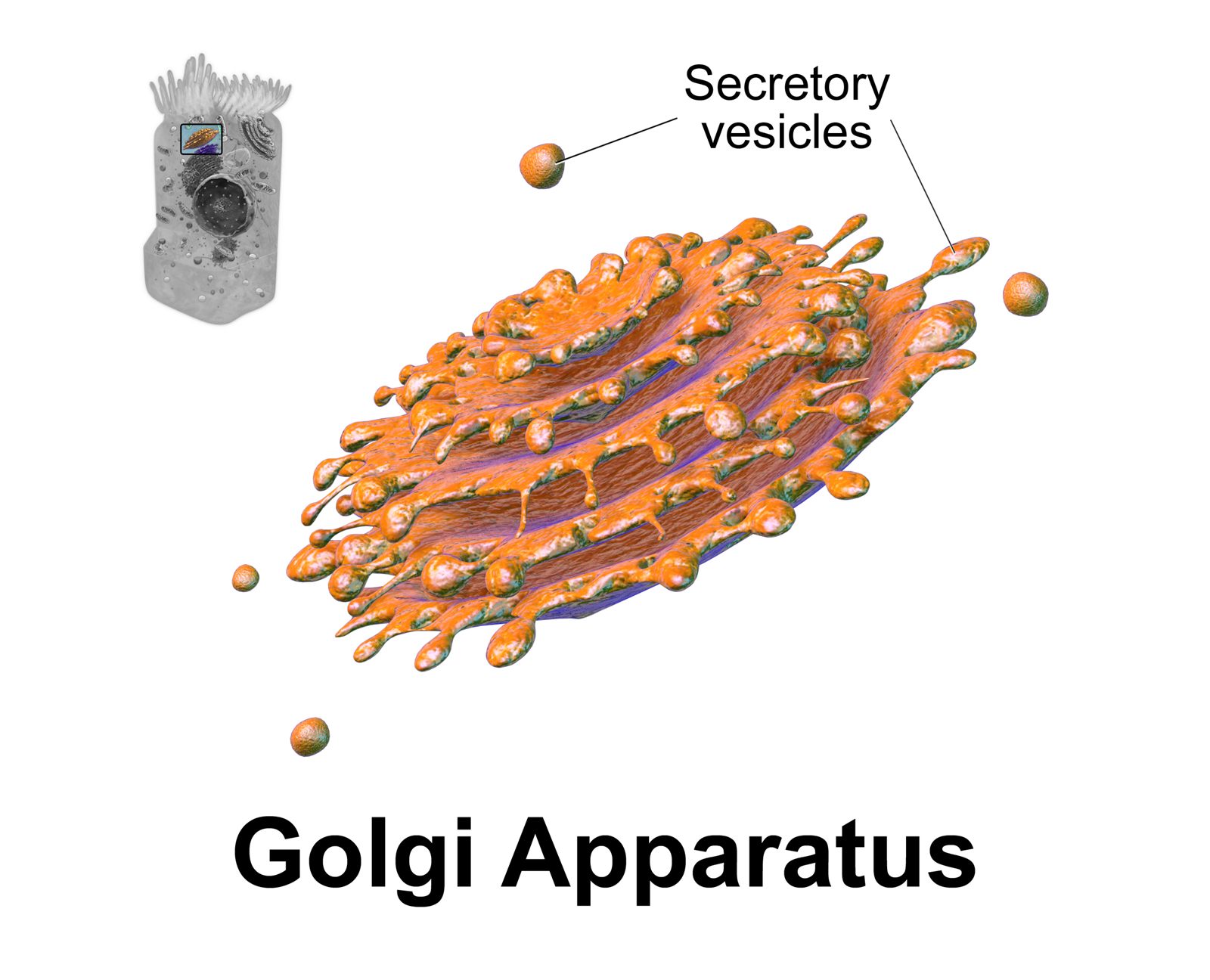
The Golgi apparatus (), also known as the Golgi complex, Golgi body, or simply the Golgi, is an organelle found in most eukaryotic cells. Part of the endomembrane system in the cytoplasm, it packages proteins into membrane-bound vesicles inside the cell before the vesicles are sent to their destination. It resides at the intersection of the secretory, lysosomal, and endocytic pathways. It is of particular importance in processing proteins for secretion, containing a set of glycosylation enzymes that attach various sugar monomers to proteins as the proteins move through the apparatus. It was identified in 1897 by the Italian scientist Camillo Golgi and was named after him in 1898. Discovery Owing to its large size and distinctive structure, the Golgi apparatus was one of the first organelles to be discovered and observed in detail. It was discovered in 1898 by Italian physician Camillo Golgi during an investigation of the nervous system. After first observing it unde ... [...More Info...] [...Related Items...] OR: [Wikipedia] [Google] [Baidu] |
Hodgkin's Lymphoma
Hodgkin lymphoma (HL) is a type of lymphoma, in which cancer originates from a specific type of white blood cell called lymphocytes, where multinucleated Reed–Sternberg cells (RS cells) are present in the patient's lymph nodes. The condition was named after the English physician Thomas Hodgkin, who first described it in 1832. Symptoms may include fever, night sweats, and weight loss. Often, nonpainful enlarged lymph nodes occur in the neck, under the arm, or in the groin. Those affected may feel tired or be itchy. The two major types of Hodgkin lymphoma are classic Hodgkin lymphoma and nodular lymphocyte-predominant Hodgkin lymphoma. About half of cases of Hodgkin lymphoma are due to Epstein–Barr virus (EBV) and these are generally the classic form. Other risk factors include a family history of the condition and having HIV/AIDS. Diagnosis is conducted by confirming the presence of cancer and identifying RS cells in lymph node biopsies. The virus-positive cases are classifi ... [...More Info...] [...Related Items...] OR: [Wikipedia] [Google] [Baidu] |
Immunohistochemistry
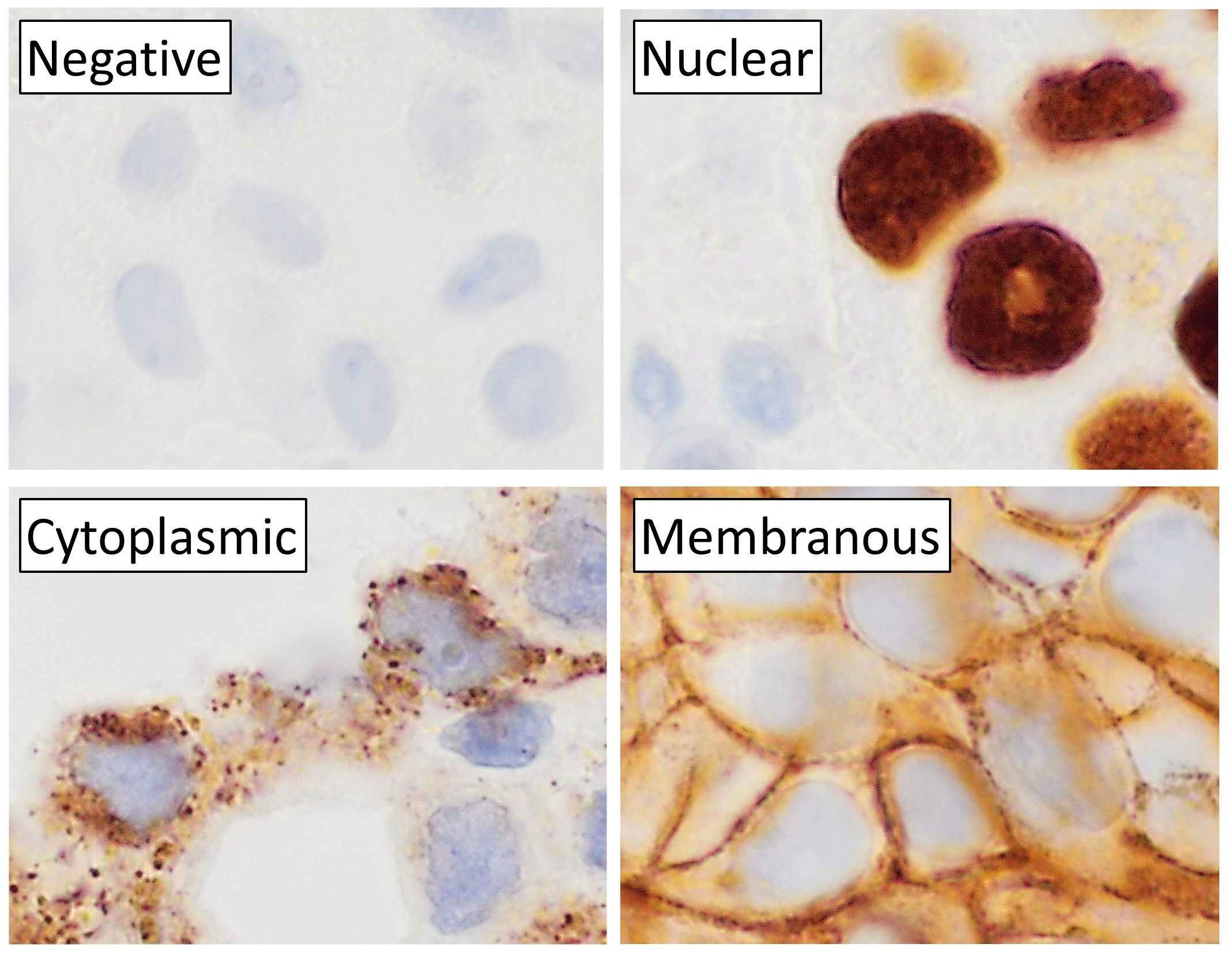
Immunohistochemistry (IHC) is the most common application of immunostaining. It involves the process of selectively identifying antigens (proteins) in cells of a tissue section by exploiting the principle of antibodies binding specifically to antigens in biological tissues. IHC takes its name from the roots "immuno", in reference to antibodies used in the procedure, and "histo", meaning tissue (compare to immunocytochemistry). Albert Coons conceptualized and first implemented the procedure in 1941. Visualising an antibody-antigen interaction can be accomplished in a number of ways, mainly either of the following: * ''Chromogenic immunohistochemistry'' (CIH), wherein an antibody is conjugated to an enzyme, such as peroxidase (the combination being termed immunoperoxidase), that can catalyse a colour-producing reaction. * ''Immunofluorescence'', where the antibody is tagged to a fluorophore, such as fluorescein or rhodamine. Immunohistochemical staining is widely used in the ... [...More Info...] [...Related Items...] OR: [Wikipedia] [Google] [Baidu] |
Reed–Sternberg Cell
Reed–Sternberg cells (also known as lacunar histiocytes for certain types) are distinctive, giant cells found with light microscopy in biopsies from individuals with Hodgkin lymphoma. They are usually derived from B lymphocytes, classically considered crippled germinal center B cells. In the vast majority of cases, the immunoglobulin genes of Reed–Sternberg cells have undergone both V(D)J recombination and somatic hypermutation, establishing an origin from a germinal center or postgerminal center B cell. Despite having the genetic signature of a B cell, the Reed–Sternberg cells of classical Hodgkin lymphoma fail to express most B-cell–specific genes, including the immunoglobulin genes. The cause of this wholesale reprogramming of gene expression has yet to be fully explained. It presumably is the result of widespread epigenetic changes of uncertain etiology, but is partly a consequence of so-called "crippling" mutations acquired during somatic hypermutation. Seen against a ... [...More Info...] [...Related Items...] OR: [Wikipedia] [Google] [Baidu] |
Chemotaxis
Chemotaxis (from '' chemo-'' + '' taxis'') is the movement of an organism or entity in response to a chemical stimulus. Somatic cells, bacteria, and other single-cell or multicellular organisms direct their movements according to certain chemicals in their environment. This is important for bacteria to find food (e.g., glucose) by swimming toward the highest concentration of food molecules, or to flee from poisons (e.g., phenol). In multicellular organisms, chemotaxis is critical to early development (e.g., movement of sperm towards the egg during fertilization) and development (e.g., migration of neurons or lymphocytes) as well as in normal function and health (e.g., migration of leukocytes during injury or infection). In addition, it has been recognized that mechanisms that allow chemotaxis in animals can be subverted during cancer metastasis. The aberrant chemotaxis of leukocytes and lymphocytes also contribute to inflammatory diseases such as atherosclerosis, asthma, and arthr ... [...More Info...] [...Related Items...] OR: [Wikipedia] [Google] [Baidu] |
Phagocytosis
Phagocytosis () is the process by which a cell uses its plasma membrane to engulf a large particle (≥ 0.5 μm), giving rise to an internal compartment called the phagosome. It is one type of endocytosis. A cell that performs phagocytosis is called a phagocyte. In a multicellular organism's immune system, phagocytosis is a major mechanism used to remove pathogens and cell debris. The ingested material is then digested in the phagosome. Bacteria, dead tissue cells, and small mineral particles are all examples of objects that may be phagocytized. Some protozoa use phagocytosis as means to obtain nutrients. History Phagocytosis was first noted by Canadian physician William Osler (1876), and later studied and named by Élie Metchnikoff (1880, 1883). In immune system Phagocytosis is one main mechanisms of the innate immune defense. It is one of the first processes responding to infection, and is also one of the initiating branches of an adaptive immune response. Although m ... [...More Info...] [...Related Items...] OR: [Wikipedia] [Google] [Baidu] |
Leukocyte Adhesion Deficiency
Leukocyte adhesion deficiency (LAD) is a rare autosomal recessive disorder characterized by immunodeficiency resulting in recurrent infections. LAD is currently divided into three subtypes: LAD1, LAD2, and the recently described LAD3, also known as LAD-1/variant. In LAD3, the immune defects are supplemented by a Glanzmann thrombasthenia-like bleeding tendency. Signs and symptoms LAD was first recognized as a distinct clinical entity in the 1970s. The classic descriptions of LAD included recurrent bacterial infections, defects in neutrophil adhesion, and a delay in umbilical cord sloughing. The adhesion defects result in poor leukocyte chemotaxis, particularly neutrophil, inability to form pus and neutrophilia. Individuals with LAD suffer from bacterial infections beginning in the neonatal period. Infections such as omphalitis, pneumonia, gingivitis, and peritonitis are common and often life-threatening due to the infant's inability to properly destroy the invading pathogens. ... [...More Info...] [...Related Items...] OR: [Wikipedia] [Google] [Baidu] |
Zona Pellucida
The zona pellucida (plural zonae pellucidae, also egg coat or pellucid zone) is a specialized extracellular matrix that surrounds the plasma membrane of mammalian oocytes. It is a vital constitutive part of the oocyte. The zona pellucida first appears in unilaminar primary oocytes. It is secreted by both the oocyte and the ovarian follicles. The zona pellucida is surrounded by the corona radiata. The corona is composed of cells that care for the egg when it is emitted from the ovary. This structure binds spermatozoa, and is required to initiate the acrosome reaction. In the mouse (the best characterised mammalian system), the zona glycoprotein, ZP3, is responsible for sperm binding, adhering to proteins on the sperm plasma membrane. ZP3 is then involved in the induction of the acrosome reaction, whereby a spermatozoon releases the contents of the acrosomal vesicle. The exact characterisation of what occurs in other species has become more complicated as further zona proteins h ... [...More Info...] [...Related Items...] OR: [Wikipedia] [Google] [Baidu] |

_mixed_cellulary_type.jpg)


