|
Sternal Angle
The sternal angle (also known as the angle of Lewis, angle of Louis, angle of Ludovic, or manubriosternal junction) is the projecting angle formed between the manubrium and body of a sternum at their junction at the manubriosternal joint. The sternal angle is a palpable and visible landmark in surface anatomy, presenting as either a slight body ridge or depression upon the upper chest wall which corresponds to the underlying manubriosternal joint. The sternal angle is palpable and often visible in young people. The sternal angle corresponds to the level of the 2nd costal cartilage on either side, and the level between the fourth and fifth thoracic vertebra. The sternal angle is used to define the Thoracic plane, transverse thoracic plane which represents the imaginary boundary between the superior mediastinum, superior and inferior mediastinum. It is also used to identify the second rib during physical examination and then the rest of the ribs by counting. Anatomy The sternal angl ... [...More Info...] [...Related Items...] OR: [Wikipedia] [Google] [Baidu] |
Costal Cartilage
Costal cartilage, also known as rib cartilage, are bars of hyaline cartilage that serve to prolong the ribs forward and contribute to the elasticity of the walls of the thorax. Costal cartilage is only found at the anterior ends of the ribs, providing medial extension. Differences from ribs 1-12 The first seven pairs are connected with the sternum; the next three are each articulated with the lower border of the cartilage of the preceding rib; the last two have pointed extremities, which end in the wall of the abdomen. Like the ribs, the costal cartilages vary in their length, breadth, and direction. They increase in length from the first to the seventh, then gradually decrease to the twelfth. Their breadth, as well as that of the intervals between them, diminishes from the first to the last. They are broad at their attachments to the ribs, and taper toward their sternal extremities, excepting the first two, which are of the same breadth throughout, and the sixth, seventh, an ... [...More Info...] [...Related Items...] OR: [Wikipedia] [Google] [Baidu] |
Pulmonary Trunk
A pulmonary artery is an artery in the pulmonary circulation that carries deoxygenated blood from the right side of the heart to the lungs. The largest pulmonary artery is the ''main pulmonary artery'' or ''pulmonary trunk'' from the heart, and the smallest ones are the arterioles, which lead to the capillaries that surround the pulmonary alveoli. Structure The pulmonary arteries are blood vessels that carry systemic venous blood from the right ventricle of the heart to the microcirculation of the lungs. Unlike in other organs where arteries supply oxygenated blood, the blood carried by the pulmonary arteries is deoxygenated, as it is venous blood returning to the heart. The main pulmonary arteries emerge from the right side of the heart and then split into smaller arteries that progressively divide and become arterioles, eventually narrowing into the capillary microcirculation of the lungs where gas exchange occurs. Pulmonary trunk In order of blood flow, the pulmonary a ... [...More Info...] [...Related Items...] OR: [Wikipedia] [Google] [Baidu] |
Pierre Charles Alexandre Louis
Pierre-Charles-Alexandre Louis (14 April 178722 August 1872) was a French physician, clinician and pathologist known for his studies on tuberculosis, typhoid fever, and pneumonia, but Louis's greatest contribution to medicine was the development of the "numerical method", forerunner to epidemiology and the modern clinical trial, paving the path for evidence-based medicine. Biography Louis was born in Ay, Champagne, the son of a wine merchant. He grew up during the French Revolution and initially thought to study law, later switching to medicine, graduating in 1813. His initial studies were in Reims, but he completed them in Paris. Louis married late in life, having a single son (Armand) who died of tuberculosis while still a boy in 1854, and Louis retired from medical practice the same year. The American politician Charles Sumner, who visited Louis and observed him teaching at the Hôtel-Dieu de Paris, described him as "a tall man, with a countenance that seems quite passive. ... [...More Info...] [...Related Items...] OR: [Wikipedia] [Google] [Baidu] |
Wilhelm Friedrich Von Ludwig
Wilhelm Frederick von Ludwig (16 September 1790 – 14 December 1865) was a German physician known for his 1836 publication on the condition now known as Ludwig's angina. Early life Ludwig was born in Uhlbach (near Stuttgart) in the state of Württemberg. His father was a clergyman and served as his childhood teacher. At the age of 10, he was sent to attend the Latin school at Markgröningen. Ludwig showed promise in medicine at an early age, and at 14, he went to Neuenburg to continue his classical studies while beginning to study medicine under a surgeon.There are several places called Neuenburg, and it is uncertain which one this was. '' ADB'' suggests either Neuenburg am Rhein or Neuenbürg. Ludwig received a certificate of proficiency in 1807, whereupon he went on to study surgery, medicine, and obstetrics at the University of Tübingen. His performance was so exemplary that he was awarded a gold medal by King Frederick I in 1809—before graduating—for the advancement o ... [...More Info...] [...Related Items...] OR: [Wikipedia] [Google] [Baidu] |
Antoine Louis
Antoine Louis () (13 February 1723 – 20 May 1792) was a French surgeon and physiologist. He was originally trained in medicine by his father, a sergeant major at a local military hospital. As a young man he moved to Paris, where he served as ''gagnant-maîtrise'' at the Salpêtrière. In 1750 he was appointed professor of physiology, a position he held for 40 years. In 1764 he was appointed lifetime secretary to the Académie Royale de Chirurgie. Louis is credited with designing a prototype of the guillotine. For a period of time after its invention, the guillotine was called a ''louisette''. However, it was later named after French physician Joseph Ignace Guillotin, whose advocacy of a more humane method of capital punishment prompted the guillotine's design. Louis published numerous articles on surgery, including several biographies of surgeons who died in his lifetime. He also published the surgical aphorisms of Dutch physician Herman Boerhaave. The "angle of Louis" is a ... [...More Info...] [...Related Items...] OR: [Wikipedia] [Google] [Baidu] |
Physical Examination
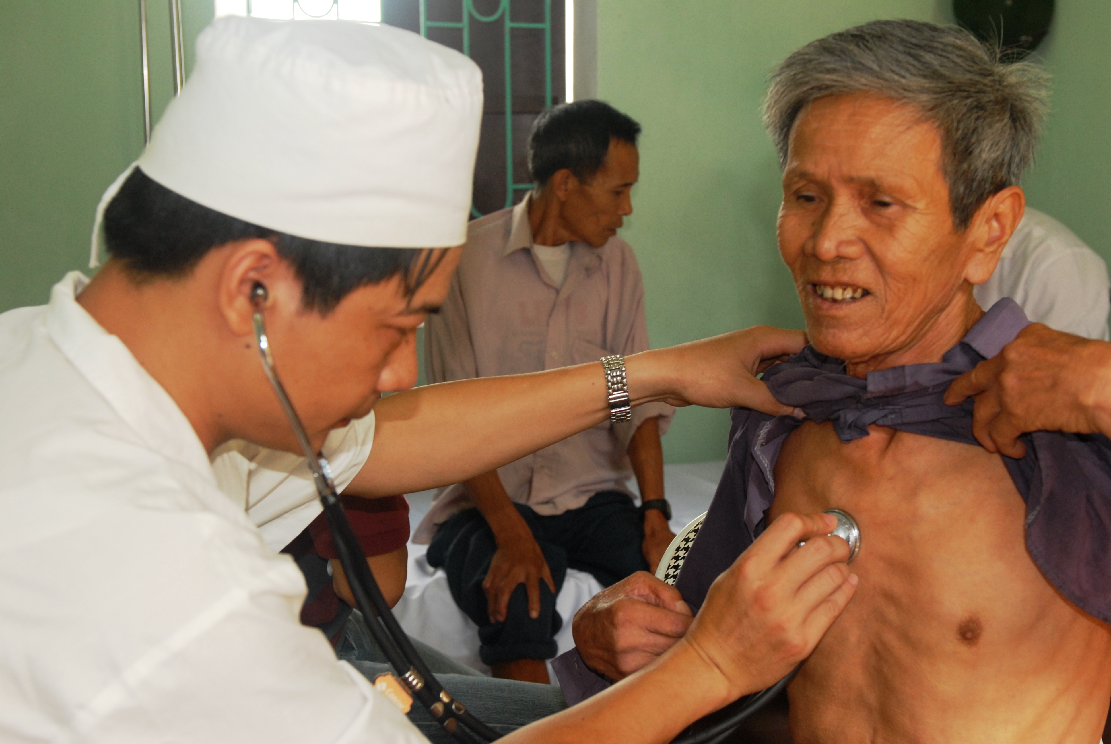
In a physical examination, medical examination, clinical examination, or medical checkup, a medical practitioner examines a patient for any possible medical signs or symptoms of a Disease, medical condition. It generally consists of a series of questions about the patient's medical history followed by an examination based on the reported symptoms. Together, the medical history and the physical examination help to determine a medical diagnosis, diagnosis and devise the treatment plan. These data then become part of the medical record. Types Routine The ''routine physical'', also known as ''general medical examination'', ''periodic health evaluation'', ''annual physical'', ''comprehensive medical exam'', ''general health check'', ''preventive health examination'', ''medical check-up'', or simply ''medical'', is a physical examination performed on an asymptomatic patient for medical screening purposes. These are normally performed by a pediatrician, family practice physician, ... [...More Info...] [...Related Items...] OR: [Wikipedia] [Google] [Baidu] |
Sternal Fracture
A sternal fracture is a bone fracture, fracture of the Human sternum, sternum (the breastbone), located in the center of the chest. The injury, which occurs in 5–8% of people who experience significant blunt chest trauma, may occur in vehicle accidents, when the still-moving chest strikes a steering wheel or dashboard or is injured by a seatbelt. Cardiopulmonary resuscitation (CPR), has also been known to cause thoracic injury, including sternum and rib fractures. Sternal fractures may also occur as a Pathologic fracture, pathological fracture, in people who have weakened bone in their sternum, due to another disease process. Sternal fracture can interfere with breathing by making it more painful; however, its primary significance is that it can indicate the presence of serious associated internal injuries, especially to the heart and lungs. Signs and symptoms Signs and symptoms include crepitus (a crunching sound made when broken bone ends rub together), pain, tenderness (med ... [...More Info...] [...Related Items...] OR: [Wikipedia] [Google] [Baidu] |
Aortic Arch
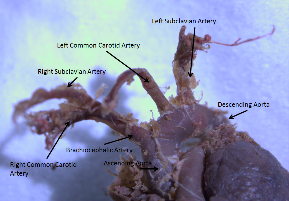
The aortic arch, arch of the aorta, or transverse aortic arch () is the part of the aorta between the ascending and descending aorta. The arch travels backward, so that it ultimately runs to the left of the trachea. Structure The aorta begins at the level of the upper border of the second/third sternocostal articulation of the right side, behind the ventricular outflow tract and pulmonary trunk. The right atrial appendage overlaps it. The first few centimeters of the ascending aorta and pulmonary trunk lies in the same pericardial sheath and runs at first upward, arches over the pulmonary trunk, right pulmonary artery, and right main bronchus to lie behind the right second coastal cartilage. The right lung and sternum lies anterior to the aorta at this point. The aorta then passes posteriorly and to the left, anterior to the trachea, and arches over left main bronchus and left pulmonary artery, and reaches to the left side of the T4 vertebral body. Apart from T4 vertebral ... [...More Info...] [...Related Items...] OR: [Wikipedia] [Google] [Baidu] |
Superior Vena Cava
The superior vena cava (SVC) is the superior of the two venae cavae, the great venous trunks that return deoxygenated blood from the systemic circulation to the right atrium of the heart. It is a large-diameter (24 mm) short length vein that receives venous return from the upper half of the body, above the diaphragm. Venous return from the lower half, below the diaphragm, flows through the inferior vena cava. The SVC is located in the anterior right superior mediastinum. It is the typical site of central venous access via a central venous catheter or a peripherally inserted central catheter. Mentions of "the cava" without further specification usually refer to the SVC. Structure The superior vena cava is formed by the left and right brachiocephalic veins, which receive blood from the upper limbs, head and neck, behind the lower border of the first right costal cartilage. It passes vertically downwards behind the first intercostal space and receives the azygos vei ... [...More Info...] [...Related Items...] OR: [Wikipedia] [Google] [Baidu] |
Azygos Vein
The azygos vein (from Ancient Greek ἄζυγος (ázugos), meaning 'unwedded' or 'unpaired') is a vein running up the right side of the thoracic vertebral column draining itself towards the superior vena cava. It connects the systems of superior vena cava and inferior vena cava and can provide an alternative path for blood to the right atrium when either of the venae cavae is blocked. Structure The azygos vein transports deoxygenated blood from the posterior walls of the thorax and abdomen into the superior vena cava. It is formed by the union of the ascending lumbar veins with the right subcostal veins at the level of the 12th thoracic vertebra, ascending to the right of the descending aorta and thoracic duct, passing behind the right crus of diaphragm, anterior to the vertebral bodies of T12 to T5 and right posterior intercostal arteries. At the level of T4 vertebrae, it arches over the root of the right lung from behind to the front to join the superior vena cava. The tra ... [...More Info...] [...Related Items...] OR: [Wikipedia] [Google] [Baidu] |
Ascending Aorta
The ascending aorta (AAo) is a portion of the aorta commencing at the upper part of the base of the left ventricle, on a level with the lower border of the third costal cartilage behind the left half of the sternum. Structure It passes obliquely upward, forward, and to the right, in the direction of the heart's axis, as high as the upper border of the second right costal cartilage, describing a slight curve in its course, and being situated, about behind the posterior surface of the sternum. The total length is about . Components The aortic root is the portion of the aorta beginning at the aortic annulus and extending to the sinotubular junction. It is sometimes regarded as a part of the ascending aorta, and sometimes regarded as a separate entity from the rest of the ascending aorta. Between each commissure of the aortic valve and opposite the cusps of the aortic valve, three small dilations called the aortic sinuses. The sinotubular junction is the point in the ascending ... [...More Info...] [...Related Items...] OR: [Wikipedia] [Google] [Baidu] |
Fibrous Pericardium
The pericardium (: pericardia), also called pericardial sac, is a double-walled sac containing the heart and the roots of the great vessels. It has two layers, an outer layer made of strong inelastic connective tissue (fibrous pericardium), and an inner layer made of serous membrane (serous pericardium). It encloses the pericardial cavity, which contains pericardial fluid, and defines the middle mediastinum. It separates the heart from interference of other structures, protects it against infection and blunt trauma, and lubricates the heart's movements. The English name originates from the Ancient Greek prefix ''peri-'' (περί) 'around' and the suffix ''-cardion'' (κάρδιον) 'heart'. Anatomy The pericardium is a tough fibroelastic sac which covers the heart from all sides except at the cardiac root (where the great vessels join the heart) and the bottom (where only the serous pericardium exists to cover the upper surface of the central tendon of diaphragm). The ... [...More Info...] [...Related Items...] OR: [Wikipedia] [Google] [Baidu] |


