|
Second Rib
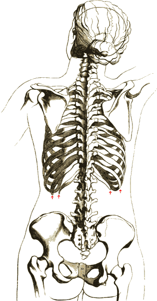
The rib cage or thoracic cage is an endoskeletal enclosure in the thorax of most vertebrates that comprises the ribs, vertebral column and sternum, which protect the vital organs of the thoracic cavity, such as the heart, lungs and great vessels and support the shoulder girdle to form the core part of the axial skeleton. A typical human thoracic cage consists of 12 pairs of ribs and the adjoining costal cartilages, the sternum (along with the manubrium and xiphoid process), and the 12 thoracic vertebrae articulating with the ribs. The thoracic cage also provides attachments for extrinsic skeletal muscles of the neck, upper limbs, upper abdomen and back, and together with the overlying skin and associated fascia and muscles, makes up the thoracic wall. In tetrapods, the rib cage intrinsically holds the muscles of respiration ( diaphragm, intercostal muscles, etc.) that are crucial for active inhalation and forced exhalation, and therefore has a major ventilatory functio ... [...More Info...] [...Related Items...] OR: [Wikipedia] [Google] [Baidu] |
Endoskeletal
An endoskeleton (From Ancient Greek ἔνδον, éndon = "within", "inner" + σκελετός, skeletos = "skeleton") is a structural frame (skeleton) — usually composed of mineralized tissue — on the inside of an animal, overlaid by soft tissues. Endoskeletons serve as structural support against gravity and mechanical loads, and provide anchoring attachment sites for skeletal muscles to transmit force and allow movements and locomotion. Vertebrates and the closely related cephalochordates are the predominant animal clade with endoskeletons (made of mostly bone and sometimes cartilage, as well as notochordal glycoprotein and collagen fibers), although invertebrates such as sponges also have evolved a form of "rebar" endoskeletons made of diffuse meshworks of calcite/silica structural elements called spicules, and echinoderms have a dermal calcite endoskeleton known as ossicles. Some coleoid cephalopods (squids and cuttlefish) have an internalized vestigial aragoni ... [...More Info...] [...Related Items...] OR: [Wikipedia] [Google] [Baidu] |
Thoracic Vertebra
In vertebrates, thoracic vertebrae compose the middle segment of the vertebral column, between the cervical vertebrae and the lumbar vertebrae. In humans, there are twelve thoracic vertebra (anatomy), vertebrae of intermediate size between the cervical and lumbar vertebrae; they increase in size going towards the lumbar vertebrae. They are distinguished by the presence of Zygapophysial joint, facets on the sides of the bodies for Articulation (anatomy), articulation with the head of rib, heads of the ribs, as well as facets on the transverse processes of all, except the eleventh and twelfth, for articulation with the tubercle (rib), tubercles of the ribs. By convention, the human thoracic vertebrae are numbered T1–T12, with the first one (T1) located closest to the skull and the others going down the spine toward the lumbar region. General characteristics These are the general characteristics of the second through eighth thoracic vertebrae. The first and ninth through twelfth v ... [...More Info...] [...Related Items...] OR: [Wikipedia] [Google] [Baidu] |
Intercostal Muscles
The intercostal muscles comprise many different groups of muscles that run between the ribs, and help form and move the chest wall. The intercostal muscles are mainly involved in the mechanical aspect of breathing by helping expand and shrink the size of the chest cavity. Structure There are three principal layers: # External intercostal muscles also known as intercostalis externus aid in quiet and forced inhalation. They originate on ribs 1–11 and have their insertion on ribs 2–12. The external intercostals are responsible for the elevation of the ribs and bending them more open, thus expanding the transverse dimensions of the thoracic cavity. The muscle fibers are directed downwards, forwards and medially in the anterior part. # Internal intercostal muscles also known as intercostalis internus aid in forced expiration (quiet expiration is a passive process). They originate on ribs 2–12 and have their insertions on ribs 1–11.Their fibers pass anterior and superior fro ... [...More Info...] [...Related Items...] OR: [Wikipedia] [Google] [Baidu] |
Thoracic Diaphragm
The thoracic diaphragm, or simply the diaphragm (; ), is a sheet of internal Skeletal striated muscle, skeletal muscle in humans and other mammals that extends across the bottom of the thoracic cavity. The diaphragm is the most important Muscles of respiration, muscle of respiration, and separates the thoracic cavity, containing the heart and lungs, from the abdominal cavity: as the diaphragm contracts, the volume of the thoracic cavity increases, creating a negative pressure there, which draws air into the lungs. Its high oxygen consumption is noted by the many mitochondria and capillaries present; more than in any other skeletal muscle. The term ''diaphragm'' in anatomy, created by Gerard of Cremona, can refer to other flat structures such as the urogenital diaphragm or Pelvic floor, pelvic diaphragm, but "the diaphragm" generally refers to the thoracic diaphragm. In humans, the diaphragm is slightly asymmetric—its right half is higher up (superior) to the left half, since th ... [...More Info...] [...Related Items...] OR: [Wikipedia] [Google] [Baidu] |
Muscles Of Respiration
The muscles of respiration are the muscles that contribute to inhalation and exhalation, by aiding in the expansion and contraction of the thoracic cavity. The diaphragm and, to a lesser extent, the intercostal muscles drive respiration during quiet breathing. The elasticity of these muscles is crucial to the health of the respiratory system and to maximize its functional capabilities. Diaphragm The diaphragm is the major muscle responsible for breathing. It is a thin, dome-shaped muscle that separates the abdominal cavity from the thoracic cavity. During inhalation, the diaphragm contracts, so that its center moves caudally (downward) and its edges move cranially (upward). This compresses the abdominal cavity, raises the ribs upward and outward and thus expands the thoracic cavity. This expansion draws air into the lungs. When the diaphragm relaxes, elastic recoil of the lungs causes the thoracic cavity to contract, forcing air out of the lungs, and returning to its dome-sh ... [...More Info...] [...Related Items...] OR: [Wikipedia] [Google] [Baidu] |
Tetrapod
A tetrapod (; from Ancient Greek :wiktionary:τετρα-#Ancient Greek, τετρα- ''(tetra-)'' 'four' and :wiktionary:πούς#Ancient Greek, πούς ''(poús)'' 'foot') is any four-Limb (anatomy), limbed vertebrate animal of the clade Tetrapoda (). Tetrapods include all Neontology#Extant taxa versus extinct taxa, extant and Extinction, extinct amphibians and amniotes, with the latter in turn Evolution, evolving into two major clades, the Sauropsida, sauropsids (reptiles, including dinosaurs and therefore birds) and synapsids (extinct pelycosaur, "pelycosaurs", therapsids and all extant mammals, including Homo sapiens, humans). Hox gene mutations have resulted in some tetrapods becoming Limbless vertebrate, limbless (snakes, legless lizards, and caecilians) or two-limbed (cetaceans, sirenians, Bipedidae, some lizards, kiwi (bird), kiwis, and the extinct moa and elephant birds). Nevertheless, they still qualify as tetrapods through their ancestry, and some retain a pair of ves ... [...More Info...] [...Related Items...] OR: [Wikipedia] [Google] [Baidu] |
Thoracic Wall
The thoracic wall or chest wall is the boundary of the thoracic cavity. Structure The bony skeletal part of the thoracic wall is the rib cage, and the rest is made up of muscle, skin, and fasciae. The chest wall has 10 layers, namely (from superficial to deep) skin (epidermis and dermis), superficial fascia, deep fascia and the invested extrinsic muscles (from the upper limbs), intrinsic muscles associated with the ribs (three layers of intercostal muscles), endothoracic fascia and parietal pleura. However, the extrinsic muscular layers vary according to the region of the chest wall. For example, the front and back sides may include attachments of large upper limb muscles like pectoralis major or latissimus dorsi, while the sides only have serratus anterior.The thoracic wall consists of a bony framework that is held together by twelve thoracic vertebrae posteriorly which give rise to ribs that encircle the lateral and anterior thoracic cavity. The first nine ribs curve a ... [...More Info...] [...Related Items...] OR: [Wikipedia] [Google] [Baidu] |
Muscle
Muscle is a soft tissue, one of the four basic types of animal tissue. There are three types of muscle tissue in vertebrates: skeletal muscle, cardiac muscle, and smooth muscle. Muscle tissue gives skeletal muscles the ability to muscle contraction, contract. Muscle tissue contains special Muscle contraction, contractile proteins called actin and myosin which interact to cause movement. Among many other muscle proteins, present are two regulatory proteins, troponin and tropomyosin. Muscle is formed during embryonic development, in a process known as myogenesis. Skeletal muscle tissue is striated consisting of elongated, multinucleate muscle cells called muscle fibers, and is responsible for movements of the body. Other tissues in skeletal muscle include tendons and perimysium. Smooth and cardiac muscle contract involuntarily, without conscious intervention. These muscle types may be activated both through the interaction of the central nervous system as well as by innervation ... [...More Info...] [...Related Items...] OR: [Wikipedia] [Google] [Baidu] |
Fascia
A fascia (; : fasciae or fascias; adjective fascial; ) is a generic term for macroscopic membranous bodily structures. Fasciae are classified as superficial, visceral or deep, and further designated according to their anatomical location. The knowledge of fascial structures is essential in surgery, as they create borders for infectious processes (for example Psoas abscess) and haematoma. An increase in pressure may result in a compartment syndrome, where a prompt fasciotomy may be necessary. For this reason, profound descriptions of fascial structures are available in anatomical literature from the 19th century. Function Fasciae were traditionally thought of as passive structures that transmit mechanical tension generated by muscular activities or external forces throughout the body. An important function of muscle fasciae is to reduce friction of muscular force. In doing so, fasciae provide a supportive and movable wrapping for nerves and blood vessels as they pass thro ... [...More Info...] [...Related Items...] OR: [Wikipedia] [Google] [Baidu] |
Skin
Skin is the layer of usually soft, flexible outer tissue covering the body of a vertebrate animal, with three main functions: protection, regulation, and sensation. Other animal coverings, such as the arthropod exoskeleton, have different developmental origin, structure and chemical composition. The adjective cutaneous means "of the skin" (from Latin ''cutis'' 'skin'). In mammals, the skin is an organ of the integumentary system made up of multiple layers of ectodermal tissue and guards the underlying muscles, bones, ligaments, and internal organs. Skin of a different nature exists in amphibians, reptiles, and birds. Skin (including cutaneous and subcutaneous tissues) plays crucial roles in formation, structure, and function of extraskeletal apparatus such as horns of bovids (e.g., cattle) and rhinos, cervids' antlers, giraffids' ossicones, armadillos' osteoderm, and os penis/ os clitoris. All mammals have some hair on their skin, even marine mammals like whales, ... [...More Info...] [...Related Items...] OR: [Wikipedia] [Google] [Baidu] |
Back
The human back, also called the dorsum (: dorsa), is the large posterior area of the human body, rising from the top of the buttocks to the back of the neck. It is the surface of the body opposite from the chest and the abdomen. The vertebral column runs the length of the back and creates a central area of recession. The breadth of the back is created by the shoulders at the top and the pelvis at the bottom. Back pain is a common medical condition, generally benign in origin. Structure The central feature of the human back is the vertebral column, specifically the length from the top of the thoracic vertebrae to the bottom of the lumbar vertebrae, which houses the spinal cord in its spinal canal, and which generally has some curvature that gives shape to the back. The ribcage extends from the spine at the top of the back (with the top of the ribcage corresponding to the T1 vertebra), more than halfway down the length of the back, leaving an area with less protection between th ... [...More Info...] [...Related Items...] OR: [Wikipedia] [Google] [Baidu] |
Upper Abdomen
In anatomy, the epigastrium (or epigastric region) is the upper central region of the abdomen. It is located between the costal margins and the subcostal plane. Pain may be referred to the epigastrium from damage to structures derived from the foregut. Structure The epigastrium is one of the nine regions of the abdomen, along with the right and left hypochondria, right and left lateral regions (lumbar areas or flanks), right and left inguinal regions (or fossae), and the umbilical and pubic regions. It is located between the costal margins and the subcostal plane. During breathing, the diaphragm contracts and flattens, displacing the viscera and producing an outward movement of the upper abdominal wall (epigastric region). It is a convergence of the diaphragm and the abdominals, so that "when both sets of muscles (diaphragm and abdominals) tense, the epigastrium pushes forward". Therefore, the epigastric region is not a muscle nor is it an organ, but it is a zone of activ ... [...More Info...] [...Related Items...] OR: [Wikipedia] [Google] [Baidu] |


