|
Scalp
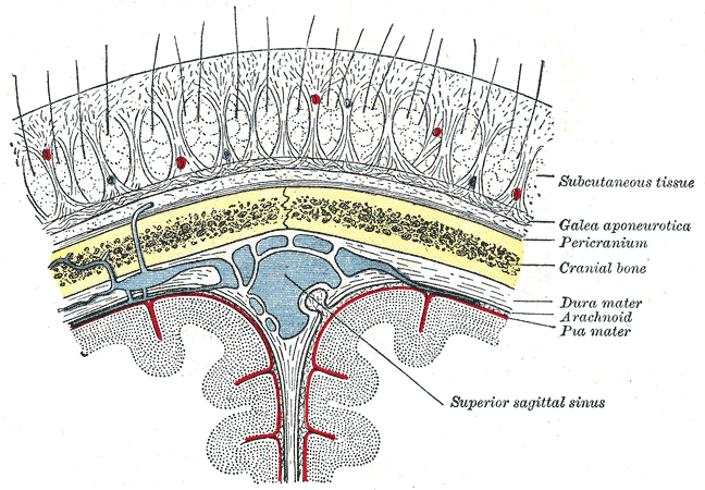
The scalp is the area of the head where head hair grows. It is made up of skin, layers of connective and fibrous tissues, and the membrane of the skull. Anatomically, the scalp is part of the epicranium, a collection of structures covering the cranium. The scalp is bordered by the face at the front, and by the neck at the sides and back. The scientific study of hair and scalp is called trichology. Structure Layers The scalp is usually described as having five layers, which can be remembered using the mnemonic 'SCALP': * S: Skin. The skin of the scalp contains numerous hair follicles and sebaceous glands. * C: Connective tissue. A dense subcutaneous layer of fat and fibrous tissue that lies beneath the skin, containing the nerves and vessels of the scalp. * A: Aponeurosis. The epicranial aponeurosis or galea aponeurotica is a tough layer of dense fibrous tissue which anchors the above layers in place. It runs from the frontalis muscle anteriorly to the occipitalis ... [...More Info...] [...Related Items...] OR: [Wikipedia] [Google] [Baidu] [Amazon] |
Hair Follicle
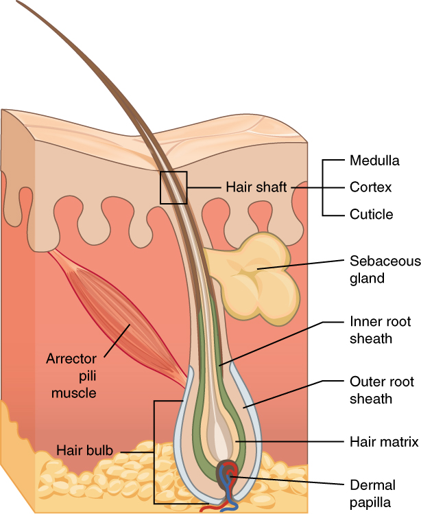
The hair follicle is an organ found in mammalian skin. It resides in the dermal layer of the skin and is made up of 20 different cell types, each with distinct functions. The hair follicle regulates hair growth via a complex interaction between hormones, neuropeptides, and immune cells. This complex interaction induces the hair follicle to produce different types of hair as seen on different parts of the body. For example, terminal hairs grow on the scalp and lanugo hairs are seen covering the bodies of fetuses in the uterus and in some newborn babies. The process of hair growth occurs in distinct sequential stages: ''anagen'' is the active growth phase, ''catagen'' is the regression of the hair follicle phase, ''telogen'' is the resting stage, ''exogen'' is the active shedding of hair phase and ''kenogen'' is the phase between the empty hair follicle and the growth of new hair. The function of hair in humans has long been a subject of interest and continues to be an impor ... [...More Info...] [...Related Items...] OR: [Wikipedia] [Google] [Baidu] [Amazon] |
Occipital Vein
The occipital vein is a vein of the scalp. It originates from a plexus around the external occipital protuberance and superior nuchal line to the back part of the vertex of the skull. It usually drains into the internal jugular vein, but may also drain into the posterior auricular vein (which joins the external jugular vein). It drains part of the scalp. Structure The occipital vein is part of the scalp. It begins as a plexus at the posterior aspect of the scalp from the external occipital protuberance and superior nuchal line to the back part of the vertex of the skull. It pierces the cranial attachment of the trapezius and, dipping into the venous plexus of the suboccipital triangle, joins the deep cervical vein and the vertebral vein. Occasionally it follows the course of the occipital artery, and ends in the internal jugular vein. Alternatively, it joins the posterior auricular vein, and ends in the external jugular vein. The parietal emissary vein connects it with the ... [...More Info...] [...Related Items...] OR: [Wikipedia] [Google] [Baidu] [Amazon] |
Trichology
Trichology is the study of the hair and scalp. The term derives from Ancient Greek θρίξ (''thríx''), "hair" and -λογία ''-logia''. In most jurisdictions the title of a ''trichologist'', not the field of trichology, is considered a para-medical discipline. The Institute of Trichologists was founded in 1902. The first journal for the field, International Journal of Trichology, was founded in 2009. Diseases *Alopecia ** Alopecia areata ** Alopecia universalis ** Frictional alopecia ** Frontal fibrosing alopecia ** Loose anagen syndrome ** Pattern hair loss (''Androgenic alopecia'') **Scarring and non scarring hair loss *Hair colour ** Canities subita ** Premature greying of hair * The hair root & hair follicle disorders ** Telogen effluvium ** Trichostasis spinulosa * Hirsutism ** Trichomegaly *Pathogenic ** Microsporum audouinii ** Piedraia hortae ** Tinea capitis ** Trichobacteriosis axillaris Diagnosis *Blood test ** Vitamin deficiency * Hair analysis **Scalp ... [...More Info...] [...Related Items...] OR: [Wikipedia] [Google] [Baidu] [Amazon] |
Meninges
In anatomy, the meninges (; meninx ; ) are the three membranes that envelop the brain and spinal cord. In mammals, the meninges are the dura mater, the arachnoid mater, and the pia mater. Cerebrospinal fluid is located in the subarachnoid space between the arachnoid mater and the pia mater. The primary function of the meninges is to protect the central nervous system. Structure Dura mater The dura mater (), is a thick, durable membrane, closest to the Human skull, skull and vertebrae. The dura mater, the outermost part, is a loosely arranged, fibroelastic layer of cells, characterized by multiple interdigitating cell processes, no extracellular collagen, and significant extracellular spaces. The middle region is a mostly fibrous portion. It consists of two layers: the endosteal layer, which lies closest to the skull, and the inner meningeal layer, which lies closer to the brain. It contains larger blood vessels that split into the capillaries in the pia mater. It is composed ... [...More Info...] [...Related Items...] OR: [Wikipedia] [Google] [Baidu] [Amazon] |
Mastoid Lymph Nodes
The mastoid lymph nodes (retroauricular lymph nodes or posterior auricular glands) are a small group of lymph nodes, usually two in number, located just beneath the ear, on the mastoid insertion of the sternocleidomastoideus muscle, beneath the posterior auricular muscle. Their mastoid lymph nodes receives lymph from the posterior part of the temporoparietal region, the upper part of the cranial surface of the visible ear and the back of the ear canal. The lymph then passes to the superior deep cervical glands. Etymology The word ''mastoid'' comes from the (, "mouth, jaws, that with which one chews"). References External links Diagramat Baylor College of Medicine The Baylor College of Medicine (BCM) is a private medical school in Houston, Texas, United States. Originally as the Baylor University College of Medicine from 1903 to 1969, the college became independent with the current name and has been se ... (listed as "retroauricular") {{Portal bar, Anatomy Ly ... [...More Info...] [...Related Items...] OR: [Wikipedia] [Google] [Baidu] [Amazon] |
Occipital Lymph Nodes
The occipital lymph nodes, one to three in number, are located on the back of the head close to the margin of the trapezius and resting on the insertion of the . Their afferent vessels drain the occipital region of the scalp The scalp is the area of the head where head hair grows. It is made up of skin, layers of connective and fibrous tissues, and the membrane of the skull. Anatomically, the scalp is part of the epicranium, a collection of structures covering th ..., while their efferents pass to the superior deep cervical glands. Additional images File:Lymph node regions.svg Etymology The word ''occipital'' comes from the ("the back of the head"). References External links * () http://www.patient.info* http://www.emedicine.com/ent/topic306.htm#section~anatomy_of_the_cervical_lymphatics Lymphatics of the head and neck {{lymphatic-stub ... [...More Info...] [...Related Items...] OR: [Wikipedia] [Google] [Baidu] [Amazon] |
Head Hair
Hair is a protein filament that grows from follicles found in the dermis. Hair is one of the defining characteristics of mammals. The human body, apart from areas of glabrous skin, is covered in follicles which produce thick terminal and fine vellus hair. Most common interest in hair is focused on hair growth, hair types, and hair care, but hair is also an important biomaterial primarily composed of protein, notably alpha-keratin. Attitudes towards different forms of hair, such as hairstyles and hair removal, vary widely across different cultures and historical periods, but it is often used to indicate a person's personal beliefs or social position, such as their age, gender, or religion. Overview Meaning The word "hair" usually refers to two distinct structures: #the part beneath the skin, called the hair follicle, or, when pulled from the skin, the bulb or root. This organ is located in the dermis and maintains stem cells, which not only re-grow the hair after it falls o ... [...More Info...] [...Related Items...] OR: [Wikipedia] [Google] [Baidu] [Amazon] |
Posterior Auricular Vein
The posterior auricular vein is a vein of the head. It begins from a plexus with the occipital vein and the superficial temporal vein, descends behind the auricle, and drains into the external jugular vein. Structure The posterior auricular vein begins upon the side of the head, in a plexus which communicates with the tributaries of the occipital vein and the superficial temporal vein. It descends behind the auricle. It joins the posterior division of the retromandibular vein. It drains into the external jugular vein. It receive the stylomastoid vein, and some tributaries from the cranial surface of the auricle. Variation The posterior auricular vein may drain into the internal jugular vein The internal jugular vein is a paired jugular vein that collects blood from the brain and the superficial parts of the face and neck. This vein runs in the carotid sheath with the common carotid artery and vagus nerve. It begins in the posteri ... or a posterior jugular v ... [...More Info...] [...Related Items...] OR: [Wikipedia] [Google] [Baidu] [Amazon] |
Neck
The neck is the part of the body in many vertebrates that connects the head to the torso. It supports the weight of the head and protects the nerves that transmit sensory and motor information between the brain and the rest of the body. Additionally, the neck is highly flexible, allowing the head to turn and move in all directions. Anatomically, the human neck is divided into four compartments: vertebral, visceral, and two vascular compartments. Within these compartments, the neck houses the cervical vertebrae, the cervical portion of the spinal cord, upper parts of the respiratory and digestive tracts, endocrine glands, nerves, arteries and veins. The muscles of the neck, which are separate from the compartments, form the boundaries of the neck triangles. In anatomy, the neck is also referred to as the or . However, when the term ''cervix'' is used alone, it often refers to the uterine cervix, the neck of the uterus. Therefore, the adjective ''cervical'' ... [...More Info...] [...Related Items...] OR: [Wikipedia] [Google] [Baidu] [Amazon] |
Occipitalis
The occipitalis muscle (occipital belly) is a muscle which covers parts of the skull. Some sources consider the occipital muscle to be a distinct muscle. However, Terminologia Anatomica currently classifies it as part of the occipitofrontalis muscle along with the frontalis muscle. The occipitalis muscle is thin and quadrilateral in form. It arises from tendinous fibers from the lateral two-thirds of the superior nuchal line of the occipital bone and from the mastoid process of the temporal and ends in the epicranial aponeurosis. The occipitalis muscle is innervated by the posterior auricular nerve (a branch of the facial nerve) and its function is to move the scalp back. The muscles receives blood from the occipital artery. Additional image File:Occipitalis muscle animation small.gif, Position of occipitalis muscle (shown in red). See also * Occipitofrontalis muscle The occipitofrontalis muscle (epicranius muscle) is a muscle which covers parts of the skull. It consists o ... [...More Info...] [...Related Items...] OR: [Wikipedia] [Google] [Baidu] [Amazon] |
Frontalis Muscle
The frontalis muscle () is a muscle which covers parts of the forehead of the skull. Some sources consider the frontalis muscle to be a distinct muscle. However, Terminologia Anatomica currently classifies it as part of the occipitofrontalis muscle along with the occipitalis muscle. In humans, the frontalis muscle only serves for facial expressions. The frontalis muscle is supplied by the facial nerve and receives blood from the supraorbital and supratrochlear arteries. Structure The frontalis muscle is thin, of a quadrilateral form, and intimately adherent to the superficial fascia. It is broader than the occipitalis and its fibers are longer and paler in color. It is located on the front of the head. The muscle has no bony attachments. Its medial fibers are continuous with those of the procerus; its intermediate fibers blend with the corrugator and orbicularis oculi muscles, thus attached to the skin of the eyebrows; and its lateral fibers are also blended with the latte ... [...More Info...] [...Related Items...] OR: [Wikipedia] [Google] [Baidu] [Amazon] |
Galea Aponeurotica
The epicranial aponeurosis (aponeurosis epicranialis, galea aponeurotica) is an aponeurosis (a tough layer of dense fibrous tissue). It covers the upper part of the skull in humans and many other animals. Structure In humans, the epicranial aponeurosis originates from the external occipital protuberance and highest nuchal lines of the occipital bone. It merges with the occipitofrontalis muscle. In front, it forms a short and narrow prolongation between its union with the frontalis muscle (the frontal part of the occipitofrontalis muscle). On either side, the epicranial aponeurosis attaches to the anterior auricular muscles and the superior auricular muscles. Here it is less aponeurotic, and is continued over the temporal fascia to the zygomatic arch as a layer of laminated areolar tissue. It is closely connected to the integument by the firm, dense, fibro-fatty layer which forms the superficial fascia of the scalp. It is attached to the pericranium by loose cellular tissue ... [...More Info...] [...Related Items...] OR: [Wikipedia] [Google] [Baidu] [Amazon] |


