|
Right Gastroepiploic Artery
The right gastroepiploic artery (or right gastro-omental artery) is one of the two terminal branches of the gastroduodenal artery. It runs from right to left along the greater curvature of the stomach, between the layers of the greater omentum, anastomosing with the left gastroepiploic artery, a branch of the splenic artery. Except at the pylorus where it is in contact with the stomach, it lies about a finger's breadth from the greater curvature. Branches This vessel gives off numerous branches: * "gastric branches": ascend to supply both surfaces of the stomach. * "omental branches": descend to supply the greater omentum and anastomose with branches of the middle colic. Use in coronary artery surgery The right gastroepiploic artery was first used as a coronary artery bypass graft (CABG) in 1984 by John Pym and colleagues at Queen's University. It has become an accepted alternative conduit, and is particularly useful in patients who do not have suitable saphenous veins to har ... [...More Info...] [...Related Items...] OR: [Wikipedia] [Google] [Baidu] |
Gastroduodenal Artery
In anatomy, the gastroduodenal artery is a small blood vessel in the abdomen. It supplies blood directly to the pylorus (distal part of the stomach) and proximal part of the duodenum. It also indirectly supplies the pancreatic head (via the anterior and posterior superior pancreaticoduodenal arteries). Structure The gastroduodenal artery most commonly arises from either the left hepatic artery or the right hepatic artery instead. It may also arise from the common hepatic artery of the coeliac trunk in a trifork arrangement with the two other arteries, but there are numerous variations of the origin.Bergman RA, Afifi AK, Miyauchi R. Variations in Origin of Gastroduodenal Artery. from Anatomy Atlases. (http://www.anatomyatlases.org/AnatomicVariants/Cardiovascular/Images0001/0017.shtml) It first gives rise to the supraduodenal artery, followed by the posterior superior pancreaticoduodenal artery. It terminates in a bifurcation when it splits into the right gastroepiploic artery a ... [...More Info...] [...Related Items...] OR: [Wikipedia] [Google] [Baidu] |
Pylorus
The pylorus ( or ), or pyloric part, connects the stomach to the duodenum. The pylorus is considered as having two parts, the ''pyloric antrum'' (opening to the body of the stomach) and the ''pyloric canal'' (opening to the duodenum). The ''pyloric canal'' ends as the ''pyloric orifice'', which marks the junction between the stomach and the duodenum. The orifice is surrounded by a sphincter, a band of muscle, called the ''pyloric sphincter''. The word ''pylorus'' comes from Greek πυλωρός, via Latin. The word ''pylorus'' in Greek means "gatekeeper", related to "gate" ( el, pyle) and is thus linguistically related to the word " pylon". Structure The pylorus is the furthest part of the stomach that connects to the duodenum. It is divided into two parts, the ''antrum'', which connects to the body of the stomach, and the ''pyloric canal'', which connects to the duodenum. Antrum The ''pyloric antrum'' is the initial portion of the pylorus. It is near the bottom of the stomach, ... [...More Info...] [...Related Items...] OR: [Wikipedia] [Google] [Baidu] |
Posterior Interventricular Artery
In the coronary circulation, the posterior interventricular artery (PIV, PIA, or PIVA), most often called the posterior descending artery (PDA), is an artery running in the posterior interventricular sulcus to the apex of the heart where it meets with the anterior interventricular artery or also known as Left Anterior Descending artery. It supplies the posterior third of the interventricular septum. The remaining anterior two-thirds is supplied by the anterior interventricular artery which is a septal branch of the left anterior descending artery, which is a branch of left coronary artery. It is typically a branch of the right coronary artery (70%, known as right dominance). Alternately, the PIV can be a branch of the circumflex coronary artery The circumflex branch of left coronary artery, or left circumflex artery or circumflex artery, is a branch of the left coronary artery. Description The left circumflex artery follows the left part of the coronary sulcus, running firs ... [...More Info...] [...Related Items...] OR: [Wikipedia] [Google] [Baidu] |
Right Coronary Artery
In the blood supply of the heart, the right coronary artery (RCA) is an artery originating above the right cusp of the aortic valve, at the right aortic sinus in the heart. It travels down the right coronary sulcus, towards the crux of the heart. It supplies the right side of the heart, and the interventricular septum. Structure The right coronary artery originates above the right aortic sinus above the aortic valve. It passes through the right coronary sulcus (right atrioventricular groove), towards the crux of the heart. It gives off many branches, including the posterior interventricular artery, the right marginal artery, the conus artery, and the sinoatrial nodal artery. Segments * Proximal: starting at RCA origin, spanning half the distance to the acute margin * Middle: from proximal segment to the acute margin * Distal: from middle segment to origination point of the posterior interventricular artery, where the posterior interventricular sulcus meets the atrioven ... [...More Info...] [...Related Items...] OR: [Wikipedia] [Google] [Baidu] |
Great Saphenous Vein
The great saphenous vein (GSV, alternately "long saphenous vein"; ) is a large, subcutaneous, superficial vein of the leg. It is the longest vein in the body, running along the length of the lower limb, returning blood from the foot, leg and thigh to the deep femoral vein at the femoral triangle. Structure The great saphenous vein originates from where the dorsal vein of the big toe (the hallux) merges with the dorsal venous arch of the foot. After passing in front of the medial malleolus (where it often can be visualized and palpated), it runs up the medial side of the leg. At the knee, it runs over the posterior border of the medial epicondyle of the femur bone. In the proximal anterior thigh inferolateral to the pubic tubercle, the great saphenous vein dives down deep through the cribriform fascia of the saphenous opening to join the femoral vein. It forms an arch, the saphenous arch, to join the common femoral vein in the region of the femoral triangle at the sapheno-femoral ... [...More Info...] [...Related Items...] OR: [Wikipedia] [Google] [Baidu] |
Queen's University At Kingston
Queen's University at Kingston, commonly known as Queen's University or simply Queen's, is a public research university in Kingston, Ontario, Canada. Queen's holds more than of land throughout Ontario and owns Herstmonceux Castle in East Sussex, England. Queen's is organized into eight faculties and schools. The Church of Scotland established Queen's College in October 1841 via a royal charter from Queen Victoria. The first classes, intended to prepare students for the ministry, were held 7 March 1842 with 13 students and two professors. In 1869, Queen's was the first Canadian university west of the Maritime provinces to admit women. In 1883, a women's college for medical education affiliated with Queen's University was established after male staff and students reacted with hostility to the admission of women to the university's medical classes. In 1912, Queen's ended its affiliation with the Presbyterian Church, and adopted its present name. During the mid-20th century, the u ... [...More Info...] [...Related Items...] OR: [Wikipedia] [Google] [Baidu] |
Coronary Artery Bypass Graft
Coronary artery bypass surgery, also known as coronary artery bypass graft (CABG, pronounced "cabbage") is a surgical procedure to treat coronary artery disease (CAD), the buildup of plaques in the arteries of the heart. It can relieve chest pain caused by CAD, slow the progression of CAD, and increase life expectancy. It aims to bypass narrowings in heart arteries by using arteries or veins harvested from other parts of the body, thus restoring adequate blood supply to the previously ischemic (deprived of blood) heart. There are two main approaches. The first uses a cardiopulmonary bypass machine, a machine which takes over the functions of the heart and lungs during surgery by circulating blood and oxygen. With the heart in arrest, harvested arteries and veins are used to connect across problematic regions—a construction known as surgical anastomosis. In the second approach, called the off-pump coronary artery bypass graft (OPCABG), these anastomoses are constructed while t ... [...More Info...] [...Related Items...] OR: [Wikipedia] [Google] [Baidu] |
Middle Colic
The middle colic artery is an artery of the abdomen; a branch of the superior mesenteric artery distributed to parts of the ascending and transverse colon. It usually divides into two terminal branches - a left one and a right one - which go on to form anastomoses with the left colic artery, and right colic artery (respectively), thus participating in the formation of the marginal artery of the colon. Parts of the artery may be removed in different types of hemicolectomy. Structure The middle colic artery supplies the superior/distal part of the ascending colon and right/proximal two-thirds of the transverse colon. Origin The middle colic artery is a branch of the superior mesenteric artery, branching off from its right aspect. Its origin is situated just inferior the neck of the pancreas. It may share a common origin with the right colic artery. Course The middle colic artery passes anterosuperiorly between the layers of the transverse mesocolon just right of the midlin ... [...More Info...] [...Related Items...] OR: [Wikipedia] [Google] [Baidu] |
Stomach Blood Supply
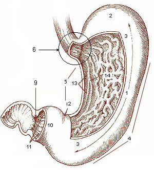
The stomach is a muscular, hollow organ in the gastrointestinal tract of humans and many other animals, including several invertebrates. The stomach has a dilated structure and functions as a vital organ in the digestive system. The stomach is involved in the gastric phase of digestion, following chewing. It performs a chemical breakdown by means of enzymes and hydrochloric acid. In humans and many other animals, the stomach is located between the oesophagus and the small intestine. The stomach secretes digestive enzymes and gastric acid to aid in food digestion. The pyloric sphincter controls the passage of partially digested food (chyme) from the stomach into the duodenum, where peristalsis takes over to move this through the rest of intestines. Structure In the human digestive system, the stomach lies between the oesophagus and the duodenum (the first part of the small intestine). It is in the left upper quadrant of the abdominal cavity. The top of the stomach lies against ... [...More Info...] [...Related Items...] OR: [Wikipedia] [Google] [Baidu] |
Right Gastroepiploic Vein
The right gastroepiploic vein (right gastroomental vein) is a blood vessel that drains blood from the greater curvature and left part of the body of the stomach into the superior mesenteric vein. It runs from left to right along the greater curvature of the stomach between the two layers of the greater omentum, along with the right gastroepiploic artery. As a tributary of the superior mesenteric vein, it is a part of the hepatic portal system In human anatomy, the hepatic portal system is the system of veins comprising the hepatic portal vein and its tributaries. It is also called the portal venous system (although it is not the only example of a portal venous system) and splanchnic .... References Veins of the torso Stomach {{cardiovascular-stub ... [...More Info...] [...Related Items...] OR: [Wikipedia] [Google] [Baidu] |
Splenic Artery
In human anatomy, the splenic artery or lienal artery is the blood vessel that supplies oxygenated blood to the spleen. It branches from the celiac artery, and follows a course superior to the pancreas. It is known for its tortuous path to the spleen. Structure The splenic artery gives off branches to the stomach and pancreas before reaching the spleen. Note that the branches of the splenic artery do not reach all the way to the lower part of the greater curvature of the stomach. Instead, that region is supplied by the right gastroepiploic artery, a branch of the gastroduodenal artery. The two gastroepiploic arteries anastomose with each other at that point. Relations The splenic artery passes between the layers of the lienorenal ligament. Along its course, it is accompanied by a similarly named vein, the splenic vein, which drains into the hepatic portal vein. Clinical significance Splenic artery aneurysms are rare, but still the third most common abdominal aneurysm, afte ... [...More Info...] [...Related Items...] OR: [Wikipedia] [Google] [Baidu] |
Left Gastroepiploic Artery
The left gastroepiploic artery (or left gastro-omental artery), the largest branch of the splenic artery, runs from left to right about a finger's breadth or more from the greater curvature of the stomach, between the layers of the greater omentum, and anastomoses with the right gastroepiploic (a branch of the right gastro-duodenal artery originating from the hepatic branch of the coeliac trunk). In its course it distributes: * "Gastric branches": several ascending branches to both surfaces of the stomach; * "Omental branches": descend to supply the greater omentum and anastomose with branches of the middle colic. Additional images File:Gray533.png, Branches of the celiac artery The celiac () artery (also spelled ''coeliac''), also known as the celiac trunk or truncus coeliacus, is the first major branch of the abdominal aorta. It is about 1.25 cm in length. Branching from the aorta at thoracic vertebra 12 (T12) in .... References External links * - "Stomach, Sp ... [...More Info...] [...Related Items...] OR: [Wikipedia] [Google] [Baidu] |


