|
Retroperitoneum
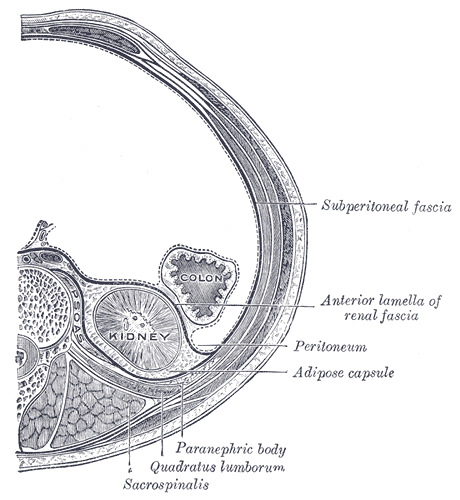
The retroperitoneal space (retroperitoneum) is the anatomical space (sometimes a potential space) behind (''retro'') the peritoneum. It has no specific delineating anatomical structures. Organs are retroperitoneal if they have peritoneum on their anterior side only. Structures that are not suspended by mesentery in the abdominal cavity and that lie between the parietal peritoneum and abdominal wall are classified as retroperitoneal. This is different from organs that are not retroperitoneal, which have peritoneum on their posterior side and are suspended by mesentery in the abdominal cavity. The retroperitoneum can be further subdivided into the following: *Perirenal (or perinephric) space *Anterior pararenal (or paranephric) space *Posterior pararenal (or paranephric) space Retroperitoneal structures Structures that lie behind the peritoneum are termed "retroperitoneal". Organs that were once suspended within the abdominal cavity by mesentery but migrated posterior to the per ... [...More Info...] [...Related Items...] OR: [Wikipedia] [Google] [Baidu] |
Mesentery
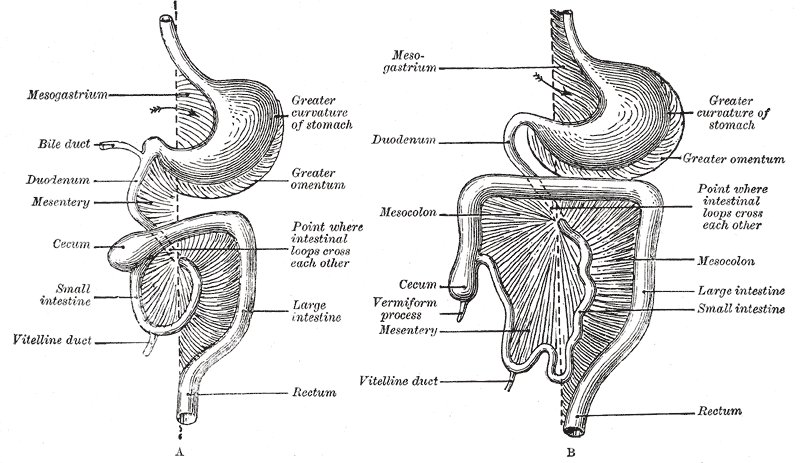
In human anatomy, the mesentery is an Organ (anatomy), organ that attaches the intestines to the posterior abdominal wall, consisting of a double fold of the peritoneum. It helps (among other functions) in storing Adipose tissue, fat and allowing blood vessels, lymphatics, and nerves to supply the intestines. The (the part of the mesentery that attaches the colon to the abdominal wall) was formerly thought to be a fragmented structure, with all named parts—the ascending, transverse, descending, and sigmoid mesocolons, the mesoappendix, and the mesorectum—separately terminating their insertion into the posterior abdominal wall. However, in 2012, new microscopy, microscopic and electron microscope, electron microscopic histology, examinations showed the mesocolon to be a single structure derived from the duodenojejunal flexure and extending to the distal mesorectal layer. Thus the mesentery is an internal organ. Structure The mesentery of the small intestine arises from th ... [...More Info...] [...Related Items...] OR: [Wikipedia] [Google] [Baidu] |
Perirenal Fat
The retroperitoneal space (retroperitoneum) is the anatomical space (sometimes a potential space) behind (''retro'') the peritoneum. It has no specific delineating anatomical structures. Organs are retroperitoneal if they have peritoneum on their anterior side only. Structures that are not suspended by mesentery in the abdominal cavity and that lie between the parietal peritoneum and abdominal wall are classified as retroperitoneal. This is different from organs that are not retroperitoneal, which have peritoneum on their posterior side and are suspended by mesentery in the abdominal cavity. The retroperitoneum can be further subdivided into the following: *Perirenal (or perinephric) space *Anterior pararenal (or paranephric) space *Posterior pararenal (or paranephric) space Retroperitoneal structures Structures that lie behind the peritoneum are termed "retroperitoneal". Organs that were once suspended within the abdominal cavity by mesentery but migrated posterior to the peri ... [...More Info...] [...Related Items...] OR: [Wikipedia] [Google] [Baidu] |
Retroperitoneal Fibrosis
Retroperitoneal fibrosis or Ormond's disease is a disease featuring the proliferation of fibrous tissue (fibrosis) in the retroperitoneum, the compartment of the body containing the kidneys, aorta, renal tract, and various other structures. It may present with lower back pain, kidney failure, hypertension, deep vein thrombosis, and other obstructive symptoms. It is named after John Kelso Ormond, who rediscovered the condition in 1948. Causes The association of idiopathic retroperitoneal fibrosis with various immune-related conditions and response to immunosuppression led to a search for an autoimmune cause of idiopathic RPF. Many of these previously idiopathic cases can now be attributed to IgG4-related disease, an autoimmune disorder proposed in 2003. Otherwise, one-third of cases are secondary to malignancy, medication ( methysergide, hydralazine, beta blockers), prior radiotherapy, or certain infections. However, emerging evidence suggests that occupational exposure to asbe ... [...More Info...] [...Related Items...] OR: [Wikipedia] [Google] [Baidu] |
Retroperitoneal Lymph Node Dissection
Retroperitoneal lymph node dissection (RPLND) is a surgical procedure to remove abdominal lymph nodes. It is used to treat testicular cancer, as well as to help establish the exact stage and type of the cancer. Indications Testicular cancer metastasizes in a predictable pattern, and lymph nodes in the retroperitoneum are typically the first place it lands. By examining the removed lymphatic tissue, a pathologist can determine whether the disease has spread. If no malignant tissue is found, the cancer can be labeled Stage I, limited to the testicle. The procedure is common in the treatment of Stage I and II non-seminomatous germ cell tumors. In seminomas, another form of testicular cancer, radiation therapy is generally preferred to the invasive RPLND procedure. Whether RPLND is needed after orchiectomy depends on the type of tumor and its stage. RPLND may be performed to remove tumor remnants that persist after chemotherapy, because these remnants might otherwise spread and ... [...More Info...] [...Related Items...] OR: [Wikipedia] [Google] [Baidu] |
Adrenal Gland
The adrenal glands (also known as suprarenal glands) are endocrine glands that produce a variety of hormones including adrenaline and the steroids aldosterone and cortisol. They are found above the kidneys. Each gland has an outer adrenal cortex, cortex which produces steroid hormones and an inner Adrenal medulla, medulla. The adrenal cortex itself is divided into three main zones: the zona glomerulosa, the zona fasciculata and the zona reticularis. The adrenal cortex produces three main types of steroid hormones: mineralocorticoids, glucocorticoids, and androgens. Mineralocorticoids (such as aldosterone) produced in the zona glomerulosa help in the regulation of blood pressure and osmoregulation, electrolyte balance. The glucocorticoids cortisol and cortisone are synthesized in the zona fasciculata; their functions include the regulation of metabolism and immune system suppression. The innermost layer of the cortex, the zona reticularis, produces androgens that are converted to ... [...More Info...] [...Related Items...] OR: [Wikipedia] [Google] [Baidu] |
Colon (anatomy)
The large intestine, also known as the large bowel, is the last part of the gastrointestinal tract and of the digestive system in tetrapods. Water is absorbed here and the remaining waste material is stored in the rectum as feces before being removed by defecation. The colon (progressing from the ascending colon to the transverse, the descending and finally the sigmoid colon) is the longest portion of the large intestine, and the terms "large intestine" and "colon" are often used interchangeably, but most sources define the large intestine as the combination of the cecum, colon, rectum, and anal canal. Some other sources exclude the anal canal. In humans, the large intestine begins in the right iliac region of the pelvis, just at or below the waist, where it is joined to the end of the small intestine at the cecum, via the ileocecal valve. It then continues as the colon ascending the abdomen, across the width of the abdominal cavity as the transverse colon, and then desce ... [...More Info...] [...Related Items...] OR: [Wikipedia] [Google] [Baidu] |
Adrenal Gland
The adrenal glands (also known as suprarenal glands) are endocrine glands that produce a variety of hormones including adrenaline and the steroids aldosterone and cortisol. They are found above the kidneys. Each gland has an outer adrenal cortex, cortex which produces steroid hormones and an inner Adrenal medulla, medulla. The adrenal cortex itself is divided into three main zones: the zona glomerulosa, the zona fasciculata and the zona reticularis. The adrenal cortex produces three main types of steroid hormones: mineralocorticoids, glucocorticoids, and androgens. Mineralocorticoids (such as aldosterone) produced in the zona glomerulosa help in the regulation of blood pressure and osmoregulation, electrolyte balance. The glucocorticoids cortisol and cortisone are synthesized in the zona fasciculata; their functions include the regulation of metabolism and immune system suppression. The innermost layer of the cortex, the zona reticularis, produces androgens that are converted to ... [...More Info...] [...Related Items...] OR: [Wikipedia] [Google] [Baidu] |
Horizontal Plane
Horizontal may refer to: *Horizontal plane, in astronomy, geography, geometry and other sciences and contexts *Horizontal coordinate system, in astronomy *Horizontalism, in monetary circuit theory *Horizontalidad, Horizontalism, in sociology *Horizontal market, in microeconomics *Horizontal (album), ''Horizontal'' (album), a 1968 album by the Bee Gees **Horizontal (song), "Horizontal" (song)" is a 1968 song by the Bee Gees See also *Horizontal and vertical *Horizontal and vertical (other) *Horizontal fissure (other), anatomical features *Horizontal bar, an apparatus used by male gymnasts in artistic gymnastics *Vertical (other) * {{disambiguation ... [...More Info...] [...Related Items...] OR: [Wikipedia] [Google] [Baidu] |
Pancreas
The pancreas (plural pancreases, or pancreata) is an Organ (anatomy), organ of the Digestion, digestive system and endocrine system of vertebrates. In humans, it is located in the abdominal cavity, abdomen behind the stomach and functions as a gland. The pancreas is a mixed or heterocrine gland, i.e., it has both an endocrine and a digestive exocrine function. Ninety-nine percent of the pancreas is exocrine and 1% is endocrine. As an endocrine gland, it functions mostly to regulate blood sugar levels, secreting the hormones insulin, glucagon, somatostatin and pancreatic polypeptide. As a part of the digestive system, it functions as an exocrine gland secreting pancreatic juice into the duodenum through the pancreatic duct. This juice contains bicarbonate, which neutralizes acid entering the duodenum from the stomach; and digestive enzymes, which break down carbohydrates, proteins and lipids, fats in food entering the duodenum from the stomach. Inflammation of the pancreas is kno ... [...More Info...] [...Related Items...] OR: [Wikipedia] [Google] [Baidu] |
Renal Blood Flow
In renal physiology, renal blood flow (RBF) is the volume of blood delivered to the kidneys per unit time. In humans, the kidneys together receive roughly 20 - 25% of cardiac output, amounting to 1.2 - 1.3 L/min in a healthy adult. It passes about 94% to the cortex. RBF is closely related to renal plasma flow (RPF), which is the volume of blood plasma delivered to the kidneys per unit time. While the terms generally apply to arterial blood delivered to the kidneys, both RBF and RPF can be used to quantify the volume of venous blood exiting the kidneys per unit time. In this context, the terms are commonly given subscripts to refer to arterial or venous blood or plasma flow, as in RBFa, RBFv, RPFa, and RPFv. Physiologically, however, the differences in these values are negligible so that arterial flow and venous flow are often assumed equal. Renal plasma flow Renal plasma flow is the volume of plasma that reaches the kidneys per unit time. Renal plasma flow is given by the F ... [...More Info...] [...Related Items...] OR: [Wikipedia] [Google] [Baidu] |
Iliopsoas Muscle
The iliopsoas muscle (; ) refers to the joined psoas major muscle, psoas major and the iliacus muscles. The two muscles are separate in the abdomen, but usually merge in the thigh. They are usually given the common name ''iliopsoas''. The iliopsoas muscle joins to the femur at the lesser trochanter. It acts as the strongest :wikt:flexion, flexor of the hip. The iliopsoas muscle is supplied by the lumbar spinal nerves Lumbar spinal nerve 1, L1–Lumbar spinal nerve 3, L3 (psoas) and parts of the femoral nerve (iliacus). Structure The iliopsoas muscle is a composite muscle formed from the psoas major muscle, and the iliacus muscle. The psoas major originates along the outer surfaces of the Body of vertebra, vertebral bodies of Thoracic spinal nerve 12, T12 and Lumbar spinal nerve 1, L1–Lumbar spinal nerve 3, L3 and their associated intervertebral discs. The iliacus originates in the iliac fossa of the pelvis. The psoas major unites with the iliacus at the level of the ... [...More Info...] [...Related Items...] OR: [Wikipedia] [Google] [Baidu] |
Duodenum
The duodenum is the first section of the small intestine in most vertebrates, including mammals, reptiles, and birds. In mammals, it may be the principal site for iron absorption. The duodenum precedes the jejunum and ileum and is the shortest part of the small intestine. In humans, the duodenum is a hollow jointed tube about long connecting the stomach to the jejunum, the middle part of the small intestine. It begins with the duodenal bulb, and ends at the duodenojejunal flexure marked by the suspensory muscle of duodenum. The duodenum can be divided into four parts: the first (superior), the second (descending), the third (transverse) and the fourth (ascending) parts. Overview The duodenum is the first section of the small intestine in most higher vertebrates, including mammals, reptiles, and birds. In fish, the divisions of the small intestine are not as clear, and the terms ''anterior intestine'' or ''proximal intestine'' may be used instead of duodenum. In mammals the d ... [...More Info...] [...Related Items...] OR: [Wikipedia] [Google] [Baidu] |


