|
Resting State FMRI
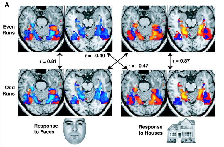
Resting state fMRI (rs-fMRI or R-fMRI), also referred to as task-independent fMRI or task-free fMRI, is a method of functional magnetic resonance imaging (fMRI) that is used in brain mapping to evaluate regional interactions that occur in a resting or task-negative state, when an explicit task is not being performed. A number of resting-state brain networks have been identified, one of which is the default mode network. These brain networks are observed through changes in Cerebral blood flow, blood flow in the brain which creates what is referred to as a blood-oxygen-level dependent (BOLD) signal that can be measured using fMRI. Because brain function, brain activity is intrinsic, present even in the absence of an externally prompted task, any brain region will have spontaneous fluctuations in BOLD signal. The resting state approach is useful to explore the brain's functional organization and to examine if it is altered in Neurological disorder, neurological or mental disorders. ... [...More Info...] [...Related Items...] OR: [Wikipedia] [Google] [Baidu] |
In Vivo
Studies that are ''in vivo'' (Latin for "within the living"; often not italicized in English) are those in which the effects of various biological entities are tested on whole, living organisms or cells, usually animals, including humans, and plants, as opposed to a tissue extract or dead organism. Examples of investigations ''in vivo'' include: the pathogenesis of disease by comparing the effects of bacterial infection with the effects of purified bacterial toxins; the development of non-antibiotics, antiviral drugs, and new drugs generally; and new surgical procedures. Consequently, animal testing and clinical trials are major elements of ''in vivo'' research. ''In vivo'' testing is often employed over ''in vitro'' because it is better suited for observing the overall effects of an experiment on a living subject. In drug discovery, for example, verification of efficacy ''in vivo'' is crucial, because ''in vitro'' assays can sometimes yield misleading results with drug c ... [...More Info...] [...Related Items...] OR: [Wikipedia] [Google] [Baidu] |
Dynamic Functional Connectivity
Dynamic functional connectivity (DFC) refers to the observed phenomenon that functional connectivity changes over a short time. Dynamic functional connectivity is a recent expansion on traditional functional connectivity analysis which typically assumes that functional networks are static in time. DFC is related to a variety of different neurological disorders, and has been suggested to be a more accurate representation of functional brain networks. The primary tool for analyzing DFC is fMRI, but DFC has also been observed with several other mediums. DFC is a recent development within the field of functional neuroimaging whose discovery was motivated by the observation of temporal variability in the rising field of steady state connectivity research. Overview and history Static connectivity Functional connectivity refers to the functionally integrated relationship between spatially separated brain regions. Unlike structural connectivity which looks for physical connections in th ... [...More Info...] [...Related Items...] OR: [Wikipedia] [Google] [Baidu] |
Motor Cortex
The motor cortex is the region of the cerebral cortex involved in the planning, motor control, control, and execution of voluntary movements. The motor cortex is an area of the frontal lobe located in the posterior precentral gyrus immediately anterior to the central sulcus. Components The motor cortex can be divided into three areas: 1. The primary motor cortex is the main contributor to generating neural impulses that pass down to the spinal cord and control the execution of movement. However, some of the other motor areas in the brain also play a role in this function. It is located on the anterior paracentral lobule on the medial surface. 2. The premotor cortex is responsible for some aspects of motor control, possibly including the preparation for movement, the sensory guidance of movement, the spatial guidance of reaching, or the direct control of some movements with an emphasis on control of proximal and trunk muscles of the body. Located anterior to the primary mo ... [...More Info...] [...Related Items...] OR: [Wikipedia] [Google] [Baidu] |
Sensory Cortex
The sensory cortex can refer sometimes to the primary somatosensory cortex, or it can be used as a term for the primary and secondary cortices of the different senses (two cortices each, on left and right hemisphere): the visual cortex on the occipital lobes, the auditory cortex on the temporal lobes, the primary olfactory cortex on the uncus of the piriform region of the temporal lobes, the gustatory cortex on the insular lobe (also referred to as the insular cortex), and the primary somatosensory cortex on the anterior parietal lobes. Just posterior to the primary somatosensory cortex lies the somatosensory association cortex or area, which integrates sensory information from the primary somatosensory cortex (temperature, pressure, etc.) to construct an understanding of the object being felt. Inferior to the frontal lobes are found the olfactory bulbs, which receive sensory input from the olfactory nerves and route those signals throughout the brain. Not all olfactory infor ... [...More Info...] [...Related Items...] OR: [Wikipedia] [Google] [Baidu] |
Biological Neural Network
A neural network, also called a neuronal network, is an interconnected population of neurons (typically containing multiple neural circuits). Biological neural networks are studied to understand the organization and functioning of nervous systems. Closely related are artificial neural networks, machine learning models inspired by biological neural networks. They consist of artificial neurons, which are mathematical functions that are designed to be analogous to the mechanisms used by neural circuits. Overview A biological neural network is composed of a group of chemically connected or functionally associated neurons. A single neuron may be connected to many other neurons and the total number of neurons and connections in a network may be extensive. Connections, called synapses, are usually formed from axons to dendrites, though dendrodendritic synapses and other connections are possible. Apart from electrical signalling, there are other forms of signalling that arise from n ... [...More Info...] [...Related Items...] OR: [Wikipedia] [Google] [Baidu] |
Dynamic Functional Connectivity
Dynamic functional connectivity (DFC) refers to the observed phenomenon that functional connectivity changes over a short time. Dynamic functional connectivity is a recent expansion on traditional functional connectivity analysis which typically assumes that functional networks are static in time. DFC is related to a variety of different neurological disorders, and has been suggested to be a more accurate representation of functional brain networks. The primary tool for analyzing DFC is fMRI, but DFC has also been observed with several other mediums. DFC is a recent development within the field of functional neuroimaging whose discovery was motivated by the observation of temporal variability in the rising field of steady state connectivity research. Overview and history Static connectivity Functional connectivity refers to the functionally integrated relationship between spatially separated brain regions. Unlike structural connectivity which looks for physical connections in th ... [...More Info...] [...Related Items...] OR: [Wikipedia] [Google] [Baidu] |
Post-traumatic Stress Disorder
Post-traumatic stress disorder (PTSD) is a mental disorder that develops from experiencing a Psychological trauma, traumatic event, such as sexual assault, domestic violence, child abuse, warfare and its associated traumas, natural disaster, traffic collision, or other threats on a person's life or well-being. Symptoms may include disturbing thoughts, feelings, or dreams related to the events, mental or physical distress (medicine), distress to Psychological trauma, trauma-related cues, attempts to avoid trauma-related cues, alterations in the way a person thinks and feels, and an increase in the fight-or-flight response. These symptoms last for more than a month after the event and can include triggers such as misophonia. Young children are less likely to show distress, but instead may express their memories through play (activity), play. Most people who experience traumatic events do not develop PTSD. People who experience interpersonal violence such as rape, other sexual ... [...More Info...] [...Related Items...] OR: [Wikipedia] [Google] [Baidu] |
Bipolar Disorder
Bipolar disorder (BD), previously known as manic depression, is a mental disorder characterized by periods of Depression (mood), depression and periods of abnormally elevated Mood (psychology), mood that each last from days to weeks, and in some cases months. If the elevated mood is severe or associated with psychosis, it is called ''mania''; if it is less severe and does not significantly affect functioning, it is called ''hypomania''. During mania, an individual behaves or feels abnormally energetic, happy, or irritable, and they often make impulsive decisions with little regard for the consequences. There is usually, but not always, a Sleep deprivation, reduced need for sleep during manic phases. During periods of depression, the individual may experience crying, have a negative outlook on life, and demonstrate poor eye contact with others. The risk of suicide is high. Over a period of 20 years, 6% of those with bipolar disorder died by suicide, with about one-third Suicide ... [...More Info...] [...Related Items...] OR: [Wikipedia] [Google] [Baidu] |
Default Network
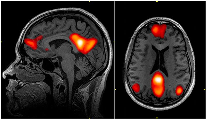
In neuroscience, the default mode network (DMN), also known as the default network, default state network, or anatomically the medial frontoparietal network (M-FPN), is a large-scale brain network primarily composed of the dorsal medial prefrontal cortex, posterior cingulate cortex, precuneus and angular gyrus. It is best known for being active when a person is not focused on the outside world and the brain is at wakeful rest, such as during daydreaming and mind-wandering. It can also be active during detailed thoughts related to external task performance. Other times that the DMN is active include when the individual is thinking about others, thinking about themselves, remembering the past, and planning for the future. The DMN creates a coherent "internal narrative" control to the construction of a sense of self. The DMN was originally noticed to be deactivated in certain goal-oriented tasks and was sometimes referred to as the task-negative network, in contrast with the tas ... [...More Info...] [...Related Items...] OR: [Wikipedia] [Google] [Baidu] |
Washington University School Of Medicine
Washington University School of Medicine (WashU Medicine) is the medical school of Washington University in St. Louis, located in the Central West End neighborhood of St. Louis, Missouri. Founded in 1891, the School of Medicine shares a campus with Barnes-Jewish Hospital, St. Louis Children's Hospital, and the Alvin J. Siteman Cancer Center. The clinical service is provided by Washington University Physicians, a comprehensive medical and surgical practice providing treatment in more than 75 medical specialties. Washington University Physicians are the medical staff of the school's two teaching hospitals – Barnes-Jewish Hospital and St. Louis Children's Hospital. They also provide inpatient and outpatient care at the St. Louis Veteran's Administration Hospital, hospitals of the BJC HealthCare system, and 35 other office locations throughout the greater St. Louis region. History Medical classes were first held at Washington University in 1891 after the St. Louis Medi ... [...More Info...] [...Related Items...] OR: [Wikipedia] [Google] [Baidu] |
Marcus Raichle
Marcus E. Raichle (born March 15, 1937) is an American neurologist at the Washington University School of Medicine in Saint Louis, Missouri. He is a professor in the Department of Radiology with joint appointments in Neurology, Neurobiology and Biomedical Engineering. His research over the past 40 years has focused on the nature of functional brain imaging signals arising from PET and fMRI and the application of these techniques to the study of the human brain in health and disease. He received the Kavli Prize in Neuroscience “for the discovery of specialized brain networks for memory and cognition", together with Brenda Milner and John O’Keefe in 2014. Career Noteworthy accomplishments of Marcus Raichle include the discovery of the relative independence of blood flow and oxygen consumption during changes in brain activity which provided the physiological basis of fMRI; the discovery of a default mode of brain function (i.e., organized intrinsic activity) and its signatur ... [...More Info...] [...Related Items...] OR: [Wikipedia] [Google] [Baidu] |


