|
Quadratojugal
The quadratojugal is a skull bone present in many vertebrates, including some living reptiles and amphibians. Anatomy and function In animals with a quadratojugal bone, it is typically found connected to the jugal (cheek) bone from the front and the squamosal bone from above. It is usually positioned at the rear lower corner of the cranium. Many modern tetrapods lack a quadratojugal bone as it has been lost or fused to other bones. Modern examples of tetrapods without a quadratojugal include salamanders, mammals, birds, and squamates (lizards and snakes). In tetrapods with a quadratojugal bone, it often forms a portion of the jaw joint. Developmentally, the quadratojugal bone is a dermal bone in the temporal series, forming the original braincase. The squamosal and quadratojugal bones together form the cheek region and may provide muscular attachments for facial muscles. In reptiles and amphibians In most modern reptiles and amphibians, the quadratojugal is a prominent, st ... [...More Info...] [...Related Items...] OR: [Wikipedia] [Google] [Baidu] |
Jugal Bone
The jugal is a skull bone found in most reptiles, amphibians and birds. In mammals, the jugal is often called the malar or zygomatic bone, zygomatic. It is connected to the quadratojugal and maxilla, as well as other bones, which may vary by species. Anatomy The jugal bone is located on either side of the skull in the Ocular scales, circumorbital region. It is the origin of several masticatory muscles in the skull. The jugal and Lacrimal bone, lacrimal bones are the only two remaining from the ancestral circumorbital series: the prefrontal, postfrontal, postorbital, jugal, and lacrimal bones. During development, the jugal bone originates from dermal bone. In dinosaurs This bone is considered key in the determination of general traits in cases in which the entire skull has not been found intact (for instance, as with dinosaurs in paleontology). In some dinosaur genera the jugal also forms part of the lower margin of either the antorbital fenestra or the infratemporal fenestr ... [...More Info...] [...Related Items...] OR: [Wikipedia] [Google] [Baidu] |
Squamosal Bone
The squamosal is a skull bone found in most reptiles, amphibians, and birds. In fishes, it is also called the pterotic bone. In most tetrapods, the squamosal and quadratojugal bones form the cheek series of the skull. The bone forms an ancestral component of the dermal roof and is typically thin compared to other skull bones. The squamosal bone lies ventral to the temporal series and otic notch, and is bordered anteriorly by the postorbital. Posteriorly, the squamosal articulates with the quadrate and pterygoid bones. The squamosal is bordered anteroventrally by the jugal and ventrally by the quadratojugal. Function in reptiles In reptiles, the quadrate and articular bones of the skull articulate to form the jaw joint. The squamosal bone lies anterior to the quadrate bone. Anatomy in synapsids Non-mammalian synapsids In non-mammalian synapsids, the jaw is composed of four bony elements and referred to as a quadro-articular jaw because the joint is between the art ... [...More Info...] [...Related Items...] OR: [Wikipedia] [Google] [Baidu] |
Rhynchocephalia
Rhynchocephalia (; ) is an order of lizard-like reptiles that includes only one living species, the tuatara (''Sphenodon punctatus'') of New Zealand. Despite its current lack of diversity, during the Mesozoic rhynchocephalians were a speciose group with high morphological and ecological diversity. The oldest record of the group is dated to the Middle Triassic around 238 to 240 million years ago, and they had achieved global distribution by the Early Jurassic. Most rhynchocephalians belong to the group Sphenodontia ('wedge-teeth'). Their closest living relatives are lizards and snakes in the order Squamata, with the two orders being grouped together in the superorder Lepidosauria. Rhynchocephalians are distinguished from squamates by a number of traits, including the retention of rib-like gastralia bones in the belly, as well as most rhynchocephalians having acrodont teeth that are fused to the crests of the jaws (the latter also found among a small number of modern lizard grou ... [...More Info...] [...Related Items...] OR: [Wikipedia] [Google] [Baidu] |
Quadrate Bone
The quadrate bone is a skull bone in most tetrapods, including amphibians, sauropsids ( reptiles, birds), and early synapsids. In most tetrapods, the quadrate bone connects to the quadratojugal and squamosal bones in the skull, and forms upper part of the jaw joint. The lower jaw articulates at the articular bone, located at the rear end of the lower jaw. The quadrate bone forms the lower jaw articulation in all classes except mammals. Evolutionarily, it is derived from the hindmost part of the primitive cartilaginous upper jaw. Function in reptiles In certain extinct reptiles, the variation and stability of the morphology of the quadrate bone has helped paleontologists in the species-level taxonomy and identification of mosasaur squamates and spinosaurine dinosaurs. In some lizards and dinosaurs, the quadrate is articulated at both ends and movable. In snakes, the quadrate bone has become elongated and very mobile, and contributes greatly to their ability to swallow ... [...More Info...] [...Related Items...] OR: [Wikipedia] [Google] [Baidu] |
Tuatara
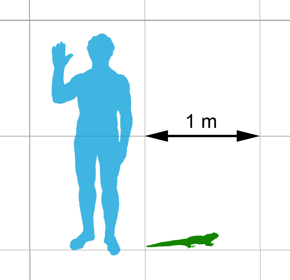
The tuatara (''Sphenodon punctatus'') is a species of reptile endemic to New Zealand. Despite its close resemblance to lizards, it is actually the only extant member of a distinct lineage, the previously highly diverse order Rhynchocephalia. The name is derived from the Māori language and means "peaks on the back". The single extant species of tuatara is the only surviving member of its order, which was highly diverse during the Mesozoic era. Rhynchocephalians first appeared in the fossil record during the Triassic, around 240 million years ago, and reached worldwide distribution and peak diversity during the Jurassic, when they represented the world's dominant group of small reptiles. Rhynchocephalians declined during the Cretaceous, with their youngest records outside New Zealand dating to the Paleocene. Their closest living relatives are squamates (lizards and snakes). Tuatara are of interest for studying the evolution of reptiles. Tuatara are greenish brown an ... [...More Info...] [...Related Items...] OR: [Wikipedia] [Google] [Baidu] |
Infratemporal Fenestra
Temporal fenestrae are openings in the temporal region of the skull of some amniotes, behind the orbit (eye socket). These openings have historically been used to track the evolution and affinities of reptiles. Temporal fenestrae are commonly (although not universally) seen in the fossilized skulls of dinosaurs and other sauropsids (the total group of reptiles, including birds). The major reptile group Diapsida, for example, is defined by the presence of two temporal fenestrae on each side of the skull. The infratemporal fenestra, also called the lateral temporal fenestra or lower temporal fenestra, is the lower of the two and is exposed primarily in lateral (side) view.The supratemporal fenestra, also called the upper temporal fenestra, is positioned above the other fenestra and is exposed primarily in dorsal (top) view. In some reptiles, particularly dinosaurs, the parts of the skull roof lying between the supratemporal fenestrae are thinned out by excavations from the adjacent ... [...More Info...] [...Related Items...] OR: [Wikipedia] [Google] [Baidu] |
Caecilian
Caecilians (; ) are a group of limbless, vermiform (worm-shaped) or serpentine (snake-shaped) amphibians with small or sometimes nonexistent eyes. They mostly live hidden in soil or in streambeds, and this cryptic lifestyle renders caecilians among the least familiar amphibians. Modern caecilians live in the tropics of South America, South and Central America, Africa, and southern Asia. Caecilians feed on small subterranean creatures, such as earthworms. The body is cylindrical and often darkly coloured, and the skull is bullet-shaped and strongly built. Caecilian heads have several unique adaptations, including fused cranial and jaw bones, a two-part system of jaw muscles, and a Chemoreceptor, chemosensory tentacle in front of the eye. The skin is slimy and bears ringlike markings or grooves and may contain scales. Modern caecilians are a clade, the Order (biology), order Gymnophiona (or Apoda ), one of the three living amphibian groups alongside Anura (frogs) and Urodela (sa ... [...More Info...] [...Related Items...] OR: [Wikipedia] [Google] [Baidu] |
Postorbital Bone
The ''postorbital'' is one of the bones in vertebrate skulls which forms a portion of the dermal skull roof and, sometimes, a ring about the orbit. Generally, it is located behind the postfrontal and posteriorly to the orbital fenestra. In some vertebrates, the postorbital is fused with the postfrontal to create a postorbitofrontal. Birds have a separate postorbital as an embryo An embryo ( ) is the initial stage of development for a multicellular organism. In organisms that reproduce sexually, embryonic development is the part of the life cycle that begins just after fertilization of the female egg cell by the male sp ..., but the bone fuses with the frontal before it hatches. References * Roemer, A. S. 1956. ''Osteology of the Reptiles''. University of Chicago Press. 772 pp. Skull bones {{Vertebrate anatomy-stub ... [...More Info...] [...Related Items...] OR: [Wikipedia] [Google] [Baidu] |
Skull
The skull, or cranium, is typically a bony enclosure around the brain of a vertebrate. In some fish, and amphibians, the skull is of cartilage. The skull is at the head end of the vertebrate. In the human, the skull comprises two prominent parts: the neurocranium and the facial skeleton, which evolved from the first pharyngeal arch. The skull forms the frontmost portion of the axial skeleton and is a product of cephalization and vesicular enlargement of the brain, with several special senses structures such as the eyes, ears, nose, tongue and, in fish, specialized tactile organs such as barbels near the mouth. The skull is composed of three types of bone: cranial bones, facial bones and ossicles, which is made up of a number of fused flat and irregular bones. The cranial bones are joined at firm fibrous junctions called sutures and contains many foramina, fossae, processes, and sinuses. In zoology, the openings in the skull are called fenestrae, the most ... [...More Info...] [...Related Items...] OR: [Wikipedia] [Google] [Baidu] |
Cranial Kinesis
Cranial kinesis is the term for significant movement of skull bones relative to each other in addition to movement at the joint between the upper and lower jaws. It is usually taken to mean relative movement between the upper jaw and the braincase. Most vertebrates have some form of a kinetic skull. Cranial kinesis, or lack thereof, is usually linked to feeding. Animals which must exert powerful bite forces, such as crocodiles, often have rigid skulls with little or no kinesis, which maximizes their strength. Animals which swallow large prey whole (snakes), which grip awkwardly shaped food items (parrots eating nuts), or, most often, which feed in the water via suction feeding often have very kinetic skulls, frequently with numerous mobile joints. In the case of mammals, which have akinetic skulls (except perhaps hares), the lack of kinesis is most likely to be related to the secondary palate, which prevents relative movement. This in turn is a consequence of the need to be able ... [...More Info...] [...Related Items...] OR: [Wikipedia] [Google] [Baidu] |
Cynodont
Cynodontia () is a clade of eutheriodont therapsids that first appeared in the Late Permian (approximately 260 Megaannum, mya), and extensively diversified after the Permian–Triassic extinction event. Mammals are cynodonts, as are their extinct ancestors and close relatives (Mammaliaformes), having evolved from advanced probainognathian cynodonts during the Late Triassic. Non-mammalian cynodonts occupied a variety of ecological niches, both as carnivores and as herbivores. Following the emergence of mammals, most other cynodont lines went extinct, with the last known non-mammaliaform cynodont group, the Tritylodontidae, having its youngest records in the Early Cretaceous. Description Early cynodonts have many of the skeletal characteristics of mammals. The teeth were fully differentiated and the braincase bulged at the back of the head. Outside of some Crown group, crown-group mammals (notably the therians), all cynodonts probably laid eggs. The temporal fenestrae#Fenestra ... [...More Info...] [...Related Items...] OR: [Wikipedia] [Google] [Baidu] |


