|
Piriform Aperture
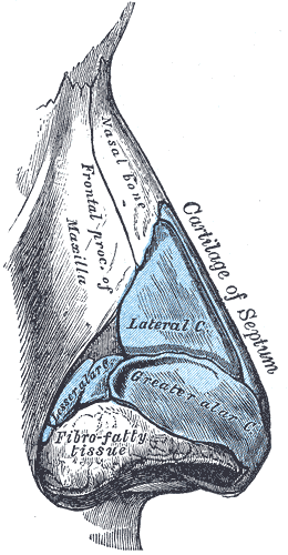
The piriform aperture, pyriform aperture, or anterior nasal aperture, is a pear-shaped opening in the human skull. Its long axis is vertical, and narrow end upward; in the recent state it is much contracted by the lateral nasal cartilage and the greater and lesser alar cartilages of the nose. It is bounded above by the inferior borders of the nasal bones; laterally by the thin, sharp margins which separate the anterior from the nasal surfaces of the maxilla; and below by the same borders, where they curve medialward to join each other at the anterior nasal spine The anterior nasal spine, or anterior nasal spine of maxilla, is a bony projection in the skull that serves as a cephalometric landmark. The anterior nasal spine is the projection formed by the fusion of the two maxillary bones at the intermaxill .... References Nose {{musculoskeletal-stub ... [...More Info...] [...Related Items...] OR: [Wikipedia] [Google] [Baidu] |
Pear
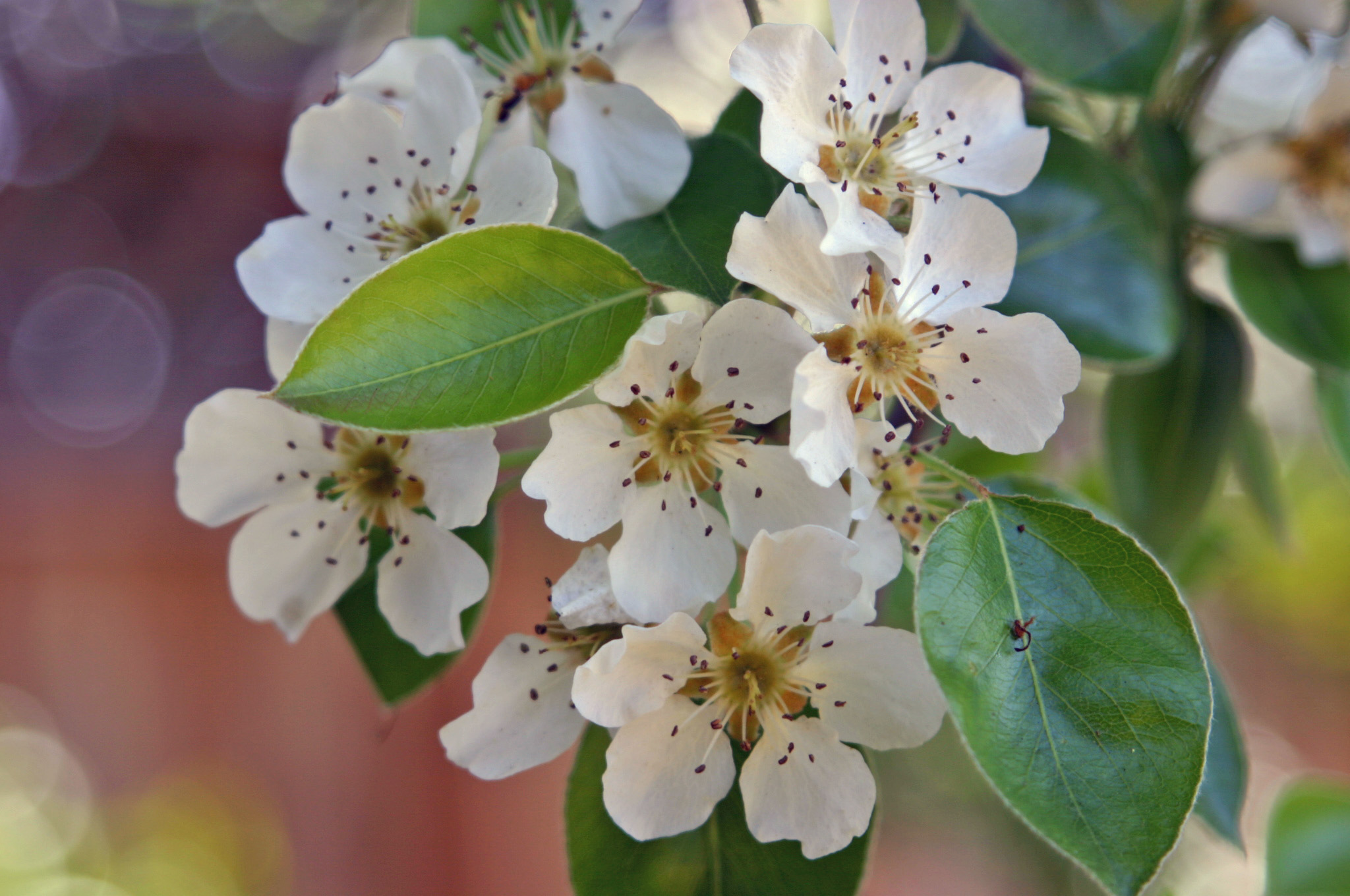
Pears are fruits produced and consumed around the world, growing on a tree and harvested in the Northern Hemisphere in late summer into October. The pear tree and shrub are a species of genus ''Pyrus'' , in the family Rosaceae, bearing the pomaceous fruit of the same name. Several species of pears are valued for their edible fruit and juices, while others are cultivated as trees. The tree is medium-sized and native to coastal and mildly temperate regions of Europe, North Africa, and Asia. Pear wood is one of the preferred materials in the manufacture of high-quality woodwind instruments and furniture. About 3,000 known varieties of pears are grown worldwide, which vary in both shape and taste. The fruit is consumed fresh, canned, as juice, or dried. Etymology The word ''pear'' is probably from Germanic ''pera'' as a loanword of Vulgar Latin ''pira'', the plural of ''pirum'', akin to Greek ''apios'' (from Mycenaean ''ápisos''), of Semitic origin (''pirâ''), meaning "fru ... [...More Info...] [...Related Items...] OR: [Wikipedia] [Google] [Baidu] |
Human Skull
The skull is a bone protective cavity for the brain. The skull is composed of four types of bone i.e., cranial bones, facial bones, ear ossicles and hyoid bone. However two parts are more prominent: the cranium and the mandible. In humans, these two parts are the neurocranium and the viscerocranium ( facial skeleton) that includes the mandible as its largest bone. The skull forms the anterior-most portion of the skeleton and is a product of cephalisation—housing the brain, and several sensory structures such as the eyes, ears, nose, and mouth. In humans these sensory structures are part of the facial skeleton. Functions of the skull include protection of the brain, fixing the distance between the eyes to allow stereoscopic vision, and fixing the position of the ears to enable sound localisation of the direction and distance of sounds. In some animals, such as horned ungulates (mammals with hooves), the skull also has a defensive function by providing the mount (on the front ... [...More Info...] [...Related Items...] OR: [Wikipedia] [Google] [Baidu] |
Lateral Nasal Cartilage
The lateral cartilage (upper lateral cartilage, lateral process of septal nasal cartilage) is situated below the inferior margin of the nasal bone, and is flattened, and triangular in shape. Its anterior margin is thicker than the posterior, and is continuous above with the septal nasal cartilage, but separated from it below by a narrow fissure; its superior margin is attached to the nasal bone and the frontal process of the maxilla; its inferior margin is connected by fibrous tissue with the greater alar cartilage The major alar cartilage (greater alar cartilage) (lower lateral cartilage) is a thin, flexible plate, situated immediately below the lateral nasal cartilage, and bent upon itself in such a manner as to form the medial wall and lateral wall of th .... Where the lateral cartilage meets the greater alar cartilage, the lateral cartilage often curls up, to join with an inward curl of the greater alar cartilage. That curl of the inferior portion of the lateral cartilage is ... [...More Info...] [...Related Items...] OR: [Wikipedia] [Google] [Baidu] |
Greater Alar Cartilage
The major alar cartilage (greater alar cartilage) (lower lateral cartilage) is a thin, flexible plate, situated immediately below the lateral nasal cartilage, and bent upon itself in such a manner as to form the medial wall and lateral wall of the nostril of its own side. The portion which forms the medial wall (crus mediale) is loosely connected with the corresponding portion of the opposite cartilage, the two forming, together with the thickened integument and subjacent tissue, the nasal septum. The part which forms the lateral wall (crus laterale) is curved to correspond with the ala of the nose; it is oval and flattened, narrow behind, where it is connected with the frontal process of the maxilla by a tough fibrous membrane, in which are found three or four small cartilaginous plates, the lesser alar cartilages (cartilagines alares minores; sesamoid cartilages). Above, it is connected by fibrous tissue to the lateral cartilage and front part of the cartilage of the septum; ... [...More Info...] [...Related Items...] OR: [Wikipedia] [Google] [Baidu] |
Lesser Alar Cartilages
In human anatomy the part of the nose which forms the lateral wall is curved to correspond with the ala of the nose; it is oval and flattened, narrow behind, where it is connected with the frontal process of the maxilla The maxilla (plural: ''maxillae'' ) in vertebrates is the upper fixed (not fixed in Neopterygii) bone of the jaw formed from the fusion of two maxillary bones. In humans, the upper jaw includes the hard palate in the front of the mouth. T ... by a tough fibrous membrane, in which are found three or four small nasal cartilages the minor alar cartilages, also referred to as lesser alar or sesamoid cartilages or accessory cartilages. ''An atlas of human anatomy f ... [...More Info...] [...Related Items...] OR: [Wikipedia] [Google] [Baidu] |
Human Nose
The human nose is the most protruding part of the face. It bears the nostrils and is the first organ of the respiratory system. It is also the principal organ in the olfactory system. The shape of the nose is determined by the nasal bones and the nasal cartilages, including the nasal septum which separates the nostrils and divides the nasal cavity into two. On average the nose of a male is larger than that of a female. The nose has an important function in breathing. The nasal mucosa lining the nasal cavity and the paranasal sinuses carries out the necessary conditioning of inhaled air by warming and moistening it. Nasal conchae, shell-like bones in the walls of the cavities, play a major part in this process. Filtering of the air by nasal hair in the nostrils prevents large particles from entering the lungs. Sneezing is a reflex to expel unwanted particles from the nose that irritate the mucosal lining. Sneezing can transmit infections, because aerosols are created in w ... [...More Info...] [...Related Items...] OR: [Wikipedia] [Google] [Baidu] |
Nasal Bones
The nasal bones are two small oblong bones, varying in size and form in different individuals; they are placed side by side at the middle and upper part of the face and by their junction, form the bridge of the upper one third of the nose. Each has two surfaces and four borders. Structure The two nasal bones are joined at the midline internasal suture and make up the bridge of the nose. Surfaces The ''outer surface'' is concavo-convex from above downward, convex from side to side; it is covered by the procerus and nasalis muscles, and perforated about its center by a foramen, for the transmission of a small vein. The ''inner surface'' is concave from side to side, and is traversed from above downward, by a groove for the passage of a branch of the nasociliary nerve. Articulations The nasal articulates with four bones: two of the cranium, the frontal and ethmoid, and two of the face, the opposite nasal and the maxilla. Other animals In primitive bony fish and tetrapod ... [...More Info...] [...Related Items...] OR: [Wikipedia] [Google] [Baidu] |
Maxilla
The maxilla (plural: ''maxillae'' ) in vertebrates is the upper fixed (not fixed in Neopterygii) bone of the jaw formed from the fusion of two maxillary bones. In humans, the upper jaw includes the hard palate in the front of the mouth. The two maxillary bones are fused at the intermaxillary suture, forming the anterior nasal spine. This is similar to the mandible (lower jaw), which is also a fusion of two mandibular bones at the mandibular symphysis. The mandible is the movable part of the jaw. Structure In humans, the maxilla consists of: * The body of the maxilla * Four processes ** the zygomatic process ** the frontal process of maxilla ** the alveolar process ** the palatine process * three surfaces – anterior, posterior, medial * the Infraorbital foramen * the maxillary sinus * the incisive foramen Articulations Each maxilla articulates with nine bones: * two of the cranium: the frontal and ethmoid * seven of the face: the nasal, zygomatic, lacrimal, inferior n ... [...More Info...] [...Related Items...] OR: [Wikipedia] [Google] [Baidu] |
Anterior Nasal Spine
The anterior nasal spine, or anterior nasal spine of maxilla, is a bony projection in the skull that serves as a cephalometric landmark. The anterior nasal spine is the projection formed by the fusion of the two maxillary bones at the intermaxillary suture. It is placed at the level of the nostrils, at the uppermost part of the philtrum and rarely fractures. Additional images File:Anterior nasal spine of maxilla - animation02.gif, Animation. Anterior nasal spine shown in red. File:Anterior nasal spine of maxilla - animation00.gif, Left maxilla. Anterior nasal spine shown in red. File:Anterior nasal spine of maxilla - skull - anterior view.png, Skull. Anterior view. Anterior nasal spine shown in red. File:Slide12hhhh.JPG, Right maxilla. Anterior nasal spine labeled at center left. See also * Posterior nasal spine The posterior nasal spine is part of the horizontal plate of the palatine bone of the skull. It is found at the medial end of its posterior border. It is paired ... [...More Info...] [...Related Items...] OR: [Wikipedia] [Google] [Baidu] |


