|
Push-up
The push-up (press-up in British English) is a common calisthenics Physical exercise, exercise beginning from the prone position. By raising and lowering the body using the arms, push-ups exercise the pectoralis major muscle, pectoral muscles, triceps, and anterior deltoid muscle, deltoids, with ancillary benefits to the rest of the deltoids, serratus anterior muscle, serratus anterior, coracobrachialis muscle, coracobrachialis and the midsection as a whole. Push-ups are a basic exercise used in civilian athletic training or physical education and commonly in Military Fitness, military physical training. It is also a common form of punishment used in the military, school sport, and some martial arts disciplines for its humiliating factor (when one fails to do a specified amount) and for its lack of equipment. Variations of push-ups, such as wide-arm push-ups, diamond push-ups target specific muscle groups and provide further challenges. Etymology The American English term '' ... [...More Info...] [...Related Items...] OR: [Wikipedia] [Google] [Baidu] [Amazon] |
Push Up (PSF)
The push-up (press-up in British English) is a common calisthenics exercise beginning from the prone position. By raising and lowering the body using the arms, push-ups exercise the pectoral muscles, triceps, and anterior deltoids, with ancillary benefits to the rest of the deltoids, serratus anterior, coracobrachialis and the midsection as a whole. Push-ups are a basic exercise used in civilian athletic training or physical education and commonly in military physical training. It is also a common form of punishment used in the military, school sport, and some martial arts disciplines for its humiliating factor (when one fails to do a specified amount) and for its lack of equipment. Variations of push-ups, such as wide-arm push-ups, diamond push-ups target specific muscle groups and provide further challenges. Etymology The American English term ''push-up'' was first used between 1905 and 1910, while the British ''press-up'' was first recorded was 1920. Body mass supported ... [...More Info...] [...Related Items...] OR: [Wikipedia] [Google] [Baidu] [Amazon] |
Erector Spinae Muscles
The erector spinae ( ) or spinal erectors is a set of muscles that straighten and rotate the back. The spinal erectors work together with the glutes ( gluteus maximus, gluteus medius and gluteus minimus) to maintain stable posture standing or sitting. Structure The erector spinae is not just one muscle, but a group of muscles and tendons which run more or less the length of the spine on the left and the right, from the sacrum, or sacral region, and hips to the base of the skull. They are also known as the sacrospinalis group of muscles. These muscles lie on either side of the spinous processes of the vertebrae and extend throughout the lumbar, thoracic, and cervical regions. The erector spinae is covered in the lumbar and thoracic regions by the thoracolumbar fascia, and in the cervical region by the nuchal ligament. This large muscular and tendinous mass varies in size and structure at different parts of the vertebral column. In the sacral region, it is narrow and ... [...More Info...] [...Related Items...] OR: [Wikipedia] [Google] [Baidu] [Amazon] |
Calisthenics
Calisthenics (American English) or callisthenics (British English) () is a form of strength training that utilizes an individual's body weight as resistance to perform multi-joint, compound movements with little or no equipment. Calisthenics solely rely on bodyweight for resistance, which naturally adapts to an individual's unique physical attributes like limb length and muscle-tendon insertion points. This allows calisthenic exercises to be more personalized and accessible for various body structures and age ranges. Calisthenics is distinct for its reliance on closed-chain movements. These exercises engage multiple joints simultaneously as the resistance moves relative to an anchored body part, promoting functional and efficient movement patterns. Calisthenics' exercises and movement patterns focuses on enhancing overall strength, stability, and coordination. The versatility that calisthenics introduces, minimizing equipment use, has made calisthenics a popular choice for encour ... [...More Info...] [...Related Items...] OR: [Wikipedia] [Google] [Baidu] [Amazon] |
Body Mass
Human body weight is a person's mass or weight. Strictly speaking, body weight is the measurement of mass without items located on the person. Practically though, body weight may be measured with clothes on, but without shoes or heavy accessories such as mobile phones and wallets, and using manual or digital weighing scales. Excess or reduced body weight is regarded as an indicator of determining a person's health, with body volume measurement providing an extra dimension by calculating the distribution of body weight. Average adult human weight varies by continent, from about in Asia and Africa to about in North America, with men on average weighing more than women. Estimation in children There are a number of methods to estimate weight in children for circumstances (such as emergencies) when actual weight cannot be measured. Most involve a parent or health care provider guessing the child's weight through weight-estimation formulas. These formulas base their findings on ... [...More Info...] [...Related Items...] OR: [Wikipedia] [Google] [Baidu] [Amazon] |
Pectoralis Minor Muscle
Pectoralis minor muscle () is a thin, triangular muscle, situated at the upper part of the chest, beneath the pectoralis major in the human body. It arises from ribs III-V; it inserts onto the coracoid process of the scapula. It is innervated by the medial pectoral nerve. Its function is to stabilise the scapula by holding it fast in position against the Thoracic wall, chest wall. Structure Attachments From the muscle's origin, the muscle's fibers pass superiorly and laterally, converging to form a flat tendon. Origin Pectoralis minor muscle arises from the upper margins and outer surfaces of the 3rd, 4th, and 5th ribs near their costal cartilages, and from the Aponeurosis, aponeuroses covering the External intercostal muscles, intercostalis. Insertion Its tendon inserts onto the medial border and upper surface of the coracoid process of the scapula. Innervation The muscle receives motor innervation from the medial pectoral nerve. Relations Pectoralis minor muscle ... [...More Info...] [...Related Items...] OR: [Wikipedia] [Google] [Baidu] [Amazon] |
Humerus Bone
The humerus (; : humeri) is a long bone in the arm that runs from the shoulder to the elbow. It connects the scapula and the two bones of the lower arm, the radius (bone), radius and ulna, and consists of three sections. The humeral upper extremity of humerus, upper extremity consists of a rounded head, a narrow neck, and two short processes (tubercles, sometimes called tuberosities). The body of humerus, body is cylindrical in its upper portion, and more prism (geometry), prismatic below. The lower extremity of humerus, lower extremity consists of 2 epicondyles, 2 processes (trochlea of the humerus, trochlea and capitulum of the humerus, capitulum), and 3 fossae (radial fossa, coronoid fossa, and olecranon fossa). As well as its true anatomical neck, the constriction below the greater and lesser tubercles of the humerus is referred to as its Surgical neck of the humerus, surgical neck due to its tendency to fracture, thus often becoming the focus of surgeons. Etymology The word ... [...More Info...] [...Related Items...] OR: [Wikipedia] [Google] [Baidu] [Amazon] |
Glenohumeral Joint
The shoulder joint (or glenohumeral joint from Greek ''glene'', eyeball, + -''oid'', 'form of', + Latin ''humerus'', shoulder) is structurally classified as a synovial ball-and-socket joint and functionally as a diarthrosis and multiaxial joint. It involves an articulation between the glenoid fossa of the scapula (shoulder blade) and the head of the humerus (upper arm bone). Due to the very loose joint capsule, it gives a limited interface of the humerus and scapula, it is the most mobile joint of the human body. Structure The shoulder joint is a ball-and-socket joint between the scapula and the humerus. The socket of the glenoid fossa of the scapula is itself quite shallow, but it is made deeper by the addition of the glenoid labrum. The glenoid labrum is a ring of cartilaginous fibre attached to the circumference of the cavity. This ring is continuous with the tendon of the biceps brachii above. Spaces Significant joint spaces are: * The normal glenohumeral space is 4� ... [...More Info...] [...Related Items...] OR: [Wikipedia] [Google] [Baidu] [Amazon] |
Scapula
The scapula (: scapulae or scapulas), also known as the shoulder blade, is the bone that connects the humerus (upper arm bone) with the clavicle (collar bone). Like their connected bones, the scapulae are paired, with each scapula on either side of the body being roughly a mirror image of the other. The name derives from the Classical Latin word for trowel or small shovel, which it was thought to resemble. In compound terms, the prefix omo- is used for the shoulder blade in medical terminology. This prefix is derived from ὦμος (ōmos), the Ancient Greek word for shoulder, and is cognate with the Latin , which in Latin signifies either the shoulder or the upper arm bone. The scapula forms the back of the shoulder girdle. In humans, it is a flat bone, roughly triangular in shape, placed on a posterolateral aspect of the thoracic cage. Structure The scapula is a thick, flat bone lying on the thoracic wall that provides an attachment for three groups of muscles: intrinsic, e ... [...More Info...] [...Related Items...] OR: [Wikipedia] [Google] [Baidu] [Amazon] |
Clavicle
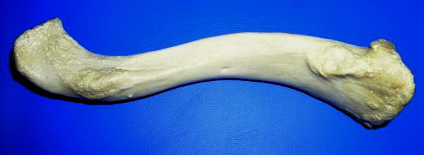
The clavicle, collarbone, or keybone is a slender, S-shaped long bone approximately long that serves as a strut between the scapula, shoulder blade and the sternum (breastbone). There are two clavicles, one on each side of the body. The clavicle is the only long bone in the body that lies horizontally. Together with the shoulder blade, it makes up the shoulder girdle. It is a palpable bone and, in people who have less fat in this region, the location of the bone is clearly visible. It receives its name from Latin ''clavicula'' 'little key' because the bone rotates along its axis like a key when the shoulder is Abduction (kinesiology), abducted. The clavicle is the most commonly fractured bone. It can easily be fractured by impacts to the shoulder from the force of falling on outstretched arms or by a direct hit. Structure The collarbone is a thin doubly curved long bone that connects the human arm, arm to the torso, trunk of the body. Located directly above the first rib, it ac ... [...More Info...] [...Related Items...] OR: [Wikipedia] [Google] [Baidu] [Amazon] |
Torso
The torso or trunk is an anatomical terminology, anatomical term for the central part, or the core (anatomy), core, of the body (biology), body of many animals (including human beings), from which the head, neck, limb (anatomy), limbs, tail and other appendages extend. The tetrapod torso — including that of a human body, human — is usually divided into the ''chest, thoracic'' segment (also known as the upper torso, where the forelimbs extend), the ''abdomen, abdominal'' segment (also known as the "mid-section" or "midriff"), and the ''pelvic'' and ''perineum, perineal'' segments (sometimes known together with the abdomen as the lower torso, where the hindlimbs extend). Anatomy Major organs In humans, most critical Organ (anatomy), organs, with the notable exception of the brain, are housed within the torso. In the upper chest, the heart and lungs are protected by the rib cage, and the abdomen contains most of the organs responsible for digestion: the stomach, which breaks ... [...More Info...] [...Related Items...] OR: [Wikipedia] [Google] [Baidu] [Amazon] |
Transversus Abdominis Muscle
The transverse abdominal muscle (TVA), also known as the transverse abdominis, transversalis muscle and transversus abdominis muscle, is a muscle layer of the anterior and lateral (front and side) abdominal wall, deep to (layered below) the internal oblique muscle. It serves to compress and retain the contents of the abdomen as well as assist in exhalation. Structure The transverse abdominal, so called for the direction of its fibers, is the innermost of the flat muscles of the abdomen. It is positioned immediately deep to the internal oblique muscle. The transverse abdominal arises as fleshy fibers, from the lateral third of the inguinal ligament, from the anterior three-fourths of the inner lip of the iliac crest, from the inner surfaces of the cartilages of the lower six ribs, interdigitating with the diaphragm, and from the thoracolumbar fascia. It ends anteriorly in a broad aponeurosis (the Spigelian fascia), the lower fibers of which curve inferomedially (medially and do ... [...More Info...] [...Related Items...] OR: [Wikipedia] [Google] [Baidu] [Amazon] |


