|
Psoas Minor Muscle
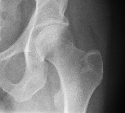
The psoas minor muscle ( or ; from ) is a long, slender skeletal muscle. When present, it is located anterior to the psoas major muscle.Tank (2005), p 93Gray (2008), p 1372 Structure The psoas minor muscle originates from the vertical fascicles inserted on the last thoracic and first lumbar vertebrae. From there, it passes down onto the medial border of the psoas major, and is inserted to the innominate line and the iliopectineal eminence. Additionally, it attaches to and stretches the deep surface of the iliac fascia and occasionally its lowermost fibers reach the inguinal ligament.Bendavid (2001), p 58 It is posteriolateral to the iliopsoas muscle. Variations occur, however, and the insertion on the iliopubic eminence sometimes radiates into the iliopectineal arch.Platzer (2004), p 234 The psoas minor muscle receives oxygenated blood from the four lumbar arteries (inferior to the subcostal artery) and the lumbar branch of the iliolumbar artery. Innervation The psoas ... [...More Info...] [...Related Items...] OR: [Wikipedia] [Google] [Baidu] |
Body Of Vertebra
Each vertebra (: vertebrae) is an irregular bone with a complex structure composed of bone and some hyaline cartilage, that make up the vertebral column or spine, of vertebrates. The proportions of the vertebrae differ according to their spinal segment and the particular species. The basic configuration of a vertebra varies; the vertebral body (also ''centrum'') is of bone and bears the load of the vertebral column. The upper and lower surfaces of the vertebra body give attachment to the intervertebral discs. The posterior part of a vertebra forms a vertebral arch, in eleven parts, consisting of two pedicles (pedicle of vertebral arch), two laminae, and seven processes. The laminae give attachment to the ligamenta flava (ligaments of the spine). There are vertebral notches formed from the shape of the pedicles, which form the intervertebral foramina when the vertebrae articulate. These foramina are the entry and exit conduits for the spinal nerves. The body of the vertebra an ... [...More Info...] [...Related Items...] OR: [Wikipedia] [Google] [Baidu] |
Iliopsoas
The iliopsoas muscle (; ) refers to the joined psoas major and the iliacus muscles. The two muscles are separate in the abdomen, but usually merge in the thigh. They are usually given the common name ''iliopsoas''. The iliopsoas muscle joins to the femur at the lesser trochanter. It acts as the strongest flexor of the hip. The iliopsoas muscle is supplied by the lumbar spinal nerves L1– L3 (psoas) and parts of the femoral nerve (iliacus). Structure The iliopsoas muscle is a composite muscle formed from the psoas major muscle, and the iliacus muscle. The psoas major originates along the outer surfaces of the vertebral bodies of T12 and L1– L3 and their associated intervertebral discs. The iliacus originates in the iliac fossa of the pelvis. The psoas major unites with the iliacus at the level of the inguinal ligament. It crosses the hip joint to insert on the lesser trochanter of the femur. The iliopsoas is classified as an "anterior hip muscle" or "inner ... [...More Info...] [...Related Items...] OR: [Wikipedia] [Google] [Baidu] |
Hip Muscles
In vertebrate anatomy, the hip, or coxaLatin ''coxa'' was used by Celsus in the sense "hip", but by Pliny the Elder in the sense "hip bone" (Diab, p 77) (: ''coxae'') in medical terminology, refers to either an anatomical region or a joint on the outer (lateral) side of the pelvis. The hip region is located lateral and anterior to the gluteal region, inferior to the iliac crest, and lateral to the obturator foramen, with muscle tendons and soft tissues overlying the greater trochanter of the femur. In adults, the three pelvic bones ( ilium, ischium and pubis) have fused into one hip bone, which forms the superomedial/deep wall of the hip region. The hip joint, scientifically referred to as the acetabulofemoral joint (''art. coxae''), is the ball-and-socket joint between the pelvic acetabulum and the femoral head. Its primary function is to support the weight of the torso in both static (e.g. standing) and dynamic (e.g. walking or running) postures. The hip joints h ... [...More Info...] [...Related Items...] OR: [Wikipedia] [Google] [Baidu] |
Hip Flexors
In anatomy, flexor is a muscle that contracts to perform flexion (from the Latin verb ''flectere'', to bend), a movement that decreases the angle between the bones converging at a joint. For example, one's elbow joint flexes when one brings their hand closer to the shoulder, thus decreasing the angle between the upper arm and the forearm. Flexors Upper limb *of the humerus bone (the bone in the upper arm) at the shoulder ** Pectoralis major ** Anterior deltoid ** Coracobrachialis **Biceps brachii * of the forearm at the elbow ** Brachialis **Brachioradialis **Biceps brachii *of carpus (the carpal bones) at the wrist **flexor carpi radialis **flexor carpi ulnaris ** palmaris longus *of the hand ** flexor pollicis longus muscle ** flexor pollicis brevis muscle **flexor digitorum profundus muscle ** flexor digitorum superficialis muscle Lower limb Hip The hip flexors are (in descending order of importance to the action of flexing the hip joint):Platzer (2004), p 246 *Collecti ... [...More Info...] [...Related Items...] OR: [Wikipedia] [Google] [Baidu] |
Thieme Medical Publishers
Thieme Medical Publishers is a German academic publishing, medical and science publisher in the Thieme Publishing Group. It produces professional journals, textbooks, atlases, monographs and reference books in both German and English covering a variety of medical specialties, including neurosurgery, orthopaedics, endocrinology, urology, radiology, anatomy, chemistry, otolaryngology, ophthalmology, audiology and speech-language pathology, complementary medicine, complementary and alternative medicine. Thieme has more than 1,000 employees and maintains offices in seven cities worldwide, including New York City, Beijing, Delhi, Stuttgart, and three other cities in Germany. History Georg Thieme Verlag was founded in 1886 in Leipzig, Germany, by Georg Thieme when he was 26 years old. Thieme remains privately held and family-owned. The company received some early success in 1896 by publishing Wilhelm Röntgen's famous picture of his wife's hand in what is still one of Thieme's and ... [...More Info...] [...Related Items...] OR: [Wikipedia] [Google] [Baidu] |
Back Pain
Back pain (Latin: ''dorsalgia'') is pain felt in the back. It may be classified as neck pain (cervical), middle back pain (thoracic), lower back pain (lumbar) or coccydynia (tailbone or sacral pain) based on the segment affected. The lumbar area is the most common area affected. An episode of back pain may be Acute (medicine), acute, subacute or Chronic condition, chronic depending on the duration. The pain may be characterized as a dull ache, shooting or piercing pain or a burning sensation. Discomfort can radiate to the arms and hands as well as the Human leg, legs or Human foot, feet, and may include Paresthesia, numbness or weakness in the legs and arms. The majority of back pain is nonspecific and Idiopathic disease, idiopathic. Common underlying mechanisms include degenerative or traumatic changes to the Intervertebral disc, discs and facet joints, which can then cause secondary pain in the muscles and nerves and referred pain to the bones, joints and extremities. Diseases ... [...More Info...] [...Related Items...] OR: [Wikipedia] [Google] [Baidu] |
Rectal Examination
Digital rectal examination (DRE), also known as a prostate exam (), is an internal examination of the rectum performed by a healthcare provider. Prior to a 2018 report from the United States Preventive Services Task Force, a digital exam was a common component of annual medical examination for older men, as it was thought to be a reliable screening test for prostate cancer. Usage This examination may be used: * for the diagnosis of prostatic disorders, benign prostatic hyperplasia and the four types of prostatitis. Chronic prostatitis/chronic pelvic pain syndrome, chronic bacterial prostatitis, acute (sudden) bacterial prostatitis, and asymptomatic inflammatory prostatitis. The DRE has a 50% specificity for benign prostatic hyperplasia. Vigorous examination of the prostate in suspected acute prostatitis can lead to seeding of septic emboli and should never be done. Its utility as a screening method for prostate cancer however is not supported by the evidence; * for the eva ... [...More Info...] [...Related Items...] OR: [Wikipedia] [Google] [Baidu] |
Horse
The horse (''Equus ferus caballus'') is a domesticated, one-toed, hoofed mammal. It belongs to the taxonomic family Equidae and is one of two extant subspecies of ''Equus ferus''. The horse has evolved over the past 45 to 55 million years from a small multi-toed creature, '' Eohippus'', into the large, single-toed animal of today. Humans began domesticating horses around 4000 BCE in Central Asia, and their domestication is believed to have been widespread by 3000 BCE. Horses in the subspecies ''caballus'' are domesticated, although some domesticated populations live in the wild as feral horses. These feral populations are not true wild horses, which are horses that have never been domesticated. There is an extensive, specialized vocabulary used to describe equine-related concepts, covering everything from anatomy to life stages, size, colors, markings, breeds, locomotion, and behavior. Horses are adapted to run, allowing them to quickly escape predator ... [...More Info...] [...Related Items...] OR: [Wikipedia] [Google] [Baidu] |
Mammal
A mammal () is a vertebrate animal of the Class (biology), class Mammalia (). Mammals are characterised by the presence of milk-producing mammary glands for feeding their young, a broad neocortex region of the brain, fur or hair, and three Evolution of mammalian auditory ossicles, middle ear bones. These characteristics distinguish them from reptiles and birds, from which their ancestors Genetic divergence, diverged in the Carboniferous Period over 300 million years ago. Around 6,640 Neontology#Extant taxon, extant species of mammals have been described and divided into 27 Order (biology), orders. The study of mammals is called mammalogy. The largest orders of mammals, by number of species, are the rodents, bats, and eulipotyphlans (including hedgehogs, Mole (animal), moles and shrews). The next three are the primates (including humans, monkeys and lemurs), the Artiodactyl, even-toed ungulates (including pigs, camels, and whales), and the Carnivora (including Felidae, ... [...More Info...] [...Related Items...] OR: [Wikipedia] [Google] [Baidu] |
Lumbar Nerves
The lumbar nerves are the five pairs of spinal nerves emerging from the lumbar vertebrae. They are divided into posterior and anterior divisions. Structure The lumbar nerves are five spinal nerves which arise from either side of the spinal cord below the thoracic spinal cord and above the sacral spinal cord. They arise from the spinal cord between each pair of lumbar spinal vertebrae and travel through the intervertebral foramina. The nerves then split into an anterior branch, which travels forward, and a posterior branch, which travels backwards and supplies the area of the back. Posterior divisions The middle divisions of the posterior branches run close to the articular processes of the vertebrae and end in the multifidus muscle. The outer branches supply the erector spinae muscles. The nerves give off branches to the skin. These pierce the aponeurosis of the greater trochanter. Anterior divisions The anterior divisions of the lumbar nerves () increase in size from ... [...More Info...] [...Related Items...] OR: [Wikipedia] [Google] [Baidu] |
Nerve
A nerve is an enclosed, cable-like bundle of nerve fibers (called axons). Nerves have historically been considered the basic units of the peripheral nervous system. A nerve provides a common pathway for the Electrochemistry, electrochemical nerve impulses called action potentials that are transmitted along each of the axons to peripheral organs or, in the case of sensory nerves, from the periphery back to the central nervous system. Each axon is an extension of an individual neuron, along with other supportive cells such as some Schwann cells that coat the axons in myelin. Each axon is surrounded by a layer of connective tissue called the endoneurium. The axons are bundled together into groups called Nerve fascicle, fascicles, and each fascicle is wrapped in a layer of connective tissue called the perineurium. The entire nerve is wrapped in a layer of connective tissue called the epineurium. Nerve cells (often called neurons) are further classified as either Sensory neuron, sens ... [...More Info...] [...Related Items...] OR: [Wikipedia] [Google] [Baidu] |
Iliolumbar Artery
The iliolumbar artery is the first branch of the posterior trunk of the internal iliac artery. Structure The iliolumbar artery is the first branch of the posterior trunk of the internal iliac artery. It turns upward behind the obturator nerve and the external iliac artery and vein, to the medial border of the psoas major muscle The psoas major ( or ; from ) is a long fusiform muscle located in the lateral lumbar region between the vertebral column and the brim of the lesser pelvis. It joins the iliacus muscle to form the iliopsoas. In other animals, this muscle is equ ..., behind which it divides into: * Lumbar branch of iliolumbar artery * Iliac branch of iliolumbar artery Anastomoses *1. Last lumbar→iliolumbar *2. Lateral sacral↔lateral sacral *3. Middle sacral→lateral sacral *4. Superior hemorrhoidal→middle hemorrhoidal *5. Medial femoral circumflex→inferior gluteal *6. Medial femoral circumflex↔obturator *7. Lateral femoral circumflex→superior gluteal *8. ... [...More Info...] [...Related Items...] OR: [Wikipedia] [Google] [Baidu] |


