|
Pseudophakic
Intraocular lens (IOL) is a lens implanted in the eye as part of a treatment for cataracts or myopia. If the natural lens is left in the eye, the IOL is known as phakic, otherwise it is a pseudophakic, or false lens. Such a lens is typically implanted during cataract surgery, after the eye's cloudy natural lens (cataract) has been removed. The pseudophakic IOL provides the same light-focusing function as the natural crystalline lens. The phakic type of IOL is placed over the existing natural lens and is used in refractive surgery to change the eye's optical power as a treatment for myopia (nearsightedness). This is an alternative to LASIK. IOLs usually consist of a small plastic lens with plastic side struts, called haptics, to hold the lens in place in the capsular bag inside the eye. IOLs were conventionally made of an inflexible material (PMMA), although this has largely been superseded by the use of flexible materials, such as silicone. Most IOLs fitted today are fixed mono ... [...More Info...] [...Related Items...] OR: [Wikipedia] [Google] [Baidu] |
Phakic Intraocular Lens
A phakic intraocular lens (PIOL) is a special kind of intraocular lens that is implanted surgically into the eye to correct myopia (nearsightedness). It is called "phakic" (meaning "having a lens") because the eye's natural lens is left untouched. Intraocular lenses that are implanted into eyes after the eye's natural lens has been removed during cataract surgery are known as pseudophakic. Phakic intraocular lenses are indicated for patients with high refractive errors when the usual laser options for surgical correction (LASIK and PRK) are contraindicated. Phakic IOLs are designed to correct high myopia ranging from −5 to −20 D if the patient has enough anterior chamber depth (ACD) of at least 3 mm. Three types of phakic IOLs are available: * Angle-supported * Iris-fixated * Sulcus-supported intraocular lens Medical uses LASIK can correct myopia up to -12 to -14 D. The higher the intended correction the thinner and flatter the cornea will be post-operatively. Fo ... [...More Info...] [...Related Items...] OR: [Wikipedia] [Google] [Baidu] |
Cataracts
A cataract is a cloudy area in the lens of the eye that leads to a decrease in vision. Cataracts often develop slowly and can affect one or both eyes. Symptoms may include faded colors, blurry or double vision, halos around light, trouble with bright lights, and trouble seeing at night. This may result in trouble driving, reading, or recognizing faces. Poor vision caused by cataracts may also result in an increased risk of falling and depression. Cataracts cause 51% of all cases of blindness and 33% of visual impairment worldwide. Cataracts are most commonly due to aging but may also occur due to trauma or radiation exposure, be present from birth, or occur following eye surgery for other problems. Risk factors include diabetes, longstanding use of corticosteroid medication, smoking tobacco, prolonged exposure to sunlight, and alcohol. The underlying mechanism involves accumulation of clumps of protein or yellow-brown pigment in the lens that reduces transmission of ... [...More Info...] [...Related Items...] OR: [Wikipedia] [Google] [Baidu] |
Phakic Intraocular Lens
A phakic intraocular lens (PIOL) is a special kind of intraocular lens that is implanted surgically into the eye to correct myopia (nearsightedness). It is called "phakic" (meaning "having a lens") because the eye's natural lens is left untouched. Intraocular lenses that are implanted into eyes after the eye's natural lens has been removed during cataract surgery are known as pseudophakic. Phakic intraocular lenses are indicated for patients with high refractive errors when the usual laser options for surgical correction (LASIK and PRK) are contraindicated. Phakic IOLs are designed to correct high myopia ranging from −5 to −20 D if the patient has enough anterior chamber depth (ACD) of at least 3 mm. Three types of phakic IOLs are available: * Angle-supported * Iris-fixated * Sulcus-supported intraocular lens Medical uses LASIK can correct myopia up to -12 to -14 D. The higher the intended correction the thinner and flatter the cornea will be post-operatively. Fo ... [...More Info...] [...Related Items...] OR: [Wikipedia] [Google] [Baidu] |
Cataract Surgery
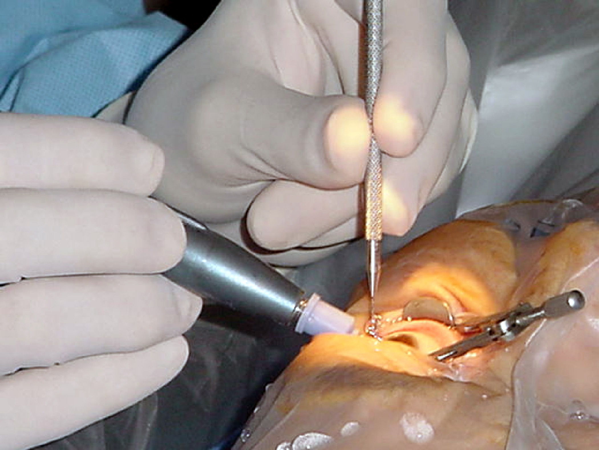
Cataract surgery, also called lens replacement surgery, is the removal of the natural lens of the eye (also called "crystalline lens") that has developed an opacification, which is referred to as a cataract, and its replacement with an intraocular lens. Metabolic changes of the crystalline lens fibers over time lead to the development of the cataract, causing impairment or loss of vision. Some infants are born with congenital cataracts, and certain environmental factors may also lead to cataract formation. Early symptoms may include strong glare from lights and small light sources at night, and reduced acuity at low light levels. During cataract surgery, a patient's cloudy natural cataract lens is removed, either by emulsification in place or by cutting it out. An artificial intraocular lens (IOL) is implanted in its place. Cataract surgery is generally performed by an ophthalmologist in an ambulatory setting at a surgical center or hospital rather than an inpatient setting. ... [...More Info...] [...Related Items...] OR: [Wikipedia] [Google] [Baidu] |
Cataract Before & After Cataract Surgery
A cataract is a cloudy area in the lens (anatomy), lens of the eye that leads to a visual impairment, decrease in vision. Cataracts often develop slowly and can affect one or both eyes. Symptoms may include faded colors, blurry or double vision, halos around light, trouble with bright lights, and Nyctalopia, trouble seeing at night. This may result in trouble driving, reading, or recognizing faces. Poor vision caused by cataracts may also result in an increased risk of Falling (accident), falling and Depression (mood), depression. Cataracts cause 51% of all cases of blindness and 33% of visual impairment worldwide. Cataracts are most commonly due to senescence, aging but may also occur due to Trauma (medicine), trauma or radiation exposure, be congenital cataract, present from birth, or occur following eye surgery for other problems. Risk factors include diabetes mellitus, diabetes, longstanding use of corticosteroid medication, smoking tobacco, prolonged exposure to sunlight ... [...More Info...] [...Related Items...] OR: [Wikipedia] [Google] [Baidu] |
Aphakia
Aphakia is the absence of the lens of the eye Eyes are organs of the visual system. They provide living organisms with vision, the ability to receive and process visual detail, as well as enabling several photo response functions that are independent of vision. Eyes detect light and conv ..., due to surgical removal, such as in cataract surgery, a perforation, perforating wound or Corneal ulcer, ulcer, or congenital anomaly. It causes a loss of accommodation (eye), accommodation, high degree of farsightedness (hyperopia), and a deep anterior chamber of eyeball, anterior chamber. Complications include detachment of the vitreous membrane, vitreous or retina, and glaucoma. Babies are rarely born with aphakia. Occurrence most often results from surgery to remove congenital cataract. Congenital cataracts usually develop as a result of infection of the fetus or genetic reasons. It is often difficult to identify the exact cause of these cataracts, especially if only one eye is aff ... [...More Info...] [...Related Items...] OR: [Wikipedia] [Google] [Baidu] |
Retinal Detachment
Retinal detachment is a disorder of the eye in which the retina peels away from its underlying layer of support tissue. Initial detachment may be localized, but without rapid treatment the entire retina may detach, leading to vision loss and blindness. It is a surgical emergency. The retina is a thin layer of light-sensitive tissue on the back wall of the eye. The optical system of the eye focuses light on the retina much like light is focused on the film in a camera. The retina translates that focused image into neural impulses and sends them to the brain via the optic nerve. Occasionally, posterior vitreous detachment, injury or trauma to the eye or head may cause a small tear in the retina. The tear allows vitreous fluid to seep through it under the retina, and peel it away like a bubble in wallpaper. Diagnosis Symptoms As the retina is responsible for vision, persons experiencing a retinal detachment have vision loss. This can be painful or painless. Imaging Ultrasou ... [...More Info...] [...Related Items...] OR: [Wikipedia] [Google] [Baidu] |
Cataract
A cataract is a cloudy area in the lens of the eye that leads to a decrease in vision. Cataracts often develop slowly and can affect one or both eyes. Symptoms may include faded colors, blurry or double vision, halos around light, trouble with bright lights, and trouble seeing at night. This may result in trouble driving, reading, or recognizing faces. Poor vision caused by cataracts may also result in an increased risk of falling and depression. Cataracts cause 51% of all cases of blindness and 33% of visual impairment worldwide. Cataracts are most commonly due to aging but may also occur due to trauma or radiation exposure, be present from birth, or occur following eye surgery for other problems. Risk factors include diabetes, longstanding use of corticosteroid medication, smoking tobacco, prolonged exposure to sunlight, and alcohol. The underlying mechanism involves accumulation of clumps of protein or yellow-brown pigment in the lens that reduces transmission of ... [...More Info...] [...Related Items...] OR: [Wikipedia] [Google] [Baidu] |
Glaucoma
Glaucoma is a group of eye diseases that result in damage to the optic nerve (or retina) and cause vision loss. The most common type is open-angle (wide angle, chronic simple) glaucoma, in which the drainage angle for fluid within the eye remains open, with less common types including closed-angle (narrow angle, acute congestive) glaucoma and normal-tension glaucoma. Open-angle glaucoma develops slowly over time and there is no pain. Peripheral vision may begin to decrease, followed by central vision, resulting in blindness if not treated. Closed-angle glaucoma can present gradually or suddenly. The sudden presentation may involve severe eye pain, blurred vision, mid-dilated pupil, redness of the eye, and nausea. Vision loss from glaucoma, once it has occurred, is permanent. Eyes affected by glaucoma are referred to as being glaucomatous. Risk factors for glaucoma include increasing age, high pressure in the eye, a family history of glaucoma, and use of steroid medicatio ... [...More Info...] [...Related Items...] OR: [Wikipedia] [Google] [Baidu] |
Endothelial Cell
The endothelium is a single layer of squamous endothelial cells that line the interior surface of blood vessels and lymphatic vessels. The endothelium forms an interface between circulating blood or lymph in the lumen and the rest of the vessel wall. Endothelial cells form the barrier between vessels and tissue and control the flow of substances and fluid into and out of a tissue. Endothelial cells in direct contact with blood are called vascular endothelial cells whereas those in direct contact with lymph are known as lymphatic endothelial cells. Vascular endothelial cells line the entire circulatory system, from the heart to the smallest capillaries. These cells have unique functions that include fluid filtration, such as in the glomerulus of the kidney, blood vessel tone, hemostasis, neutrophil recruitment, and hormone trafficking. Endothelium of the interior surfaces of the heart chambers is called endocardium. An impaired function can lead to serious health issu ... [...More Info...] [...Related Items...] OR: [Wikipedia] [Google] [Baidu] |
Lens (optics)
A lens is a transmissive optical device which focuses or disperses a light beam by means of refraction. A simple lens consists of a single piece of transparent material, while a compound lens consists of several simple lenses (''elements''), usually arranged along a common axis. Lenses are made from materials such as glass or plastic, and are ground and polished or molded to a desired shape. A lens can focus light to form an image, unlike a prism, which refracts light without focusing. Devices that similarly focus or disperse waves and radiation other than visible light are also called lenses, such as microwave lenses, electron lenses, acoustic lenses, or explosive lenses. Lenses are used in various imaging devices like telescopes, binoculars and cameras. They are also used as visual aids in glasses to correct defects of vision such as myopia and hypermetropia. History The word '' lens'' comes from '' lēns'', the Latin name of the lentil (a seed of a ... [...More Info...] [...Related Items...] OR: [Wikipedia] [Google] [Baidu] |
Sulcus (anatomy)
In biological morphology and anatomy, a sulcus (pl. ''sulci'') is a furrow or fissure (Latin ''fissura'', plural ''fissurae''). It may be a groove, natural division, deep furrow, elongated cleft, or tear in the surface of a limb or an organ, most notably on the surface of the brain, but also in the lungs, certain muscles (including the heart), as well as in bones, and elsewhere. Many sulci are the product of a surface fold or junction, such as in the gums, where they fold around the neck of the tooth. In invertebrate zoology, a sulcus is a fold, groove, or boundary, especially at the edges of sclerites or between segments. In pollen a grain that is grooved by a sulcus is termed sulcate. Examples in anatomy Liver * Ligamentum teres hepatis fissure * Ligamentum venosum fissure * Portal fissure, found in the under-surface of the liver * Transverse fissure of liver, found in the lower surface of the liver * Umbilical fissure, found in front of the liver Lung *Azygos fissur ... [...More Info...] [...Related Items...] OR: [Wikipedia] [Google] [Baidu] |

_PHIL_4284_lores.jpg)


