|
Pontine Tegmentum
The pontine tegmentum, or dorsal pons, is the dorsal part of the pons located within the brainstem. The ventral part or ventral pons is known as the basilar part of the pons, or basilar pons. Along with the dorsal surface of the medulla oblongata, it forms part of the rhomboid fossa – the floor of the fourth ventricle. The pontine tegmentum is all the material dorsal from the basilar pons to the fourth ventricle, and includes the reticulotegmental nucleus, the pedunculopontine nucleus, the laterodorsal tegmental nucleus, and several cranial nerve nuclei. It also houses the pontine respiratory group of the respiratory center which includes the pneumotaxic centre, and the apneustic centre. Anatomy The pontine tegmentum contains nuclei of the cranial nerves ( trigeminal (5th), abducens (6th), facial (7th), and vestibulocochlear (8th) and their associated fibre tracts. The dorsal pons also contains the reticulotegmental nucleus, the mesopontine cholinergic system c ... [...More Info...] [...Related Items...] OR: [Wikipedia] [Google] [Baidu] |
Pons
The pons (from Latin , "bridge") is part of the brainstem that in humans and other mammals, lies inferior to the midbrain, superior to the medulla oblongata and anterior to the cerebellum. The pons is also called the pons Varolii ("bridge of Varolius"), after the Italian anatomist and surgeon Costanzo Varolio (1543–75). This region of the brainstem includes neural pathways and tracts that conduct signals from the brain down to the cerebellum and medulla, and tracts that carry the sensory signals up into the thalamus. Structure The pons in humans measures about in length. It is the part of the brainstem situated between the midbrain and the medulla oblongata. The horizontal ''medullopontine sulcus'' demarcates the boundary between the pons and medulla oblongata on the ventral aspect of the brainstem, and the roots of cranial nerves VI/VII/VIII emerge from the brainstem along this groove. The junction of pons, medulla oblongata, and cerebellum forms the cerebellopontine ... [...More Info...] [...Related Items...] OR: [Wikipedia] [Google] [Baidu] |
Facial Nerve
The facial nerve, also known as the seventh cranial nerve, cranial nerve VII, or simply CN VII, is a cranial nerve that emerges from the pons of the brainstem, controls the muscles of facial expression, and functions in the conveyance of taste sensations from the anterior two-thirds of the tongue. The nerve typically travels from the pons through the facial canal in the temporal bone and exits the skull at the stylomastoid foramen. It arises from the brainstem from an area posterior to the cranial nerve VI (abducens nerve) and anterior to cranial nerve VIII (vestibulocochlear nerve). The facial nerve also supplies preganglionic parasympathetic fibers to several head and neck ganglia. The facial and intermediate nerves can be collectively referred to as the nervus intermediofacialis. The path of the facial nerve can be divided into six segments: # intracranial (cisternal) segment (from brainstem pons to internal auditory canal) # meatal (canalicular) segment (with ... [...More Info...] [...Related Items...] OR: [Wikipedia] [Google] [Baidu] |
Muscles Of Facial Expression
The facial muscles are a group of striated skeletal muscles supplied by the facial nerve (cranial nerve VII) that, among other things, control facial expression. These muscles are also called mimetic muscles. They are only found in mammals, although they derive from neural crest cells found in all vertebrates. They are the only muscles that attach to the dermis. Structure The facial muscles are just under the skin ( subcutaneous) muscles that control facial expression. They generally originate from the surface of the skull bone (rarely the fascia), and insert on the skin of the face. When they contract, the skin moves. These muscles also cause wrinkles at right angles to the muscles’ action line. Nerve supply The facial muscles are supplied by the facial nerve (cranial nerve VII), with each nerve serving one side of the face. In contrast, the nearby masticatory muscles are supplied by the mandibular nerve, a branch of the trigeminal nerve (cranial nerve V). List of muscle ... [...More Info...] [...Related Items...] OR: [Wikipedia] [Google] [Baidu] |
Superior Salivary Nucleus
The salivatory nuclei are two general visceral efferent nuclei located in the caudal pons, dorsal and lateral to the facial nucleus. Their neurons give rise to preganglionic parasympathetic nerve fibers in the control of salivation.Digital version The superior salivatory nucleus supplies fibers to the intermediate nerve (part of the facial nerve (CN VII). The inferior salivatory nucleus supplies fibers to the glossopharyngeal nerve (CN IX). The nuclei may also be involved in parasympathetic control of (extracranial and intracranial) head vasculature. Superior salivatory nucleus The superior salivatory nucleus (or nucleus salivatorius superior) is a visceral motor cranial nerve nucleus of the facial nerve (CN VII). It is located in the pontine tegmentum. It projects pre-ganglionic visceral motor parasympathetic efferents (via CN VII) to the pterygopalatine ganglion, and submandibular ganglion. The term "lacrimal nucleus" is sometimes used to refer to a portion of the superi ... [...More Info...] [...Related Items...] OR: [Wikipedia] [Google] [Baidu] |
Facial Motor Nucleus
The facial motor nucleus is a collection of neurons in the brainstem that belong to the facial nerve (cranial nerve VII). These lower motor neurons innervate the muscles of facial expression and the stapedius. Structure The nucleus is situated in the caudal portion of the ventrolateral pontine tegmentum. Its axons take an unusual course, traveling dorsally and looping around the abducens nucleus, then traveling ventrally to exit the ventral pons medial to the spinal trigeminal nucleus. These axons form the motor component of the facial nerve, with parasympathetic and sensory components forming the intermediate nerve. The nucleus has a dorsal and ventral region, with neurons in the dorsal region innervating muscles of the upper face and neurons in the ventral region innervating muscles of the lower face. Function Because it innervates muscles derived from pharyngeal arches, the facial motor nucleus is considered part of the special visceral efferent (SVE) cell column, whic ... [...More Info...] [...Related Items...] OR: [Wikipedia] [Google] [Baidu] |
Abducens Nucleus
The abducens nucleus is the originating nucleus from which the abducens nerve (VI) emerges—a cranial nerve nucleus. This nucleus is located beneath the fourth ventricle in the Anatomical terms of location#Rostral, cranial, and caudal, caudal portion of the pons near the midline, Anatomical terms of location, medial to the sulcus limitans. The abducens nucleus along with the internal genu of the facial nerve make up the facial colliculus, a hump at the caudal end of the medial eminence on the dorsal aspect of the pons. Structure Two primary neuron types are located in the abducens nucleus: Motor neuron, motoneurons and Interneuron, interneurons. The former directly drive the contraction of the ipsilateral lateral rectus muscle via the abducens nerve (sixth cranial nerve); contraction of this muscle rotates the eye outward (abduction). The latter relay signals from the abducens nucleus to the contralateral oculomotor nucleus, where motoneurons drive the contraction of the ipsilate ... [...More Info...] [...Related Items...] OR: [Wikipedia] [Google] [Baidu] |
Spinal Nerve
A spinal nerve is a mixed nerve, which carries Motor neuron, motor, Sensory neuron, sensory, and Autonomic nervous system, autonomic signals between the spinal cord and the body. In the human body there are 31 pairs of spinal nerves, one on each side of the vertebral column. These are grouped into the corresponding cervical vertebrae, cervical, thoracic vertebrae, thoracic, lumbar vertebrae, lumbar, sacral vertebrae, sacral and coccygeal vertebrae, coccygeal regions of the spine. There are eight pairs of cervical nerves, twelve pairs of thoracic nerves, five pairs of lumbar nerves, five pairs of sacral nerves, and one pair of coccygeal nerves. The spinal nerves are part of the peripheral nervous system. Structure Each spinal nerve is a mixed nerve, formed from the combination of nerve root axon, fibers from its Dorsal root of spinal nerve, dorsal and Ventral root of spinal nerve, ventral roots. The dorsal root is the afferent nerve fiber, afferent sensory root and carries sen ... [...More Info...] [...Related Items...] OR: [Wikipedia] [Google] [Baidu] |
Sleep
Sleep is a state of reduced mental and physical activity in which consciousness is altered and certain Sensory nervous system, sensory activity is inhibited. During sleep, there is a marked decrease in muscle activity and interactions with the surrounding environment. While sleep differs from wakefulness in terms of the ability to react to Stimulus (physiology), stimuli, it still involves active Human brain, brain patterns, making it more reactive than a coma or disorders of consciousness. Sleep occurs in sleep cycle, repeating periods, during which the body alternates between two distinct modes: rapid eye movement sleep (REM) and Non-rapid eye movement sleep, non-REM sleep. Although REM stands for "rapid eye movement", this mode of sleep has many other aspects, including virtual Rapid eye movement sleep#Muscle, paralysis of the body. Dreams are a succession of images, ideas, emotions, and sensations that usually occur involuntarily in the mind during certain stages of sleep. ... [...More Info...] [...Related Items...] OR: [Wikipedia] [Google] [Baidu] |
Dorsal Respiratory Group
The respiratory center is located in the medulla oblongata and pons, in the brainstem. The respiratory center is made up of three major respiratory groups of neurons, two in the medulla and one in the pons. In the medulla they are the dorsal respiratory group, and the ventral respiratory group. In the pons, the pontine respiratory group includes two areas known as the pneumotaxic center and the apneustic center. The respiratory center is responsible for generating and maintaining the rhythm of respiration, and also of adjusting this in homeostatic response to physiological changes. The respiratory center receives input from chemoreceptors, mechanoreceptors, the cerebral cortex, and the hypothalamus in order to regulate the rate and depth of breathing. Input is stimulated by altered levels of oxygen, carbon dioxide, and blood pH, by hormonal changes relating to stress and anxiety from the hypothalamus, and also by signals from the cerebral cortex to give a conscious control of ... [...More Info...] [...Related Items...] OR: [Wikipedia] [Google] [Baidu] |
Locus Coeruleus
The locus coeruleus () (LC), also spelled locus caeruleus or locus ceruleus, is a nucleus in the pons of the brainstem involved with physiological responses to stress and panic. It is a part of the reticular activating system in the reticular formation. The locus coeruleus, which in Latin means "blue spot", is the principal site for brain synthesis of norepinephrine (noradrenaline). The locus coeruleus and the areas of the body affected by the norepinephrine it produces are described collectively as the locus coeruleus-noradrenergic system or LC-NA system. Norepinephrine may also be released directly into the blood from the adrenal medulla. Anatomy The locus coeruleus (LC) is located in the posterior area of the rostral pons in the lateral floor of the fourth ventricle. It is composed of mostly medium-size neurons. Melanin granules inside the neurons contribute to its blue colour. Thus, it is also known as the ''blue nucleus'', or the ''nucleus pigmentosus pontis'' (hea ... [...More Info...] [...Related Items...] OR: [Wikipedia] [Google] [Baidu] |
Raphe Nuclei
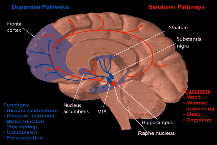
The raphe nuclei (, "seam") are a moderate-size cluster of nuclei found in the brain stem. They have 5-HT1 receptors which are coupled with Gi/Go-protein-inhibiting adenyl cyclase. They function as autoreceptors in the brain and decrease the release of serotonin. The anxiolytic drug Buspirone acts as partial agonist against these receptors. Selective serotonin reuptake inhibitor (SSRI) antidepressants are believed to act in these nuclei, as well as at their targets. Anatomy The raphe nuclei are traditionally considered to be the medial portion of the reticular formation, and appear as a ridge of cells in the center and most medial portion of the brain stem. In order from caudal to rostral, the raphe nuclei are known as the '' nucleus raphe obscurus'', the '' nucleus raphe pallidus'', the '' nucleus raphe magnus'', the '' nucleus raphe pontis'', the '' median raphe nucleus'', ''dorsal raphe nucleus'', ''caudal linear nucleus''. In the first systematic examination of the raph ... [...More Info...] [...Related Items...] OR: [Wikipedia] [Google] [Baidu] |
Pontine Nuclei
The pontine nuclei (or griseum pontis) are all the neurons of the ventral pons. Corticopontine fibres project from the primary motor cortex to the ipsilateral pontine nucleus; pontocerebellar fibers then relay the information to the contralateral cerebellum via the middle cerebellar peduncle. They are involved in motor function: the pontine nuclei are involved in adjusting movements according to their outcome (movement error correction), and are therefore important in learning motor skills. Anatomy The pontine nuclei encompass all of the about 20 million neurons scattered throughout the basilar part of pons. The pontine nuclei nuclei extend caudally into the medulla oblongata as the arcuate nucleus which is functionally homologous with the pontine nuclei. Afferents Corticopontine fibres arise primarily from the neocortex layer V of the premotor, somatosensory, non- striate visual, posterior parietal, and cingulate cerebral cortex; there are also a few fibers originating ... [...More Info...] [...Related Items...] OR: [Wikipedia] [Google] [Baidu] |


