|
Pneumatized
Skeletal pneumaticity is the presence of air spaces within bones. It is generally produced during development by excavation of bone by pneumatic diverticula (air sacs) from an air-filled space, such as the lungs or nasal cavity. Pneumatization is highly variable between individuals, and bones not normally pneumatized can become pneumatized in pathological development. Cranial pneumaticity Pneumatization occurs in the skulls of mammals, crocodilians and birds among extant tetrapods. Pneumatization has been documented in extinct archosaurs including dinosaurs and pterosaurs. Pneumatic spaces include the paranasal sinuses and some of the mastoid cells. Postcranial pneumaticity Postcranial pneumaticity is found largely in certain archosaur groups, namely saurischian dinosaurs, pterosaurs, and birds. Vertebral pneumatization is widespread among saurischian dinosaurs, and some theropods have quite widespread pneumatization, for example '' Aerosteon riocoloradensis'' has pneumatization ... [...More Info...] [...Related Items...] OR: [Wikipedia] [Google] [Baidu] |
Bird Anatomy
Bird anatomy, or the physiological structure of birds' bodies, shows many unique adaptations, mostly aiding flight. Birds have a light skeletal system and light but powerful musculature which, along with circulatory and respiratory systems capable of very high metabolic rates and oxygen supply, permit the bird to fly. The development of a beak has led to evolution of a specially adapted digestive system. Skeletal system Birds have many bones that are hollow ( pneumatized) with criss-crossing struts or trusses for structural strength. The number of hollow bones varies among species, though large gliding and soaring birds tend to have the most. Respiratory air sacs often form air pockets within the semi-hollow bones of the bird's skeleton. The bones of diving birds are often less hollow than those of non-diving species. Penguins, loons, puffins, and kiwis are without pneumatized bones entirely. Flightless birds, such as ostriches and emus, have pneumatized femurs and, in ... [...More Info...] [...Related Items...] OR: [Wikipedia] [Google] [Baidu] |
Sauropod
Sauropoda (), whose members are known as sauropods (; from '' sauro-'' + '' -pod'', 'lizard-footed'), is a clade of saurischian ('lizard-hipped') dinosaurs. Sauropods had very long necks, long tails, small heads (relative to the rest of their body), and four thick, pillar-like legs. They are notable for the enormous sizes attained by some species, and the group includes the largest animals to have ever lived on land. Well-known genera include '' Apatosaurus'', '' Argentinosaurus'', '' Alamosaurus'', ''Brachiosaurus'', '' Camarasaurus'', '' Diplodocus,'' and '' Mamenchisaurus''. The oldest known unequivocal sauropod dinosaurs are known from the Early Jurassic. '' Isanosaurus'' and '' Antetonitrus'' were originally described as Triassic sauropods, but their age, and in the case of ''Antetonitrus'' also its sauropod status, were subsequently questioned. Sauropod-like sauropodomorph tracks from the Fleming Fjord Formation (Greenland) might, however, indicate the occurrence of the g ... [...More Info...] [...Related Items...] OR: [Wikipedia] [Google] [Baidu] |
Paranasal Sinuses
Paranasal sinuses are a group of four paired air-filled spaces that surround the nasal cavity. The maxillary sinuses are located under the eyes; the frontal sinuses are above the eyes; the ethmoidal sinuses are between the eyes and the sphenoidal sinuses are behind the eyes. The sinuses are named for the facial bones and sphenoid bone in which they are located. Their role is disputed. Structure Humans possess four pairs of paranasal sinuses, divided into subgroups that are named according to the bones within which the sinuses lie. They are all innervated by branches of the trigeminal nerve (CN V). * The maxillary sinuses, the largest of the paranasal sinuses, are under the eyes, in the maxillary bones (open in the back of the semilunar hiatus of the nose). They are innervated by the maxillary nerve (CN V2). * The frontal sinuses, superior to the eyes, in the frontal bone, which forms the hard part of the forehead. They are innervated by the ophthalmic nerve (CN V1 ... [...More Info...] [...Related Items...] OR: [Wikipedia] [Google] [Baidu] |
Bird Feet And Legs
The anatomy of bird legs and feet is diverse, encompassing many accommodations to perform a wide variety of functions. Most birds are classified as digitigrade animals, meaning they walk on their toes rather than the entire foot. Some of the lower bones of the foot (the distals and most of the metatarsal) are fused to form the tarsometatarsus – a third segment of the leg, specific to birds. The upper bones of the foot ( proximals), in turn, are fused with the tibia to form the tibiotarsus, as over time the centralia disappeared. The fibula also reduced.; ; The legs are attached to a strong assembly consisting of the pelvic girdle extensively fused with the uniform spinal bone (also specific to birds) called the '' synsacrum'', built from some of the fused bones. Hindlimbs Birds are generally digitigrade animals ( toe-walkers), which affects the structure of their leg skeleton. They use only their hindlimbs to walk (bipedalism). Their forelimbs evolved to become ... [...More Info...] [...Related Items...] OR: [Wikipedia] [Google] [Baidu] |
Mastoid Cells
The mastoid cells (also called air cells of Lenoir or mastoid cells of Lenoir) are air-filled cavities within the mastoid process of the temporal bone of the cranium. The mastoid cells are a form of skeletal pneumaticity. Infection in these cells is called mastoiditis. The term ''cells'' here refers to enclosed spaces, not cells as living, biological units. Anatomy The mastoid air cells vary greatly in number, shape, and size; they may be extensive or minimal or even absent. The cells are typically interconnected and their walls lined by mucosa that is continuous with that of the mastoid antrum and tympanic cavity. Extent They may excavate the mastoid process to its tip, and be separated from the posterior cranial fossa and sigmoid sinus by a mere slip of bone or not at all. They may extend into the squamous part of temporal bone, petrous part of the temporal bone zygomatic process of temporal bone, and - rarely - the jugular process of occipital bone; they may thus c ... [...More Info...] [...Related Items...] OR: [Wikipedia] [Google] [Baidu] |
Paranasal Sinuses Numbers
Paranasal sinuses are a group of four paired air-filled spaces that surround the nasal cavity. The maxillary sinuses are located under the eyes; the frontal sinuses are above the eyes; the ethmoidal sinuses are between the eyes and the sphenoidal sinuses are behind the eyes. The sinuses are named for the facial bones and sphenoid bone in which they are located. Their role is disputed. Structure Humans possess four pairs of paranasal sinuses, divided into subgroups that are named according to the bones within which the sinuses lie. They are all innervated by branches of the trigeminal nerve (CN V). * The maxillary sinuses, the largest of the paranasal sinuses, are under the eyes, in the maxillary bones (open in the back of the semilunar hiatus of the nose). They are innervated by the maxillary nerve (CN V2). * The frontal sinuses, superior to the eyes, in the frontal bone, which forms the hard part of the forehead. They are innervated by the ophthalmic nerve (CN V1). * The ethmo ... [...More Info...] [...Related Items...] OR: [Wikipedia] [Google] [Baidu] |
Alouatta
Howler monkeys (genus ''Alouatta'', monotypic in subfamily Alouattinae) are the most widespread primate genus in the Neotropics and are among the largest of the platyrrhines along with the muriquis (''Brachyteles''), the spider monkeys (''Ateles'') and woolly monkeys (''Lagotrix''). The monkeys are native to South and Central American forests. They are famous for their howls, which can be heard from a distance through dense rain forest. Fifteen species are recognized. Previously classified in the family Cebidae, they are now placed in the family Atelidae. They are primarily folivores but also significant frugivores, acting as seed dispersal agents through their digestive system and their locomotion. Threats include human predation, habitat destruction, illegal wildlife trade, and capture for pets or zoo animals. Classification Anatomy and physiology Howler monkeys have short snouts and wide-set, round nostrils. Their noses are very keen, and they can smell out food (pri ... [...More Info...] [...Related Items...] OR: [Wikipedia] [Google] [Baidu] |
Loon
Loons (North American English) or divers (British English, British / Irish English) are a group of aquatic birds found in much of North America and northern Eurasia. All living species of loons are members of the genus ''Gavia'', family (biology), family Gaviidae and order (biology), order Gaviiformes. Description Loons, which are the size of large ducks or small goose, geese, resemble these birds in shape when swimming. Like ducks and geese, but unlike coots (which are Rallidae) and grebes (Podicipedidae), the loon's toes are connected by Bird feet and legs#Webbing and lobation, webbing. The loons may be confused with the cormorants (Phalacrocoracidae), but can be distinguished from them by their distinct call. Cormorants are not-too-distant relatives of loons, and like them are heavy-set birds whose bellies, unlike those of ducks and geese, are submerged when swimming. Loons in flight resemble plump geese with seagulls' wings that are relatively small in proportion to their bu ... [...More Info...] [...Related Items...] OR: [Wikipedia] [Google] [Baidu] |
Lung
The lungs are the primary Organ (biology), organs of the respiratory system in many animals, including humans. In mammals and most other tetrapods, two lungs are located near the Vertebral column, backbone on either side of the heart. Their function in the respiratory system is to extract oxygen from the atmosphere and transfer it into the bloodstream, and to release carbon dioxide from the bloodstream into the atmosphere, in a process of gas exchange. Respiration is driven by different muscular systems in different species. Mammals, reptiles and birds use their musculoskeletal systems to support and foster breathing. In early tetrapods, air was driven into the lungs by the pharyngeal muscles via buccal pumping, a mechanism still seen in amphibians. In humans, the primary muscle that drives breathing is the Thoracic diaphragm, diaphragm. The lungs also provide airflow that makes Animal communication#Auditory, vocalisation including speech possible. Humans have two lungs, a ri ... [...More Info...] [...Related Items...] OR: [Wikipedia] [Google] [Baidu] |
Atlas Vertebra
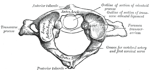
In anatomy, the atlas (C1) is the most superior (first) cervical vertebra of the spine and is located in the neck. The bone is named for Atlas of Greek mythology, just as Atlas bore the weight of the heavens, the first cervical vertebra supports the head. However, the term atlas was first used by the ancient Romans for the seventh cervical vertebra (C7) due to its suitability for supporting burdens. In Greek mythology, Atlas was condemned to bear the weight of the heavens as punishment for rebelling against Zeus. Ancient depictions of Atlas show the globe of the heavens resting at the base of his neck, on C7. Sometime around 1522, anatomists decided to call the first cervical vertebra the atlas. Scholars believe that by switching the designation atlas from the seventh to the first cervical vertebra Renaissance anatomists were commenting that the point of man's burden had shifted from his shoulders to his head—that man's true burden was not a physical load, but rather, his min ... [...More Info...] [...Related Items...] OR: [Wikipedia] [Google] [Baidu] |
JAMA Otolaryngology–Head & Neck Surgery
''JAMA Otolaryngology—Head & Neck Surgery'' is a monthly peer-reviewed medical journal published by the American Medical Association and covering all aspects of prevention, diagnosis, and treatment of diseases of the head, neck, ear, nose, and throat. The editor-in-chief is Jay F. Piccirillo (Washington University School of Medicine). It was established in 1925 as the ''Archives of Otolaryngology'' and renamed ''A.M.A. Archives of Otolaryngology'' in 1950, then renamed ''Archives of Otolaryngology–Head & Neck Surgery'' in 1960, before obtaining its current name in 2013. According to the ''Journal Citation Reports'', the journal has a 2021 impact factor of 8.961, ranking it 1st out of 43 titles in the category "Otorhinolaryngology". Naming history The journal has undergone several name changes in its history: *''JAMA Otorhinolaryngology—Head & Neck Surgery'' (2013-present, ) *''Archives of Otorhinolaryngology—Head & Neck Surgery'' (1986-2012, ) *''Archives of Otolaryngology ... [...More Info...] [...Related Items...] OR: [Wikipedia] [Google] [Baidu] |


