|
Peritoneal Cavity
The peritoneal cavity is a potential space located between the two layers of the peritoneum—the parietal peritoneum, the serous membrane that lines the abdominal wall, and visceral peritoneum, which surrounds the internal organs. While situated within the abdominal cavity, the term ''peritoneal cavity'' specifically refers to the potential space enclosed by these peritoneal membranes. The cavity contains a thin layer of lubricating serous fluid that enables the organs to move smoothly against each other, facilitating the movement and expansion of internal organs during digestion. The parietal and visceral peritonea are named according to their location and function. The peritoneal cavity, derived from the coelomic cavity in the embryo, is one of several body cavities, including the pleural cavities surrounding the lungs and the pericardial cavity around the heart. The peritoneal cavity is the largest serosal sac and fluid-filled cavity in the body, it secretes approximately ... [...More Info...] [...Related Items...] OR: [Wikipedia] [Google] [Baidu] [Amazon] |
Intraembryonic Coelom
In the development of the human embryo, the intraembryonic coelom (or somatic coelom) is a portion of the conceptus forming in the mesoderm during the third week of development. During the third week of development, the lateral plate mesoderm splits into a dorsal somatic mesoderm ( somatopleure) and a ventral splanchnic mesoderm ( splanchnopleure). The resulting cavity between the somatopleure and splanchnopleure is called the intraembryonic coelom. This space will give rise to the thoracic and abdominal cavities. The coelomic spaces in the lateral mesoderm and cardiogenic area are isolated. The isolated coelom The coelom (or celom) is the main body cavity in many animals and is positioned inside the body to surround and contain the digestive tract and other organs. In some animals, it is lined with mesothelium. In other animals, such as molluscs, i ... begins to organize into a horseshoe shape. The spaces soon join together and form a single horseshoe-shaped cavity: the int ... [...More Info...] [...Related Items...] OR: [Wikipedia] [Google] [Baidu] [Amazon] |
Omental Foramen
In human anatomy, the omental foramen (epiploic foramen, foramen of Winslow after the anatomist Jacob B. Winslow, or uncommonly aditus; ) is the passage of communication, or foramen, between the greater sac, and the lesser sac of the peritoneal cavity. Borders It has the following borders: * ''anterior'': the free border of the lesser omentum, known as the hepatoduodenal ligament. This has two layers and within these layers are the common bile duct, hepatic artery, and hepatic portal vein. * ''posterior'': the peritoneum covering the inferior vena cava * ''superior'': the peritoneum covering the caudate lobe of the liver * ''inferior'': the peritoneum covering the commencement of the duodenum and the hepatic artery, the latter passing forward below the foramen before ascending between the two layers of the lesser omentum. * ''left lateral'': gastrosplenic ligament and splenorenal ligament As the portal vein is the most posterior structure in the hepatoduodenal ligament, ... [...More Info...] [...Related Items...] OR: [Wikipedia] [Google] [Baidu] [Amazon] |
Ascites
Ascites (; , meaning "bag" or "sac") is the abnormal build-up of fluid in the abdomen. Technically, it is more than 25 ml of fluid in the peritoneal cavity, although volumes greater than one liter may occur. Symptoms may include increased abdominal size, increased weight, abdominal discomfort, and shortness of breath. Complications can include spontaneous bacterial peritonitis. In the developed world, the most common cause is liver cirrhosis. Other causes include cancer, heart failure, tuberculosis, pancreatitis, and blockage of the hepatic vein. In cirrhosis, the underlying mechanism involves high blood pressure in the portal system and dysfunction of blood vessels. Diagnosis is typically based on an examination together with ultrasound or a CT scan. Testing the fluid can help in determining the underlying cause. Treatment often involves a low-salt diet, medication such as diuretics, and draining the fluid. A transjugular intrahepatic portosystemic shunt (TIPS) may be ... [...More Info...] [...Related Items...] OR: [Wikipedia] [Google] [Baidu] [Amazon] |
Interstitium
In anatomy, the interstitium is a contiguous fluid-filled space existing between a structural barrier, such as a cell membrane or the skin, and internal structures, such as organs, including muscles and the circulatory system. The fluid in this space is called interstitial fluid, comprises water and solutes which drains into the lymph system. The interstitial compartment is composed of connective and supporting tissues within the body called the ( extracellular matrix) that are situated outside the blood and lymphatic vessels and the parenchyma of organs. The role of the interstitium in solute concentration, protein transport and hydrostatic pressure impacts human pathology and physiological responses such as edema, inflammation and shock. Structure The non-fluid parts of the interstitium are predominantly collagen types I, III, and V; elastin; and glycosaminoglycans, such as hyaluronan and proteoglycans, that are cross-linked to form a honeycomb-like reticulum. Collagen bundles ... [...More Info...] [...Related Items...] OR: [Wikipedia] [Google] [Baidu] [Amazon] |
Capillary Pressure
In fluid statics, capillary pressure () is the pressure between two immiscible fluids in a thin tube (see capillary action), resulting from the interactions of forces between the fluids and solid walls of the tube. Capillary pressure can serve as both an opposing or driving force for fluid transport and is a significant property for research and industrial purposes (namely microfluidic design and oil extraction from porous rock). It is also observed in natural phenomena. Definition Capillary pressure is defined as: :p_c=p_-p_ where: :p_ is the capillary pressure :p_ is the pressure of the non-wetting phase :p_ is the pressure of the wetting phase The wetting phase is identified by its ability to preferentially diffuse across the capillary walls before the non-wetting phase. The "wettability" of a fluid depends on its surface tension, the forces that drive a fluid's tendency to take up the minimal amount of space possible, and it is determined by the contact angle of the fluid.F ... [...More Info...] [...Related Items...] OR: [Wikipedia] [Google] [Baidu] [Amazon] |
Peritoneal Dialysis
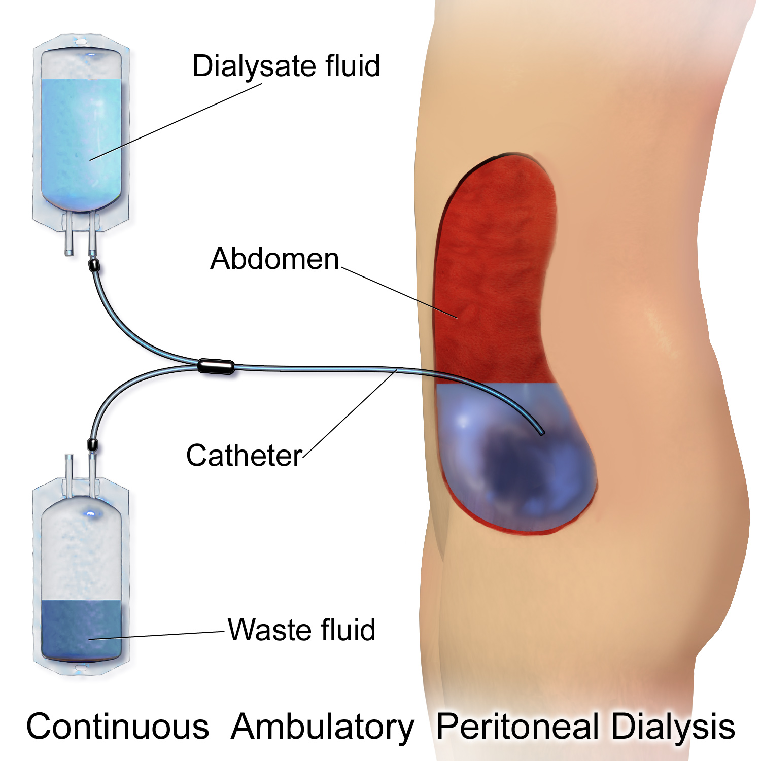
Peritoneal dialysis (PD) is a type of kidney dialysis, dialysis that uses the peritoneum in a person's abdomen as the membrane through which fluid and dissolved substances are exchanged with the blood. It is used to remove excess fluid, correct electrolyte problems, and remove toxins in those with kidney failure. Peritoneal dialysis has better outcomes than hemodialysis during the first two years. Other benefits include greater flexibility and better tolerability in those with significant heart disease. Side effects Complications may include peritonitis, infections within the abdomen, hernias, high blood sugar, bleeding in the abdomen, and blockage of the catheter. Peritoneal dialysis is not possible in those with significant prior abdominal surgery or inflammatory bowel disease. It requires some degree of technical skill to be done properly. Mechanism In peritoneal dialysis, a specific solution is introduced and then removed through a permanent tube in the lower abdomen. Th ... [...More Info...] [...Related Items...] OR: [Wikipedia] [Google] [Baidu] [Amazon] |
Chemotherapy Drug
Chemotherapy (often abbreviated chemo, sometimes CTX and CTx) is the type of cancer treatment that uses one or more anti-cancer drugs (chemotherapeutic agents or alkylating agents) in a standard regimen. Chemotherapy may be given with a curative intent (which almost always involves combinations of drugs), or it may aim only to prolong life or to reduce symptoms (palliative chemotherapy). Chemotherapy is one of the major categories of the medical discipline specifically devoted to pharmacotherapy for cancer, which is called ''medical oncology''. The term ''chemotherapy'' now means the non-specific use of intracellular poisons to inhibit mitosis (cell division) or to induce DNA damage (so that DNA repair can augment chemotherapy). This meaning excludes the more-selective agents that block extracellular signals (signal transduction). Therapies with specific molecular or genetic targets, which inhibit growth-promoting signals from classic endocrine hormones (primarily estrogens ... [...More Info...] [...Related Items...] OR: [Wikipedia] [Google] [Baidu] [Amazon] |
Intraperitoneal Injections
Intraperitoneal injection or IP injection is the injection of a substance into the peritoneum (body cavity). It is more often applied to non-human animals than to humans. In general, it is preferred when large amounts of blood replacement fluids are needed or when low blood pressure or other problems prevent the use of a suitable blood vessel for intravenous injection. In humans, the method is widely used to administer chemotherapy drugs to treat some cancers, particularly ovarian cancer. Although controversial, intraperitoneal use in ovarian cancer has been recommended as a standard of care. Fluids are injected intraperitoneally in infants, also used for peritoneal dialysis. Intraperitoneal injections are a way to administer therapeutics and drugs through a peritoneal route (body cavity). They are one of the few ways drugs can be administered through injection, and have uses in research involving animals, drug administration to treat ovarian cancers, and much more. Understandin ... [...More Info...] [...Related Items...] OR: [Wikipedia] [Google] [Baidu] [Amazon] |
Paracolic Gutter
The paracolic gutters (paracolic sulci, paracolic recesses) are peritoneal recesses – spaces between the colon and the abdominal wall. Structure There are two paracolic gutters: * The right lateral paracolic gutter. * The left medial paracolic gutter. The right and left paracolic gutters are peritoneal recesses on the posterior abdominal wall lying alongside the ascending and descending colon. The main paracolic gutter lies lateral to the colon on each side. A less obvious medial paracolic gutter may be formed, especially on the right side, if the colon possesses a short mesentery for part of its length. The right (lateral) paracolic gutter runs from the superiolateral aspect of the hepatic flexure of the colon, down the lateral aspect of the ascending colon, and around the cecum. It is continuous with the peritoneum as it descends into the pelvis over the pelvic brim. Superiorly, it is continuous with the peritoneum which lines the hepatorenal pouch and, through the epiploic fo ... [...More Info...] [...Related Items...] OR: [Wikipedia] [Google] [Baidu] [Amazon] |
Descending Colon
In the anatomy of humans and homologous primates, the descending colon is the part of the colon extending from the left colic flexure to the level of the iliac crest (whereupon it transitions into the sigmoid colon). The function of the descending colon in the digestive system is to store the remains of digested food that will be emptied into the rectum. The descending colon is on the left side of the body (barring any malformations). The term left colon is hypernymous to ''descending colon'' in precise use; many casual mentions of the left colon chiefly concern the descending colon. Structure The descending colon extends from the left colic flexure at the upper left part of the abdomen inferior-ward through the left hypochondrium and lumbar regions, along the outer border of the left kidney, ending at the level of the iliac crest at the lower left part of the abdomen, being continued thenceforth as the sigmoid colon. It is usually retroperitoneal (being lined by perit ... [...More Info...] [...Related Items...] OR: [Wikipedia] [Google] [Baidu] [Amazon] |
Small Intestine
The small intestine or small bowel is an organ (anatomy), organ in the human gastrointestinal tract, gastrointestinal tract where most of the #Absorption, absorption of nutrients from food takes place. It lies between the stomach and large intestine, and receives bile and pancreatic juice through the pancreatic duct to aid in digestion. The small intestine is about long and folds many times to fit in the abdomen. Although it is longer than the large intestine, it is called the small intestine because it is narrower in diameter. The small intestine has three distinct regions – the duodenum, jejunum, and ileum. The duodenum, the shortest, is where preparation for absorption through small finger-like protrusions called intestinal villus, intestinal villi begins. The jejunum is specialized for the absorption through its lining by enterocytes: small nutrient particles which have been previously digested by enzymes in the duodenum. The main function of the ileum is to absorb vitami ... [...More Info...] [...Related Items...] OR: [Wikipedia] [Google] [Baidu] [Amazon] |
Spleen
The spleen (, from Ancient Greek '' σπλήν'', splḗn) is an organ (biology), organ found in almost all vertebrates. Similar in structure to a large lymph node, it acts primarily as a blood filter. The spleen plays important roles in regard to red blood cells (erythrocytes) and the immune system. It removes old red blood cells and holds a reserve of blood, which can be valuable in case of Shock (circulatory), hemorrhagic shock, and also Human iron metabolism, recycles iron. As a part of the mononuclear phagocyte system, it metabolizes hemoglobin removed from senescent red blood cells. The globin portion of hemoglobin is degraded to its constitutive amino acids, and the heme portion is metabolized to bilirubin, which is removed in the liver. The spleen houses antibody-producing lymphocytes in its white pulp and monocytes which remove antibody-coated bacteria and antibody-coated blood cells by way of blood and lymph node circulation. These monocytes, upon moving to injured ... [...More Info...] [...Related Items...] OR: [Wikipedia] [Google] [Baidu] [Amazon] |


