|
Orbital Part Of Inferior Frontal Gyrus
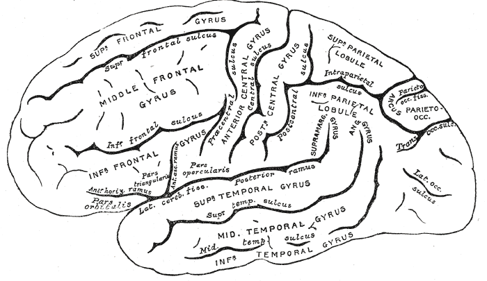
The orbital part of inferior frontal gyrus also known as the pars orbitalis is the orbital part of the inferior frontal gyrus. In humans, this region is bordered by the triangular part of the inferior frontal gyrus (pars triangularis) and, surrounding the anterior horizontal limb of the lateral sulcus, a portion of the opercular part of inferior frontal gyrus The inferior frontal gyrus (IFG; also gyrus frontalis inferior) is the lowest positioned gyrus of the frontal gyri, of the frontal lobe, and is part of the prefrontal cortex. Its superior border is the inferior frontal sulcus (which divides it ... (pars opercularis). Bounded caudally by the anterior ascending limb of the lateral sulcus, it borders on the insula in the depth of the lateral sulcus. It is bordered anteriorly/inferiorly by the lateral orbital sulcus. Cytoarchitectonically it is most closely represented by Brodmann area 47 (BA47).Brodmann, K. (1909). Vergleichende Lokalisationslehre der Grosshirnrind ... [...More Info...] [...Related Items...] OR: [Wikipedia] [Google] [Baidu] |
Orbit (anatomy)
In anatomy Anatomy () is the branch of morphology concerned with the study of the internal structure of organisms and their parts. Anatomy is a branch of natural science that deals with the structural organization of living things. It is an old scien ..., the orbit is the Body cavity, cavity or socket/hole of the skull in which the eye and Accessory visual structures, its appendages are situated. "Orbit" can refer to the bony socket, or it can also be used to imply the contents. In the adult human, the volume of the orbit is about , of which the eye occupies . The orbital contents comprise the eye, the Orbital fascia, orbital and retrobulbar fascia, extraocular muscles, cranial nerves optic nerve, II, oculomotor nerve, III, trochlear nerve, IV, trigeminal nerve, V, and abducens nerve, VI, blood vessels, fat, the lacrimal gland with its Lacrimal sac, sac and nasolacrimal duct, duct, the eyelids, Medial palpebral ligament, medial and Lateral palpebral raphe, lateral palpebr ... [...More Info...] [...Related Items...] OR: [Wikipedia] [Google] [Baidu] |
Inferior Frontal Gyrus
The inferior frontal gyrus (IFG; also gyrus frontalis inferior) is the lowest positioned gyrus of the frontal gyri, of the frontal lobe, and is part of the prefrontal cortex. Its superior border is the inferior frontal sulcus (which divides it from the middle frontal gyrus), its inferior border is the lateral sulcus (which divides it from the superior temporal gyrus) and its posterior border is the inferior precentral sulcus. Above it is the middle frontal gyrus, behind it is the precentral gyrus. The inferior frontal gyrus contains Broca's area, which is involved in language processing and speech production. Structure The inferior frontal gyrus is highly convoluted and has three cytoarchitecturally diverse regions. The three subdivisions are an opercular part, a triangular part, and an orbital part. These divisions are marked by two rami arising from the lateral sulcus. The ascending ramus separates the opercular and triangular parts. The anterior (horizontal) ramus ... [...More Info...] [...Related Items...] OR: [Wikipedia] [Google] [Baidu] |
Lateral Sulcus
The lateral sulcus (or lateral fissure, also called Sylvian fissure, after Franciscus Sylvius) is the most prominent sulcus (neuroanatomy), sulcus of each cerebral hemisphere in the human brain. The lateral sulcus (neuroanatomy), sulcus is a deep fissure (anatomy), fissure in each hemisphere that separates the frontal lobe, frontal and parietal lobes from the temporal lobe. The insular cortex lies deep within the lateral sulcus. Anatomy The lateral sulcus divides both the frontal lobe and parietal lobe above from the temporal lobe below. It is in both Cerebral hemisphere, hemispheres of the brain. The lateral Sulcus (neuroanatomy), sulcus is one of the earliest-developing sulci of the human brain, appearing around the fourteenth week of gestational age. The insular cortex lies deep within the lateral sulcus. The lateral sulcus has a number of side branches. Two of the most prominent and most regularly found are the ascending (also called vertical) ramus and the horizontal ramus ... [...More Info...] [...Related Items...] OR: [Wikipedia] [Google] [Baidu] |
Cytoarchitecture
Cytoarchitecture (from Greek κύτος 'cell' and ἀρχιτεκτονική 'architecture'), also known as cytoarchitectonics, is the study of the cellular composition of the central nervous system's tissues under the microscope. Cytoarchitectonics is one of the ways to parse the brain, by obtaining sections of the brain using a microtome and staining them with chemical agents which reveal where different neurons are located. The study of the parcellation of ''nerve fibers'' (primarily axons) into layers forms the subject of myeloarchitectonics (from Greek μυελός 'marrow' and ἀρχιτεκτονική 'architecture'), an approach complementary to cytoarchitectonics. History of the cerebral cytoarchitecture Defining cerebral cytoarchitecture began with the advent of histology—the science of slicing and staining brain slices for examination. It is credited to the Viennese psychiatrist Theodor Meynert (1833–1892), who in 1867 noticed regional variations in the ... [...More Info...] [...Related Items...] OR: [Wikipedia] [Google] [Baidu] |
Brodmann Area 47
Brodmann area 47, or BA47, is part of the frontal cortex in the human brain. It curves from the lateral surface of the frontal lobe into the ventral (orbital) frontal cortex. It is inferior to BA10 and BA45, and lateral to BA11. This cytoarchitectonic region most closely corresponds to the gyral region the orbital part of inferior frontal gyrus, although these regions are not equivalent. Pars orbitalis is not based on cytoarchitectonic distinctions, and rather is defined according to gross anatomical landmarks. Despite a clear distinction, these two terms are often used liberally in peer-reviewed research journals. BA47 is also known as orbital area 47. In the human, on the orbital surface it surrounds the caudal portion of the orbital sulcus (H) from which it extends laterally into the orbital part of inferior frontal gyrus (H). Cytoarchitectonically it is bounded caudally by the triangular area 45, medially by the prefrontal area 11 of Brodmann-1909, and rostrally by ... [...More Info...] [...Related Items...] OR: [Wikipedia] [Google] [Baidu] |
Gyrus
In neuroanatomy, a gyrus (: gyri) is a ridge on the cerebral cortex. It is generally surrounded by one or more sulci (depressions or furrows; : sulcus). Gyri and sulci create the folded appearance of the brain in humans and other mammals. Structure The gyri are part of a system of folds and ridges that create a larger surface area for the human brain and other mammalian brains. Because the brain is confined to the skull, brain size is limited. Ridges and depressions create folds allowing a larger cortical surface area, and greater cognitive function, to exist in the confines of a smaller cranium. Development The human brain undergoes gyrification during fetal and neonatal development. In embryonic development, all mammalian brains begin as smooth structures derived from the neural tube. A cerebral cortex without surface convolutions is lissencephalic, meaning 'smooth-brained'. As development continues, gyri and sulci begin to take shape on the fetal brain, with deepe ... [...More Info...] [...Related Items...] OR: [Wikipedia] [Google] [Baidu] |
Broca's Region
Broca's area, or the Broca area (, also , ), is a region in the frontal lobe of the dominant hemisphere, usually the left, of the brain with functions linked to speech production. Language processing has been linked to Broca's area since Pierre Paul Broca reported impairments in two patients. They had lost the ability to speak after injury to the posterior inferior frontal gyrus (pars triangularis) (BA45) of the brain. Since then, the approximate region he identified has become known as Broca's area, and the deficit in language production as Broca's aphasia, also called expressive aphasia. Broca's area is now typically defined in terms of the pars opercularis and pars triangularis of the inferior frontal gyrus, represented in Brodmann's cytoarchitectonic map as Brodmann area 44 and Brodmann area 45 of the dominant hemisphere. Functional magnetic resonance imaging (fMRI) has shown language processing to also involve the third part of the inferior frontal gyrus the pars orbitali ... [...More Info...] [...Related Items...] OR: [Wikipedia] [Google] [Baidu] |
Gyri
In neuroanatomy, a gyrus (: gyri) is a ridge on the cerebral cortex. It is generally surrounded by one or more sulcus (neuroanatomy), sulci (depressions or furrows; : sulcus). Gyri and sulci create the folded appearance of the brain in humans and other mammals. Structure The gyri are part of a system of folds and ridges that create a larger surface area for the human brain and other mammalian brains. Because the brain is confined to the skull, brain size is limited. Ridges and depressions create folds allowing a larger cortical surface area, and greater cognitive function, to exist in the confines of a smaller cranium. Development The human brain undergoes gyrification during fetal and neonatal development. In embryonic development, all mammalian brains begin as smooth structures derived from the neural tube. A cerebral cortex without surface convolutions is Lissencephaly, lissencephalic, meaning 'smooth-brained'. As development continues, gyri and Sulcus (neuroanatomy), ... [...More Info...] [...Related Items...] OR: [Wikipedia] [Google] [Baidu] |


