|
Mesovarium
The mesovarium is the portion of the broad ligament of the uterus that suspends the ovaries. The ovary is not covered by the mesovarium; rather, it is covered by germinal epithelium. At first the mesonephros and genital ridge are suspended by a common mesentery, but as the embryo grows the genital ridge gradually becomes pinched off from the mesonephros, with which it is at first continuous, though it still remains connected to the remnant of this body by a fold of peritoneum. In the male this is the mesorchium, and in the female, this is the mesovarium. See also * Mesometrium * Mesosalpinx The mesosalpinx is part of the lining of the abdominal cavity in higher vertebrates, specifically the portion of the broad ligament that stretches from the ovary to the level of the fallopian tube. See also * Mesometrium * Mesovarium * Salpin ... External links * - "The Female Pelvis: The Broad ligament" * - "Posterior view of the broad ligament of the uterus, on the left side." * ... [...More Info...] [...Related Items...] OR: [Wikipedia] [Google] [Baidu] |
Broad Ligament
The broad ligament of the uterus is the wide fold of peritoneum that connects the sides of the uterus to the walls and floor of the pelvis. Structure Subdivisions Contents The contents of the broad ligament include the following: * Reproductive ** uterine tubes (or Fallopian tube) ** ovary (some sources consider the ovary to be on the broad ligament, but not in it.) * vessels ** ovarian artery (in the suspensory ligament) ** uterine artery (in reality, travels in the cardinal ligament) * ligaments ** ovarian ligament ** round ligament of uterus ** suspensory ligament of the ovary (Some sources consider it a part of the broad ligament, while other sources just consider it a "termination" of the ligament.) Relations The peritoneum surrounds the uterus like a flat sheet that folds over its fundus, covering it anteriorly and posteriorly; on the sides of the uterus, this sheet of peritoneum comes in direct contact with itself, forming the double layer of peritoneum known ... [...More Info...] [...Related Items...] OR: [Wikipedia] [Google] [Baidu] |
Mesentery
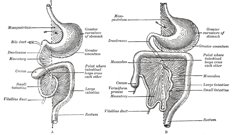
The mesentery is an organ that attaches the intestines to the posterior abdominal wall in humans and is formed by the double fold of peritoneum. It helps in storing fat and allowing blood vessels, lymphatics, and nerves to supply the intestines, among other functions. The mesocolon was thought to be a fragmented structure, with all named parts—the ascending, transverse, descending, and sigmoid mesocolons, the mesoappendix, and the mesorectum—separately terminating their insertion into the posterior abdominal wall. However, in 2012, new microscopic and electron microscopic examinations showed the mesocolon to be a single structure derived from the duodenojejunal flexure and extending to the distal mesorectal layer. Thus, the mesentery is an internal organ. Structure The mesentery of the small intestine arises from the root of the mesentery (or mesenteric root) and is the part connected with the structures in front of the vertebral column. The root is narrow, about ... [...More Info...] [...Related Items...] OR: [Wikipedia] [Google] [Baidu] |
Peritoneum
The peritoneum is the serous membrane forming the lining of the abdominal cavity or coelom in amniotes and some invertebrates, such as annelids. It covers most of the intra-abdominal (or coelomic) organs, and is composed of a layer of mesothelium supported by a thin layer of connective tissue. This peritoneal lining of the cavity supports many of the abdominal organs and serves as a conduit for their blood vessels, lymphatic vessels, and nerves. The abdominal cavity (the space bounded by the vertebrae, abdominal muscles, diaphragm, and pelvic floor) is different from the intraperitoneal space (located within the abdominal cavity but wrapped in peritoneum). The structures within the intraperitoneal space are called "intraperitoneal" (e.g., the stomach and intestines), the structures in the abdominal cavity that are located behind the intraperitoneal space are called "retroperitoneal" (e.g., the kidneys), and those structures below the intraperitoneal space are called ... [...More Info...] [...Related Items...] OR: [Wikipedia] [Google] [Baidu] |
Mesosalpinx
The mesosalpinx is part of the lining of the abdominal cavity in higher vertebrates, specifically the portion of the broad ligament that stretches from the ovary to the level of the fallopian tube. See also * Mesometrium * Mesovarium The mesovarium is the portion of the broad ligament of the uterus that suspends the ovaries. The ovary is not covered by the mesovarium; rather, it is covered by germinal epithelium. At first the mesonephros and genital ridge are suspended by a ... * Salpinx in anatomy References External links * - "Posterior view of the broad ligament of the uterus, on the left side." * - "The Female Pelvis - The Broad Ligament" * * * (, ) * Mammal female reproductive system {{ligament-stub ... [...More Info...] [...Related Items...] OR: [Wikipedia] [Google] [Baidu] |
Broad Ligament
The broad ligament of the uterus is the wide fold of peritoneum that connects the sides of the uterus to the walls and floor of the pelvis. Structure Subdivisions Contents The contents of the broad ligament include the following: * Reproductive ** uterine tubes (or Fallopian tube) ** ovary (some sources consider the ovary to be on the broad ligament, but not in it.) * vessels ** ovarian artery (in the suspensory ligament) ** uterine artery (in reality, travels in the cardinal ligament) * ligaments ** ovarian ligament ** round ligament of uterus ** suspensory ligament of the ovary (Some sources consider it a part of the broad ligament, while other sources just consider it a "termination" of the ligament.) Relations The peritoneum surrounds the uterus like a flat sheet that folds over its fundus, covering it anteriorly and posteriorly; on the sides of the uterus, this sheet of peritoneum comes in direct contact with itself, forming the double layer of peritoneum known ... [...More Info...] [...Related Items...] OR: [Wikipedia] [Google] [Baidu] |
Embryo
An embryo is an initial stage of development of a multicellular organism. In organisms that reproduce sexually, embryonic development is the part of the life cycle that begins just after fertilization of the female egg cell by the male sperm cell. The resulting fusion of these two cells produces a single-celled zygote that undergoes many cell divisions that produce cells known as blastomeres. The blastomeres are arranged as a solid ball that when reaching a certain size, called a morula, takes in fluid to create a cavity called a blastocoel. The structure is then termed a blastula, or a blastocyst in mammals. The mammalian blastocyst hatches before implantating into the endometrial lining of the womb. Once implanted the embryo will continue its development through the next stages of gastrulation, neurulation, and organogenesis. Gastrulation is the formation of the three germ layers that will form all of the different parts of the body. Neurulation forms the ner ... [...More Info...] [...Related Items...] OR: [Wikipedia] [Google] [Baidu] |
Uterus
The uterus (from Latin ''uterus'', plural ''uteri'') or womb () is the organ in the reproductive system of most female mammals, including humans that accommodates the embryonic and fetal development of one or more embryos until birth. The uterus is a hormone-responsive sex organ that contains glands in its lining that secrete uterine milk for embryonic nourishment. In the human, the lower end of the uterus, is a narrow part known as the isthmus that connects to the cervix, leading to the vagina. The upper end, the body of the uterus, is connected to the fallopian tubes, at the uterine horns, and the rounded part above the openings to the fallopian tubes is the fundus. The connection of the uterine cavity with a fallopian tube is called the uterotubal junction. The fertilized egg is carried to the uterus along the fallopian tube. It will have divided on its journey to form a blastocyst that will implant itself into the lining of the uterus – the endometrium, w ... [...More Info...] [...Related Items...] OR: [Wikipedia] [Google] [Baidu] |
Ovary
The ovary is an organ in the female reproductive system that produces an ovum. When released, this travels down the fallopian tube into the uterus, where it may become fertilized by a sperm. There is an ovary () found on each side of the body. The ovaries also secrete hormones that play a role in the menstrual cycle and fertility. The ovary progresses through many stages beginning in the prenatal period through menopause. It is also an endocrine gland because of the various hormones that it secretes. Structure The ovaries are considered the female gonads. Each ovary is whitish in color and located alongside the lateral wall of the uterus in a region called the ovarian fossa. The ovarian fossa is the region that is bounded by the external iliac artery and in front of the ureter and the internal iliac artery. This area is about 4 cm x 3 cm x 2 cm in size.Daftary, Shirish; Chakravarti, Sudip (2011). Manual of Obstetrics, 3rd Edition. Elsevier. pp. 1-16. . The ... [...More Info...] [...Related Items...] OR: [Wikipedia] [Google] [Baidu] |
Mesonephros
The mesonephros ( el, middle kidney) is one of three excretory organs that develop in vertebrates. It serves as the main excretory organ of aquatic vertebrates and as a temporary kidney in reptiles, birds, and mammals. The mesonephros is included in the Wolffian body after Caspar Friedrich Wolff who described it in 1759. (The Wolffian body is composed of: mesonephros + paramesonephrotic blastema) Structure The mesonephros acts as a structure similar to the kidney that, in humans, functions between the sixth and tenth weeks of embryological life. Despite the similarity in structure, function, and terminology, however, the mesonephric nephrons do not form any part of the mature kidney or nephrons. In humans, the mesonephros consists of units which are similar in structure and function to nephrons of the adult kidney. Each of these consists of a glomerulus, a tuft of capillaries which arises from lateral branches of dorsal aorta and drains into the inferior cardinal vein; a Bowman ... [...More Info...] [...Related Items...] OR: [Wikipedia] [Google] [Baidu] |
Genital Ridge
The genital ridge (or gonadal ridge) is the precursor to the gonads. The genital ridge initially consists mainly of mesenchyme and cells of underlying mesonephric origin. Once oogonia enter this area they attempt to associate with these somatic cells. Development proceeds and the oogonia become fully surrounded by a layer of cells (pre- granulosa cells). The genital ridge appears at approximately five weeks, and gives rise to the sex cords. Associated genes Genes associated with the developing gonad can be categorized into those that form the sexually indifferent gonad, those that determine whether the indifferent gonad will differentiate as male or female, and those that promote differentiation into male or female parts. Genes that form the sexually indifferent gonad are '' SF1'' and '' WT1''. Genes that determine sex are '' SRY'', '' SOX9'', and ''DAX1''. Genes driving the differentiation into male or female structures are ''SF1'', ''WT1'', and '' WNT4''. SRY is the only ge ... [...More Info...] [...Related Items...] OR: [Wikipedia] [Google] [Baidu] |
Mesorchium
The testes, at an early period of foetal life, are placed at the back part of the abdominal cavity, behind the peritoneum, and each is attached by a peritoneal fold, the mesorchium, to the mesonephros. See also * mesentery The mesentery is an organ that attaches the intestines to the posterior abdominal wall in humans and is formed by the double fold of peritoneum. It helps in storing fat and allowing blood vessels, lymphatics, and nerves to supply the intesti ... * mesovarium Mesorchium is the fibrous sheath which attaches vascular and avascular structures of spermatic cord together. References External links * - "Inguinal Region, Scrotum and Testes: Coverings of the Testis" * Animal developmental biology {{Portal bar, Anatomy ... [...More Info...] [...Related Items...] OR: [Wikipedia] [Google] [Baidu] |


