|
Melanosis Coli
Melanosis coli, also pseudomelanosis coli, is a disorder of pigmentation of the wall of the colon, often identified at the time of colonoscopy. It is benign and may have no significant correlation with disease. The brown pigment is lipofuscin in macrophages, not melanin. Cause The most common cause of melanosis coli is the extended use of laxatives, and commonly anthraquinone containing laxatives such as senna, aloe vera, and other plant glycosides. The anthranoid laxatives pass through the gastrointestinal tract unabsorbed until they reach the large intestine, where they are changed into their active forms. The resulting active compounds cause damage to the cells in the lining of the intestine and leads to apoptosis (a form of cell death). The damaged (apoptotic) cells appear as darkly pigmented bodies that may be taken up by scavenger cells known as macrophages. When enough cells have been damaged, the characteristic pigmentation of the bowel wall develops. The condition can ... [...More Info...] [...Related Items...] OR: [Wikipedia] [Google] [Baidu] |
Colonoscopy
Colonoscopy () or coloscopy () is the endoscopic examination of the large bowel and the distal part of the small bowel with a CCD camera or a fiber optic camera on a flexible tube passed through the anus. It can provide a visual diagnosis (''e.g.,'' ulceration, polyps) and grants the opportunity for biopsy or removal of suspected colorectal cancer lesions. Colonoscopy can remove polyps smaller than one millimeter. Once polyps are removed, they can be studied with the aid of a microscope to determine if they are precancerous or not. Colonoscopy is similar to sigmoidoscopy—the difference being related to which parts of the colon each can examine. A colonoscopy allows an examination of the entire colon (1,200–1,500mm in length). A sigmoidoscopy allows an examination of the distal portion (about 600mm) of the colon, which may be sufficient because benefits to cancer survival of colonoscopy have been limited to the detection of lesions in the distal portion of the colon. [...More Info...] [...Related Items...] OR: [Wikipedia] [Google] [Baidu] |
Biopsy
A biopsy is a medical test commonly performed by a surgeon, interventional radiologist, or an interventional cardiologist. The process involves extraction of sample cells or tissues for examination to determine the presence or extent of a disease. The tissue is then fixed, dehydrated, embedded, sectioned, stained and mounted before it is generally examined under a microscope by a pathologist; it may also be analyzed chemically. When an entire lump or suspicious area is removed, the procedure is called an excisional biopsy. An incisional biopsy or core biopsy samples a portion of the abnormal tissue without attempting to remove the entire lesion or tumor. When a sample of tissue or fluid is removed with a needle in such a way that cells are removed without preserving the histological architecture of the tissue cells, the procedure is called a needle aspiration biopsy. Biopsies are most commonly performed for insight into possible cancerous or inflammatory conditions. History T ... [...More Info...] [...Related Items...] OR: [Wikipedia] [Google] [Baidu] |
Colostomy
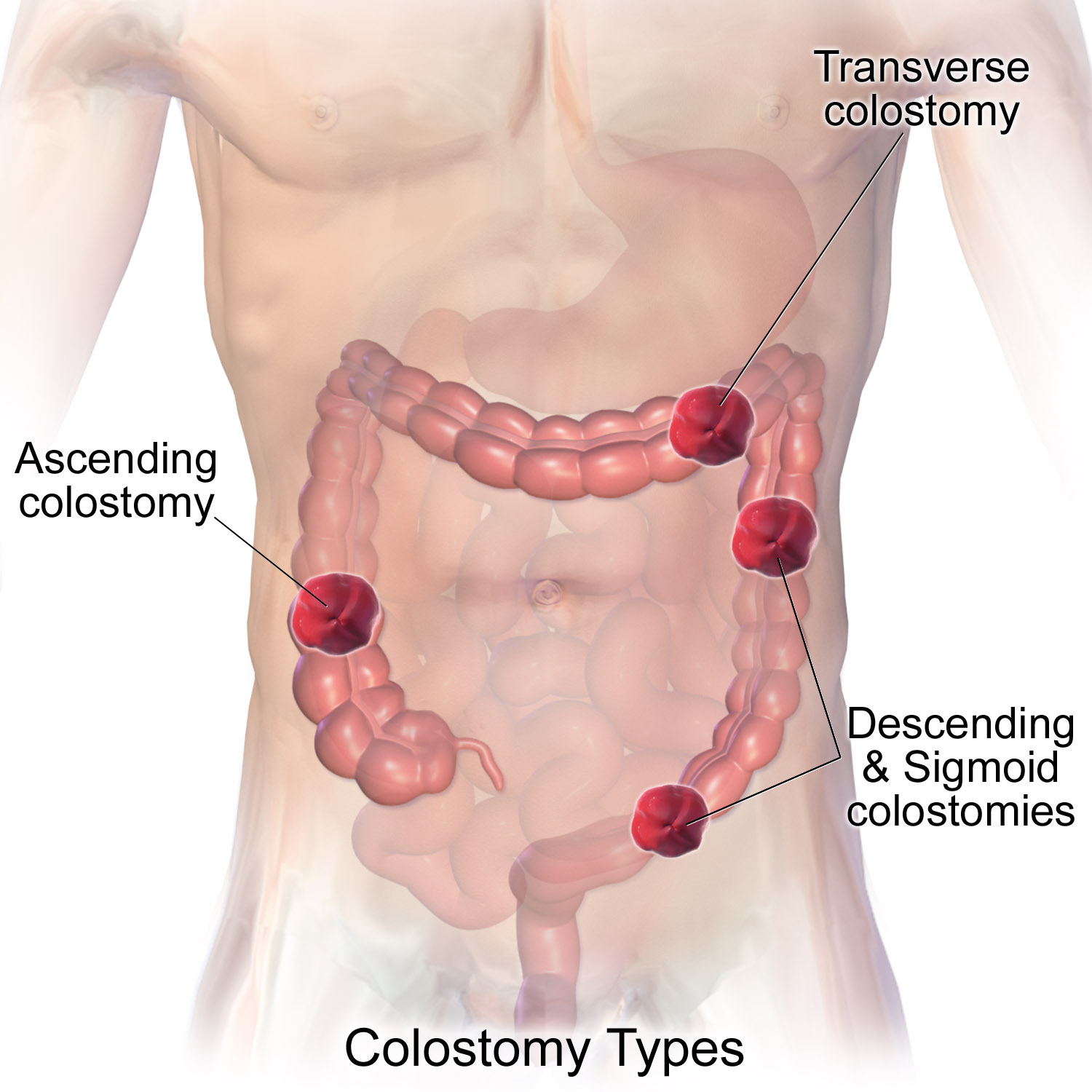
A colostomy is an opening ( stoma) in the large intestine (colon), or the surgical procedure that creates one. The opening is formed by drawing the healthy end of the colon through an incision in the anterior abdominal wall and suturing it into place. This opening, often in conjunction with an attached ostomy system, provides an alternative channel for feces to leave the body. Thus if the natural anus is unavailable for that function (for example, in cases where it has been removed in the fight against colorectal cancer or ulcerative colitis), an artificial anus takes over. It may be reversible or irreversible, depending on the circumstances. Uses There are many reasons for this procedure. Some common reasons are: * A part of the colon has been removed, e.g. due to colon cancer requiring a total mesorectal excision, diverticulitis, injury, etc., so that it is no longer possible for feces to exit via the anus. * A part of the colon has been operated upon and needs to be 'r ... [...More Info...] [...Related Items...] OR: [Wikipedia] [Google] [Baidu] |
Digestive Tract
The gastrointestinal tract (GI tract, digestive tract, alimentary canal) is the tract or passageway of the digestive system that leads from the mouth to the anus. The GI tract contains all the major organs of the digestive system, in humans and other animals, including the esophagus, stomach, and intestines. Food taken in through the mouth is digested to extract nutrients and absorb energy, and the waste expelled at the anus as feces. ''Gastrointestinal'' is an adjective meaning of or pertaining to the stomach and intestines. Most animals have a "through-gut" or complete digestive tract. Exceptions are more primitive ones: sponges have small pores ( ostia) throughout their body for digestion and a larger dorsal pore (osculum) for excretion, comb jellies have both a ventral mouth and dorsal anal pores, while cnidarians and acoels have a single pore for both digestion and excretion. The human gastrointestinal tract consists of the esophagus, stomach, and intestines, and is ... [...More Info...] [...Related Items...] OR: [Wikipedia] [Google] [Baidu] |
Smoker's Melanosis
Smoker's melanosis is seen with the naked eye as a brown to black pigmentation of the oral tissue i.e. the gums, cheeks or palate Axéll T, Hedin CA: Epidemiologic study of excessive oral melanin pigmentation with special reference to the influence of tobacco habits. Scand J Dent Res 1982; 90:434-442. as well as in larynx. It is most often seen in the lower labial gingiva of tobacco users. Most easily it is found in Caucasian race, Caucasians, due to their lack of a genetically caused melanin pigmentation.Hedin CA: Smoker's Melanosis. Occurrence and localization in the attached gingiva. Arch Dermatol 1977; 113:1533-1538. The brown to black colour is melanin. In skin, melanin prevents harmful UV-light from reaching deeper, sensible parts of the tissue. If UV-light penetrates deep, some of the toxic substances due to the UV-light damage to the cells, are bound to melanin in the Epithelium, epithelial cells and travel with the ageing cells to the skin surface, where they are expelle ... [...More Info...] [...Related Items...] OR: [Wikipedia] [Google] [Baidu] |
Peutz–Jeghers Syndrome
Peutz–Jeghers syndrome (often abbreviated PJS) is an autosomal dominant genetic disorder characterized by the development of benign hamartomatous polyps in the gastrointestinal tract and hyperpigmented macules on the lips and oral mucosa (melanosis). This syndrome can be classed as one of various hereditary intestinal polyposis syndromes and one of various hamartomatous polyposis syndromes. It has an incidence of approximately 1 in 25,000 to 300,000 births. Signs and symptoms The risks associated with this syndrome include a substantial risk of cancer, especially of the breast and gastrointestinal tracts. Colorectal is the most common malignancy, with a lifetime risk of 39 percent, followed by breast cancer in females with a lifetime risk of 32 to 54 percent. Patients with the syndrome also have an increased risk of developing carcinomas of the liver, lungs, breast, ovaries, uterus, testes, and other organs. Specifically, it is associated with an increased risk of sex-cord strom ... [...More Info...] [...Related Items...] OR: [Wikipedia] [Google] [Baidu] |
H&E Stain
Hematoxylin and eosin stain ( or haematoxylin and eosin stain or hematoxylin-eosin stain; often abbreviated as H&E stain or HE stain) is one of the principal tissue stains used in histology. It is the most widely used stain in medical diagnosis and is often the gold standard. For example, when a pathologist looks at a biopsy of a suspected cancer, the histological section is likely to be stained with H&E. H&E is the combination of two histological stains: hematoxylin and eosin. The hematoxylin stains cell nuclei a purplish blue, and eosin stains the extracellular matrix and cytoplasm pink, with other structures taking on different shades, hues, and combinations of these colors. Hence a pathologist can easily differentiate between the nuclear and cytoplasmic parts of a cell, and additionally, the overall patterns of coloration from the stain show the general layout and distribution of cells and provides a general overview of a tissue sample's structure. Thus, pattern recogniti ... [...More Info...] [...Related Items...] OR: [Wikipedia] [Google] [Baidu] |
Lamina Propria
The lamina propria is a thin layer of connective tissue that forms part of the moist linings known as mucous membranes or mucosae, which line various tubes in the body, such as the respiratory tract, the gastrointestinal tract, and the urogenital tract. The lamina propria is a thin layer of loose (areolar) connective tissue, which lies beneath the epithelium, and together with the epithelium and basement membrane constitutes the mucosa. As its Latin name indicates, it is a characteristic component of the mucosa, or the mucosa's "own special layer." Thus, the term mucosa or mucous membrane refers to the combination of the epithelium and the lamina propria. The connective tissue of the lamina propria is loose and rich in cells. The cells of the lamina propria are variable and can include fibroblasts, lymphocytes, plasma cells, macrophages, eosinophilic leukocytes, and mast cells. It provides support and nutrition to the epithelium, as well as the means to bind to the underl ... [...More Info...] [...Related Items...] OR: [Wikipedia] [Google] [Baidu] |
Macrophages
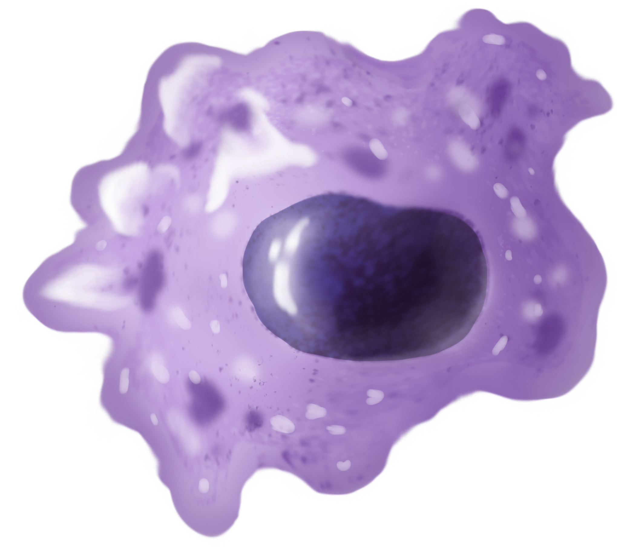
Macrophages (abbreviated as M φ, MΦ or MP) ( el, large eaters, from Greek ''μακρός'' (') = large, ''φαγεῖν'' (') = to eat) are a type of white blood cell of the immune system that engulfs and digests pathogens, such as cancer cells, microbes, cellular debris, and foreign substances, which do not have proteins that are specific to healthy body cells on their surface. The process is called phagocytosis, which acts to defend the host against infection and injury. These large phagocytes are found in essentially all tissues, where they patrol for potential pathogens by amoeboid movement. They take various forms (with various names) throughout the body (e.g., histiocytes, Kupffer cells, alveolar macrophages, microglia, and others), but all are part of the mononuclear phagocyte system. Besides phagocytosis, they play a critical role in nonspecific defense (innate immunity) and also help initiate specific defense mechanisms (adaptive immunity) by recruiting other immun ... [...More Info...] [...Related Items...] OR: [Wikipedia] [Google] [Baidu] |
Micrograph
A micrograph or photomicrograph is a photograph or digital image taken through a microscope or similar device to show a magnified image of an object. This is opposed to a macrograph or photomacrograph, an image which is also taken on a microscope but is only slightly magnified, usually less than 10 times. Micrography is the practice or art of using microscopes to make photographs. A micrograph contains extensive details of microstructure. A wealth of information can be obtained from a simple micrograph like behavior of the material under different conditions, the phases found in the system, failure analysis, grain size estimation, elemental analysis and so on. Micrographs are widely used in all fields of microscopy. Types Photomicrograph A light micrograph or photomicrograph is a micrograph prepared using an optical microscope, a process referred to as ''photomicroscopy''. At a basic level, photomicroscopy may be performed simply by connecting a camera to a microscope, th ... [...More Info...] [...Related Items...] OR: [Wikipedia] [Google] [Baidu] |
Hemosiderin-laden Macrophage
A siderophage is a hemosiderin-containing macrophage. Heart failure cells are siderophages generated in the alveoli of the lungs of people with left heart failure or chronic pulmonary edema, when the high pulmonary blood pressure causes red blood cells to pass through the vascular wall. Siderophages are not specific of heart failure. They are present wherever red blood cells encounter macrophages, such as pulmonary hemorrhage. In left heart failure, the left ventricle can not keep pace with the incoming blood from the pulmonary veins. The resulting backup causes increased pressure on the alveolar capillaries, and red blood cells leak out. Alveolar macrophages (dust cells) engulf the red blood cells, and become engorged with brownish hemosiderin. In chronic pulmonary edema, alveolar septa become thick and fibrous, again increasing pressure on alveolar capillaries and resulting in leakage of red blood cells which undergo phagocytosis by alveolar macrophage An alveolar macroph ... [...More Info...] [...Related Items...] OR: [Wikipedia] [Google] [Baidu] |
Differential Diagnosis
In healthcare, a differential diagnosis (abbreviated DDx) is a method of analysis of a patient's history and physical examination to arrive at the correct diagnosis. It involves distinguishing a particular disease or condition from others that present with similar clinical features. Differential diagnostic procedures are used by clinicians to diagnose the specific disease in a patient, or, at least, to consider any imminently life-threatening conditions. Often, each individual option of a possible disease is called a differential diagnosis (e.g., acute bronchitis could be a differential diagnosis in the evaluation of a cough, even if the final diagnosis is common cold). More generally, a differential diagnostic procedure is a systematic diagnostic method used to identify the presence of a disease entity where multiple alternatives are possible. This method may employ algorithms, akin to the process of elimination, or at least a process of obtaining information that shrinks the "p ... [...More Info...] [...Related Items...] OR: [Wikipedia] [Google] [Baidu] |


