|
Melanonychia
Melanonychia is a black or brown pigmentation of the normal nail plate, and may be present as a normal finding on many digits in Afro-Caribbeans, as a result of trauma, systemic disease, or medications, or as a postinflammatory event from such localized events as lichen planus or fixed drug eruption.James, William; Berger, Timothy; Elston, Dirk (2005). ''Andrews' Diseases of the Skin: Clinical Dermatology''. (10th ed.). Saunders. .Freedberg, et al. (2003). ''Fitzpatrick's Dermatology in General Medicine''. (6th ed.). McGraw-Hill. . There are two types, longitudinal and transverse melanonychia. Longitudinal melanonychia may be a sign of subungual melanoma (acral lentiginous melanoma),Baran, Robert, et al. 2008. ''Baran & Dawber's Diseases of the Nails and Their Management''. Oxford: Blackwell, p. 516. although there are other diagnoses such as chronic paronychia, onychomycosis, subungual hematoma, pyogenic granuloma, glomus tumour, subungual verruca, mucous cyst, subungual fibro ... [...More Info...] [...Related Items...] OR: [Wikipedia] [Google] [Baidu] |
List Of Cutaneous Conditions
Many skin conditions affect the human integumentary system—the organ system covering the entire surface of the body and composed of skin, hair, nails, and related muscle and glands. The major function of this system is as a barrier against the external environment. The skin weighs an average of four kilograms, covers an area of two square metres, and is made of three distinct layers: the epidermis, dermis, and subcutaneous tissue. The two main types of human skin are: glabrous skin, the hairless skin on the palms and soles (also referred to as the "palmoplantar" surfaces), and hair-bearing skin.Burns, Tony; ''et al''. (2006) ''Rook's Textbook of Dermatology CD-ROM''. Wiley-Blackwell. . Within the latter type, the hairs occur in structures called pilosebaceous units, each with hair follicle, sebaceous gland, and associated arrector pili muscle. In the embryo, the epidermis, hair, and glands form from the ectoderm, which is chemically influenced by the underlying meso ... [...More Info...] [...Related Items...] OR: [Wikipedia] [Google] [Baidu] |
Lichen Planus
Lichen planus (LP) is a chronic inflammatory and immune-mediated disease that affects the skin, nails, hair, and mucous membranes. It is not an actual lichen, and is only named that because it looks like one. It is characterized by polygonal, flat-topped, violaceous papules and plaques with overlying, reticulated, fine white scale ( Wickham's striae), commonly affecting dorsal hands, flexural wrists and forearms, trunk, anterior lower legs and oral mucosa. The hue may be gray-brown in people with darker skin. Although there is a broad clinical range of LP manifestations, the skin and oral cavity remain as the major sites of involvement. The cause is unknown, but it is thought to be the result of an autoimmune process with an unknown initial trigger. There is no cure, but many different medications and procedures have been used in efforts to control the symptoms. The term lichenoid reaction (lichenoid eruption or lichenoid lesion) refers to a lesion of similar or identical histopa ... [...More Info...] [...Related Items...] OR: [Wikipedia] [Google] [Baidu] |
Fixed Drug Eruption
Fixed drug reactions, are common and so named because they recur at the same site with each exposure to a particular medication. Medications inducing fixed drug eruptions are usually those taken intermittently. Signs and symptoms A painful and itchy reddish/purple patch of skin that occurs in the same location with repeated exposures to the culprit drug is the classic presentation of a fixed drug reaction. The lips, genitals, and hands are often involved. Cause Medications that are commonly implicated as a cause of fixed drug eruptions include the following: *Cetirizine *Ciprofloxacin *Clarithromycin *Cotrimoxazole * Doxycycline *Fluconazole * NSAIDs (e.g., ibuprofen, etoricoxib, naproxen) *Phenytoin * Pseudoephedrine *Trimethoprim See also * Drug eruption * List of cutaneous conditions * List of human leukocyte antigen alleles associated with cutaneous conditions There are many human leukocyte antigen (HLA) alleles associated with conditions of or affecting the human inte ... [...More Info...] [...Related Items...] OR: [Wikipedia] [Google] [Baidu] |
Acral Lentiginous Melanoma
Acral lentiginous melanoma is an aggressive type of skin cancer that is not caused by sunlight. Melanoma is a group of serious skin cancers that arise from pigment cells (melanocytes); acral lentiginous melanoma is a kind of lentiginous skin melanoma. Acral lentiginous melanoma is the most common subtype in people with darker skins and is rare in people with lighter skin types. It is not caused by exposure to sunlight or UV radiation, and wearing sunscreen does not protect against it. Acral lentiginous melanoma is commonly found on the palms, soles, under the nails, and in the oral mucosa. It occurs on non-hair-bearing surfaces of the body, which have not necessarily been exposed to sunlight. It is also found on mucous membranes. The absolute incidence of ALM is the same for people of all skin colors, and has not changed significantly for decades. However, because rates of other melanomas are low in non-white populations, ALM is the most common form of melanoma diagnosed am ... [...More Info...] [...Related Items...] OR: [Wikipedia] [Google] [Baidu] |
Chronic Paronychia
Paronychia is an inflammation of the skin around the nail, which can occur suddenly, when it is usually due to the bacterium '' Staphylococcus aureus'', or gradually when it is commonly caused by the fungus ''Candida albicans''. The term is from el, παρωνυχία from ''para'' 'around', ''onyx'' 'nail', and the abstract noun suffix '' -ia''. Risk factors include repeatedly washing hands and trauma to the cuticle such as may occur from repeated nail biting or hangnails. Treatment includes antibiotics and antifungals, and if pus is present, the consideration of incision and drainage. Paronychia is commonly misapplied as a synonym for herpetic whitlow or felon. Definition and etymology Paronychia is an inflammation of the skin around the nail, which can occur suddenly (acute), when it is usually due to the bacterium ''Staphylococcus aureus'', or gradually (chronic) when it is commonly caused by ''Candida albicans''. The term is from el, παρωνυχία from ''par ... [...More Info...] [...Related Items...] OR: [Wikipedia] [Google] [Baidu] |
Onychomycosis
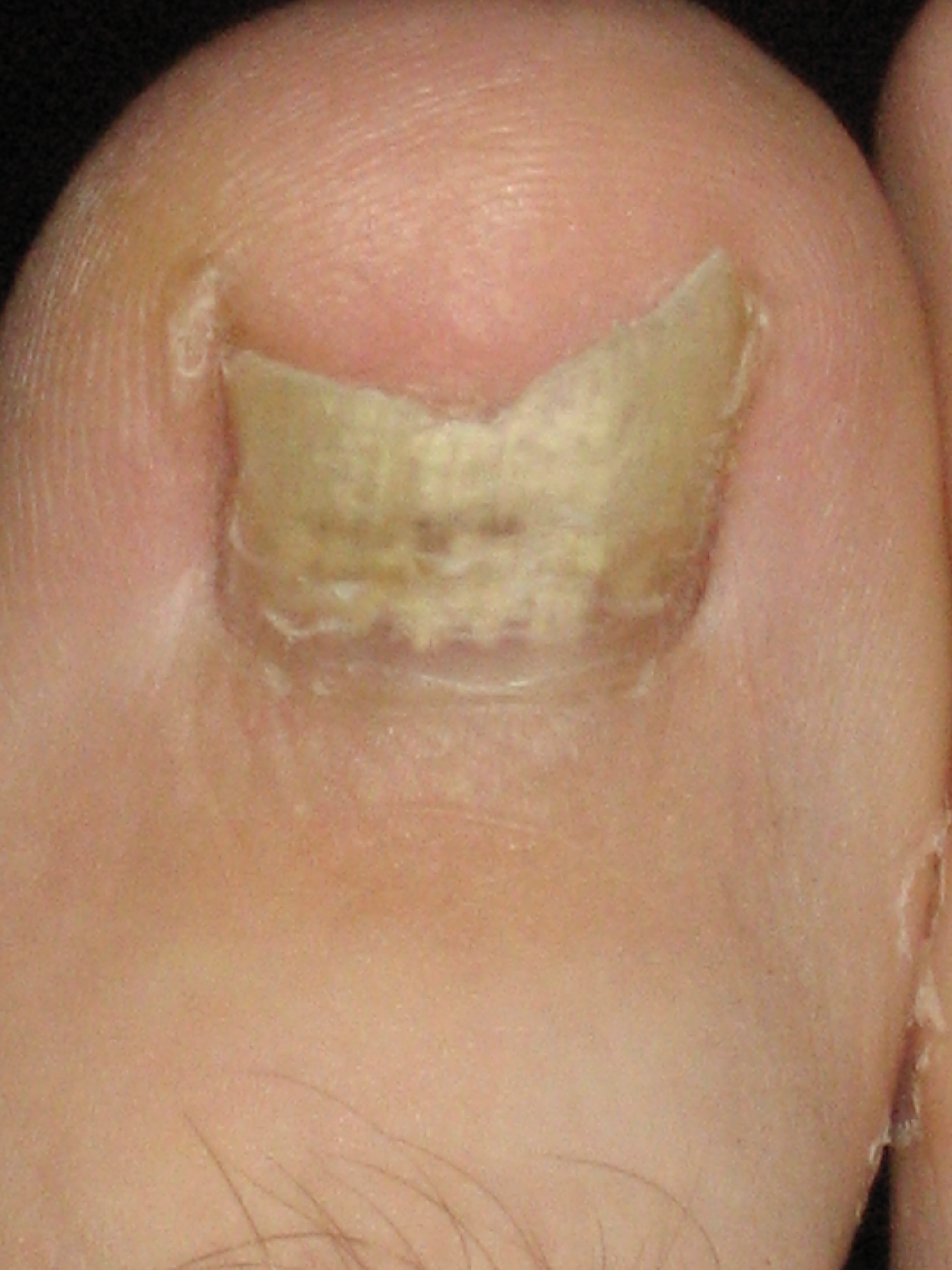
Onychomycosis, also known as tinea unguium, is a fungal infection of the nail. Symptoms may include white or yellow nail discoloration, thickening of the nail, and separation of the nail from the nail bed. Toenails or fingernails may be affected, but it is more common for toenails. Complications may include cellulitis of the lower leg. A number of different types of fungus can cause onychomycosis, including dermatophytes and ''Fusarium''. Risk factors include athlete's foot, other nail diseases, exposure to someone with the condition, peripheral vascular disease, and poor immune function. The diagnosis is generally suspected based on the appearance and confirmed by laboratory testing. Onychomycosis does not necessarily require treatment. The antifungal medication terbinafine taken by mouth appears to be the most effective but is associated with liver problems. Trimming the affected nails when on treatment also appears useful. There is a ciclopirox-containing nail polish, but ... [...More Info...] [...Related Items...] OR: [Wikipedia] [Google] [Baidu] |
Subungual Hematoma
A subungual hematoma is a collection of blood (hematoma) underneath a toenail or fingernail. It can be extremely painful for an injury of its size, although otherwise it is not a serious medical condition. Nature A laceration of the nail bed causes bleeding into the constricted area underneath the hard nail plate. The blood pools under the nail, giving a reddish, brownish, blueish, or grey/blackish discoloration. The blood puts pressure to the nailbed causing pain which can be throbbing in quality and disappears when the pressure on the nail bed is relieved. Subungual hematomas typically heal without incident, though infection may occur. The pressure of the blood blister may cause separation of nail plate from the nail bed (onycholysis), but the nail should not be pulled off, as this can cause scarring of the nailbed and deformed nails. Nail discolouration may last some months. The nail plate may also become thicker and more brittle as a result of the injury ( onychochauxis) ... [...More Info...] [...Related Items...] OR: [Wikipedia] [Google] [Baidu] |
Pyogenic Granuloma
A pyogenic granuloma or lobular capillary hemangioma is a vascular tumor that occurs on both mucosa and skin, and appears as an overgrowth of tissue due to irritation, physical trauma, or hormonal factors. It is often found to involve the gums, skin, or nasal septum, and has also been found far from the head, such as in the thigh. Pyogenic granulomas may be seen at any age, and are more common in females than males. In pregnant women, lesions may occur in the first trimester with an increasing incidence until the seventh month, and are often seen on the gums. Signs and symptoms The appearance of pyogenic granuloma is usually a color ranging from red/pink to purple, grows rapidly, and can be smooth or mushroom-shaped. Younger lesions are more likely to be red because of their high number of blood vessels. Older lesions begin to change into a pink color. Size commonly ranges from a few millimeters to centimeters, though smaller or larger lesions may occur. A pyogenic granuloma ... [...More Info...] [...Related Items...] OR: [Wikipedia] [Google] [Baidu] |
Glomus Tumour
:''Glomus tumor was also the name formerly (and incorrectly) used for a tumor now called a paraganglioma.'' A glomus tumor (also known as a "solitary glomus tumor," "solid glomus tumor,") is a rare neoplasm arising from the glomus body and mainly found under the nail, on the fingertip or in the foot.Freedberg, et al. (2003). ''Fitzpatrick's Dermatology in General Medicine''. (6th ed.). McGraw-Hill. . They account for less than 2% of all soft tissue tumors. The majority of glomus tumors are benign, but they can also show malignant features. Glomus tumors were first described by Hoyer in 1877 while the first complete clinical description was given by Masson in 1924. Histologically, glomus tumors are made up of an afferent arteriole, anastomotic vessel, and collecting venule. Glomus tumors are modified smooth muscle cells that control the thermoregulatory function of dermal glomus bodies. As stated above, these lesions should not be confused with paragangliomas, which were formerl ... [...More Info...] [...Related Items...] OR: [Wikipedia] [Google] [Baidu] |
Mucous Cyst Of The Oral Mucosa
Oral mucocele (also mucous extravasation cyst, mucous cyst of the oral mucosa, and mucous retention and extravasation phenomena.) is a condition caused by two related phenomena - mucus extravasation phenomenon and mucous retention cyst. Mucous extravasation phenomenon is a swelling of connective tissue consisting of a collection of fluid called mucus. This occurs because of a ruptured salivary gland duct usually caused by local trauma (damage) in the case of mucous extravasation phenomenon and an obstructed or ruptured salivary duct in the case of a mucus retention cyst. The mucocele has a bluish, translucent color, and is more commonly found in children and young adults. Although these lesions are often called cysts, mucoceles are not true cysts because they have no epithelial lining. Rather, they are polyps. Signs and symptoms The size of oral mucoceles vary from 1 mm to several centimeters and they usually are slightly transparent with a blue tinge. On palpation, muc ... [...More Info...] [...Related Items...] OR: [Wikipedia] [Google] [Baidu] |
Keratoacanthoma
Keratoacanthoma (KA) is a common low-grade (unlikely to metastasize or invade) rapidly-growing skin tumour that is believed to originate from the hair follicle (pilosebaceous unit) and can resemble squamous cell carcinoma. The defining characteristic of a keratoacanthoma is that it is dome-shaped, symmetrical, surrounded by a smooth wall of inflamed skin, and capped with keratin scales and debris. It grows rapidly, reaching a large size within days or weeks, and if untreated for months will almost always starve itself of nourishment, necrose (die), slough, and heal with scarring. Keratoacanthoma is commonly found on sun-exposed skin, often face, forearms and hands. It is rarely found at a mucocutaneous junction or on mucous membranes. Keratoacanthoma may be difficult to distinguish visually from a skin cancer. Under the microscope, keratoacanthoma very closely resembles squamous cell carcinoma. In order to differentiate between the two, almost the entire structure needs to be re ... [...More Info...] [...Related Items...] OR: [Wikipedia] [Google] [Baidu] |
Nail (anatomy)
A nail is a claw-like plate found at the tip of the fingers and toes on most primates. Nails correspond to the claws found in other animals. Fingernails and toenails are made of a tough protective protein called alpha-keratin, which is a polymer. Alpha-keratin is found in the hooves, claws, and horns of vertebrates. Structure The nail consists of the nail plate, the nail matrix and the nail bed below it, and the grooves surrounding it. Parts of the nail The matrix, sometimes called the ''matrix unguis'', keratogenous membrane, nail matrix, or onychostroma, is the active tissue (or germinal matrix) that generates cells, which harden as they move outward from the nail root to the nail plate. It is the part of the nail bed that is beneath the nail and contains nerves, lymph and blood vessels. The matrix produces cells that become the nail plate. The width and thickness of the nail plate is determined by the size, length, and thickness of the matrix, while the shape of the fing ... [...More Info...] [...Related Items...] OR: [Wikipedia] [Google] [Baidu] |


