|
Metacarpophalangeal Joint
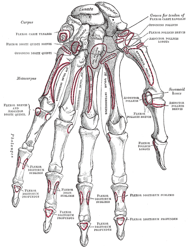
The metacarpophalangeal joints (MCP) are situated between the metacarpal bones and the proximal phalanges of the fingers. These joints are of the condyloid kind, formed by the reception of the rounded heads of the metacarpal bones into shallow cavities on the proximal ends of the proximal phalanges. Being condyloid, they allow the movements of flexion, extension, abduction, adduction and circumduction (see anatomical terms of motion) at the joint. Structure Ligaments Each joint has: * palmar ligaments of metacarpophalangeal articulations * collateral ligaments of metacarpophalangeal articulations Dorsal surfaces The dorsal surfaces of these joints are covered by the expansions of the Extensor tendons, together with some loose areolar tissue which connects the deep surfaces of the tendons to the bones. Function The movements which occur in these joints are flexion, extension, adduction, abduction, and circumduction; the movements of abduction and adduction are very ... [...More Info...] [...Related Items...] OR: [Wikipedia] [Google] [Baidu] |
Epiphyses
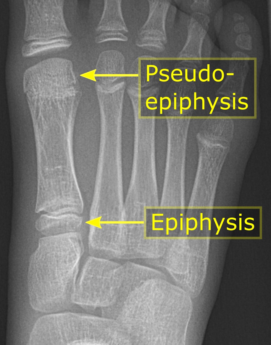
An epiphysis (; : epiphyses) is one of the rounded ends or tips of a long bone that ossify from one or more secondary centers of ossification. Between the epiphysis and diaphysis (the long midsection of the long bone) lies the metaphysis, including the epiphyseal plate (growth plate). During formation of the secondary ossification center, vascular canals (epiphysial canals) stemming from the perichondrium invade the epiphysis, supplying nutrients to the developing secondary centers of ossification. At the joint, the epiphysis is covered with articular cartilage; below that covering is a zone similar to the epiphyseal plate, known as subchondral bone. The epiphysis is mostly found in mammals but it is also present in some lizards. However, the secondary center of ossification may have evolved multiple times, having been found in the Jurassic sphenodont '' Sapheosaurus'' as well as in the therapsid '' Niassodon mfumukasi.'' The epiphysis is filled with red bone marrow, which ... [...More Info...] [...Related Items...] OR: [Wikipedia] [Google] [Baidu] |
Gray's Anatomy
''Gray's Anatomy'' is a reference book of human anatomy written by Henry Gray, illustrated by Henry Vandyke Carter and first published in London in 1858. It has had multiple revised editions, and the current edition, the 42nd (October 2020), remains a standard reference, often considered "the doctors' bible". Earlier editions were called ''Anatomy: Descriptive and Surgical'', ''Anatomy of the Human Body'' and ''Gray's Anatomy: Descriptive and Applied'', but the book's name is commonly shortened to, and later editions are titled, ''Gray's Anatomy''. The book is widely regarded as an extremely influential work on the subject. Publication history Origins The English anatomist Henry Gray was born in 1827. He studied the development of the endocrine glands and spleen and in 1853 was appointed Lecturer on Anatomy at St George's Hospital Medical School in London. In 1855, he approached his colleague Henry Vandyke Carter with his idea to produce an inexpensive and access ... [...More Info...] [...Related Items...] OR: [Wikipedia] [Google] [Baidu] |
Extensor Pollicis Brevis Muscle
In human anatomy, the extensor pollicis brevis (EPB) is a skeletal muscle on the dorsal side of the forearm. It lies on the medial side of, and is closely connected with, the abductor pollicis longus. The extensor pollicis brevis belongs to the deep group of the posterior fascial compartment of the forearm. It is a part of the lateral border of the anatomical snuffbox. Structure The extensor pollicis brevis arises from the ulna distal to the abductor pollicis longus, from the interosseous membrane, and from the dorsal surface of the radius. Its direction is similar to that of the abductor pollicis longus, its tendon passing the same groove on the lateral side of the lower end of the radius, to be inserted into the base of the first phalanx of the thumb. Variation Absence; fusion of tendon with that of the extensor pollicis longus or abductor pollicis longus muscle. Function In a close relationship to the abductor pollicis longus, the extensor pollicis brevis both ex ... [...More Info...] [...Related Items...] OR: [Wikipedia] [Google] [Baidu] |
Extensor Pollicis Longus Muscle
In human anatomy, the extensor pollicis longus muscle (EPL) is a skeletal muscle located dorsally on the forearm. It is much larger than the extensor pollicis brevis, the origin of which it partly covers and acts to stretch the thumb together with this muscle. Structure The extensor pollicis longus arises from the dorsal surface of the ulna and from the interosseous membrane, next to the origins of abductor pollicis longus and extensor pollicis brevis. Passing through the third tendon compartment, lying in a narrow, oblique groove on the back of the lower end of the radius,''Gray's Anatomy'' 1918, see infobox it crosses the wrist close to the dorsal midline before turning towards the thumb using Lister's tubercle on the distal end of the radius as a pulley. It obliquely crosses the tendons of the extensores carpi radialis longus and brevis, and is separated from the extensor pollicis brevis by a triangular interval, the anatomical snuff box in which the radial artery is foun ... [...More Info...] [...Related Items...] OR: [Wikipedia] [Google] [Baidu] |
Flexor Pollicis Brevis Muscle
The flexor pollicis brevis is a muscle in the hand that flexes the thumb. It is one of three thenar muscles. It has both a superficial part and a deep part. Origin and insertion The muscle's superficial head arises from the distal edge of the flexor retinaculum and the tubercle of the trapezium, the most lateral bone in the distal row of carpal bones. It passes along the radial side of the tendon of the flexor pollicis longus. The deeper (and medial) head "varies in size and may be absent."Gray's 37th British Edition, p. 630" It arises from the trapezoid and capitate bones on the floor of the carpal tunnel, as well as the ligaments of the distal carpal row. Both heads become tendinous and insert together into the radial side of the base of the proximal phalanx of the thumb; at the junction between the tendinous heads there is a sesamoid bone.''Gray's Anatomy'' 1918, see infobox Innervation The superficial head is usually innervated by the lateral terminal branch of the med ... [...More Info...] [...Related Items...] OR: [Wikipedia] [Google] [Baidu] |
Flexor Pollicis Longus Muscle
The flexor pollicis longus (; FPL, Latin ''flexor'', bender; ''pollicis'', of the thumb; ''longus'', long) is a muscle in the forearm and hand that flexes the thumb. It lies in the same plane as the flexor digitorum profundus. This muscle is unique to humans, being either rudimentary or absent in other primates. A meta-analysis indicated accessory flexor pollicis longus is present in around 48% of the population. Human anatomy Origin and insertion It arises from the grooved anterior (side of palm) surface of the body of the radius, extending from immediately below the radial tuberosity and oblique line to within a short distance of the pronator quadratus muscle.Gray 1918, ''Flexor Pollicis Longus'', paras 20, 25 An occasionally present accessory long head of the flexor pollicis longus muscle is called 'Gantzer's muscle'. It may cause compression of the anterior interosseous nerve. It arises also from the adjacent part of the interosseous membrane of the forearm, and generall ... [...More Info...] [...Related Items...] OR: [Wikipedia] [Google] [Baidu] |
Extensor Digiti Minimi Muscle
The extensor digiti minimi (extensor digiti quinti proprius) is a slender muscle of the forearm, placed on the ulnar side of the extensor digitorum communis, with which it is generally connected. It arises from the common extensor tendon by a thin tendinous slip and frequently from the intermuscular septa between it and the adjacent muscles. Its tendon passes through a compartment of the extensor retinaculum, posterior to distal radio-ulnar joint, then divides into two as it crosses the dorsum of the hand, and finally joins the extensor digitorum tendon. All three tendons attach to the dorsal digital expansion of the fifth digit (little finger). There may be a slip of tendon to the fourth digit. Variations * An additional fibrous slip from the lateral epicondyle: The tendon of insertion may not divide or may send a slip to the ring finger The ring finger, third finger, fourth finger, leech finger, or annulary is the fourth digit of the human hand, located between the m ... [...More Info...] [...Related Items...] OR: [Wikipedia] [Google] [Baidu] |
Extensor Indicis Proprius
In human anatomy, the extensor indicis (proprius) is a narrow, elongated skeletal muscle in the deep layer of the dorsal forearm, placed medial to, and parallel with, the extensor pollicis longus. Its tendon goes to the index finger, which it extends. Structure It arises from the distal third of the dorsal part of the body of the ulna and from the interosseous membrane. It runs through the fourth tendon compartment together with the extensor digitorum, from where it projects into the dorsal aponeurosis of the index finger. Opposite the head of the second metacarpal bone, it joins the ulnar side of the tendon of the extensor digitorum which belongs to the index finger. Like the extensor digiti minimi (i.e. the extensor of the little finger), the tendon of the extensor indicis runs and inserts on the ulnar side of the tendon of the common extensor digitorum. The extensor indicis lacks the juncturae tendinum interlinking the tendons of the extensor digitorum on the dorsal side o ... [...More Info...] [...Related Items...] OR: [Wikipedia] [Google] [Baidu] |
Extensor Digitorum Communis
In anatomy, extension is a movement of a joint that increases the angle between two bones or body surfaces at a joint. Extension usually results in straightening of the bones or body surfaces involved. For example, extension is produced by extending the flexed (bent) elbow. Straightening of the arm would require extension at the elbow joint. If the head is tilted all the way back, the neck is said to be extended. Extensor muscles Upper limb *of arm at shoulder **Axilla and shoulder ***Latissimus dorsi *** Posterior fibres of deltoid *** Teres major *of forearm at elbow **Posterior compartment of the arm ***Triceps brachii *** Anconeus *of hand at wrist **Posterior compartment of the forearm *** Extensor carpi radialis longus ***Extensor carpi radialis brevis *** Extensor carpi ulnaris *** Extensor digitorum *of phalanges, at all joints **Posterior compartment of the forearm *** Extensor digitorum *** Extensor digiti minimi (little finger only) ***Extensor indicis (index finger ... [...More Info...] [...Related Items...] OR: [Wikipedia] [Google] [Baidu] |
Flexor Digiti Minimi Brevis (hand)
The flexor digiti minimi brevis is a hypothenar muscle in the hand that flexes the little finger (digit V) at the metacarpophalangeal joint. It lies lateral to the abductor digiti minimi when the hand is in anatomical position. Structure The flexor digiti minimi brevis arises from the hamulus of the hamate bone and the palmar surface of the flexor retinaculum of the hand. It is inserted into the medial side of the base of the proximal phalanx of digit V. It is separated from the abductor digiti minimi, at its origin, by the deep branches of the ulnar artery and the ulnar nerve. The flexor digiti minimi brevis is sometimes not present; in these cases, the abductor digiti minimi is usually larger than normal. The flexor digiti minimi brevis is one of three muscles in the hypothenar muscle group. These three muscles form the fleshy mass at the base of the little finger, and are solely concerned with the movement of digit V. The other two muscles that make up the hypothenar ... [...More Info...] [...Related Items...] OR: [Wikipedia] [Google] [Baidu] |
Little Finger
The little finger or pinkie, also known as the baby finger, fifth digit, or pinky finger, is the most ulnar and smallest digit of the human hand, and next to the ring finger. Etymology The word "pinkie" is derived from the Dutch word ''pink'', meaning "little finger". The earliest recorded use of the term "pinkie" is from Scotland in 1808. The term (sometimes spelled "pinky") is common in Scottish English and American English, and is also used extensively in other Commonwealth countries such as New Zealand, Canada, and Australia. Nerves and muscles There are nine muscles that control the fifth digit: Three in the hypothenar eminence, two extrinsic flexors, two extrinsic extensors, and two more intrinsic muscles: * Hypothenar eminence: ** Opponens digiti minimi muscle ** Abductor minimi digiti muscle (adduction from third palmar interossei) ** Flexor digiti minimi brevis (the "longus" is absent in most humans) * Two extrinsic flexors: ** Flexor digitorum superficialis ** ... [...More Info...] [...Related Items...] OR: [Wikipedia] [Google] [Baidu] |
Interossei
{{short description, Muscles between certain bones Interossei refer to muscles between certain bones. There are many interossei in a human body. Specific interossei include: On the hands * Dorsal interossei muscles of the hand * Palmar interossei muscles In human anatomy, the palmar or volar interossei (interossei volares in older literature) are four muscles, one on the thumb that is occasionally missing, and three small, unipennate, central muscles in the hand that lie between the Metacarpus, me ... File:Gray428.png, Dorsal interossei muscles of the hand File:Gray429.png, Palmar interossei muscles On the feet * Dorsal interossei muscles of the foot * Plantar interossei muscles File:Gray446.png, Dorsal interossei muscles of the foot File:Gray447.png, Plantar interossei muscles Muscular system ... [...More Info...] [...Related Items...] OR: [Wikipedia] [Google] [Baidu] |

