|
Jugular Surface
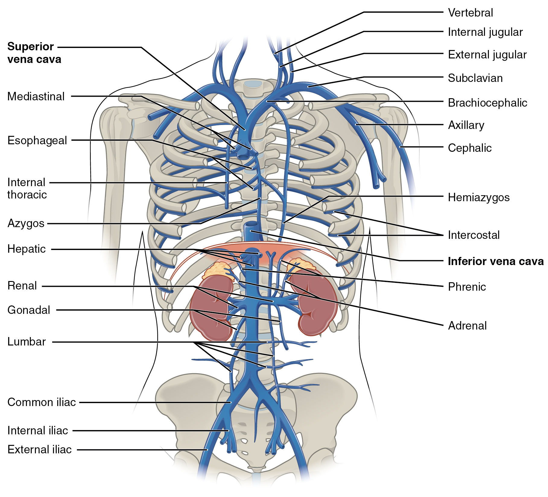
The jugular veins are veins that take deoxygenated blood from the head back to the heart via the superior vena cava. The internal jugular vein descends next to the internal carotid artery and continues posteriorly to the sternocleidomastoid muscle. Structure and Function There are two sets of jugular veins: external and internal. The left and right external jugular veins drain into the subclavian veins. The internal jugular veins join with the subclavian veins more medially to form the brachiocephalic veins. Finally, the left and right brachiocephalic veins join to form the superior vena cava, which delivers deoxygenated blood to the right atrium of the heart. The Jugular veins help carry blood from the heart to and from the brain. An average human brain weighs about 3 pounds, and gets about 15%-20% of the blood that the heart pumps out. It is important for the brain to get enough blood for many reasons. The jugular ... [...More Info...] [...Related Items...] OR: [Wikipedia] [Google] [Baidu] |
Brachiocephalic Vein
The left and right brachiocephalic veins (previously called innominate veins) are major veins in the upper chest, formed by the union of each corresponding internal jugular vein and subclavian vein. This is at the level of the sternoclavicular joint. The left brachiocephalic vein is nearly always longer than the right. These veins merge to form the superior vena cava, a great vessel, posterior to the junction of the first costal cartilage with the manubrium of the sternum. The brachiocephalic veins are the major veins returning blood to the superior vena cava. Tributaries The brachiocephalic vein is formed by the confluence of the subclavian and internal jugular veins. In addition it receives drainage from: * Left and right internal thoracic vein (Also called internal mammary veins): drain into the inferior border of their corresponding vein * Left and right inferior thyroid veins: drain into the superior aspect of their corresponding veins near the confluence * Left and righ ... [...More Info...] [...Related Items...] OR: [Wikipedia] [Google] [Baidu] |
Occipital Vein
The occipital vein is a vein of the scalp. It originates from a plexus around the external occipital protuberance and superior nuchal line to the back part of the vertex of the skull. It usually drains into the internal jugular vein, but may also drain into the posterior auricular vein (which joins the external jugular vein). It drains part of the scalp. Structure The occipital vein is part of the scalp. It begins as a plexus at the posterior aspect of the scalp from the external occipital protuberance and superior nuchal line to the back part of the vertex of the skull. It pierces the cranial attachment of the trapezius and, dipping into the venous plexus of the suboccipital triangle, joins the deep cervical vein and the vertebral vein. Occasionally it follows the course of the occipital artery, and ends in the internal jugular vein. Alternatively, it joins the posterior auricular vein, and ends in the external jugular vein. The parietal emissary vein connects it with the su ... [...More Info...] [...Related Items...] OR: [Wikipedia] [Google] [Baidu] |
Jugular Venous Pressure
The jugular venous pressure (JVP, sometimes referred to as ''jugular venous pulse'') is the indirectly observed pressure over the venous system via visualization of the internal jugular vein. It can be useful in the differentiation of different forms of heart and lung disease. Classically three upward deflections and two downward deflections have been described. * The upward deflections are the "a" (atrial contraction), "c" (ventricular contraction and resulting bulging of tricuspid into the right atrium during isovolumetric systole) and "v" (venous filling). * The downward deflections of the wave are the "x" descent (the atrium relaxes and the tricuspid valve moves downward) and the "y" descent (filling of ventricle after tricuspid opening). Method Visualization The patient is positioned at a 45° incline, and the filling level of the external jugular vein determined. The internal jugular vein is visualised when looking for the pulsation. In healthy people, the filling level of ... [...More Info...] [...Related Items...] OR: [Wikipedia] [Google] [Baidu] |
Jugular Venous Pulse
The jugular veins are veins that take deoxygenated blood from the head back to the heart via the superior vena cava. The internal jugular vein descends next to the internal carotid artery and continues posteriorly to the sternocleidomastoid muscle. Structure and Function There are two sets of jugular veins: external and internal. The left and right external jugular veins drain into the subclavian veins. The internal jugular veins join with the subclavian veins more medially to form the brachiocephalic veins. Finally, the left and right brachiocephalic veins join to form the superior vena cava, which delivers deoxygenated blood to the right atrium of the heart. The Jugular veins help carry blood from the heart to and from the brain. An average human brain weighs about 3 pounds, and gets about 15%-20% of the blood that the heart pumps out. It is important for the brain to get enough blood for many reasons. The jugular ... [...More Info...] [...Related Items...] OR: [Wikipedia] [Google] [Baidu] |
Anterior Jugular Vein
The anterior jugular vein is a vein in the neck. Structure The anterior jugular vein lies lateral to the cricothyroid ligament. It begins near the hyoid bone by the confluence of several superficial veins from the submandibular region. Its tributaries are some laryngeal veins, and occasionally a small thyroid vein. It descends between the median line and the anterior border of the sternocleidomastoid muscle, and, at the lower part of the neck, passes beneath that muscle to open into the termination of the external jugular vein, or, in some instances, into the subclavian vein. Just above the sternum the two anterior jugular veins communicate by a transverse trunk, the venous jugular arch, which receive tributaries from the inferior thyroid veins; each also communicates with the internal jugular. There are no valves in this vein. The pretracheal lymph nodes follow the anterior jugular vein on each side of the midline. Variation The anterior jugular vein varies considerably ... [...More Info...] [...Related Items...] OR: [Wikipedia] [Google] [Baidu] |
Sternocleidomastoid
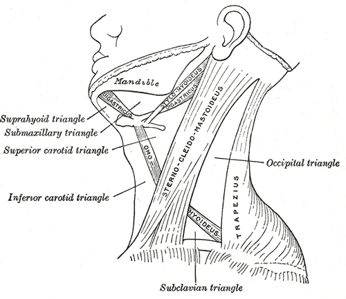
The sternocleidomastoid muscle is one of the largest and most superficial cervical muscles. The primary actions of the muscle are rotation of the head to the opposite side and flexion of the neck. The sternocleidomastoid is innervated by the accessory nerve. Etymology and location It is given the name ''sternocleidomastoid'' because it originates at the manubrium of the sternum (''sterno-'') and the clavicle (''cleido-'') and has an insertion at the mastoid process of the temporal bone of the skull. Structure The sternocleidomastoid muscle originates from two locations: the manubrium of the sternum and the clavicle. It travels obliquely across the side of the neck and inserts at the mastoid process of the temporal bone of the skull by a thin aponeurosis. The sternocleidomastoid is thick and narrow at its centre, and broader and thinner at either end. The sternal head is a round fasciculus, tendinous in front, fleshy behind, arising from the upper part of the front of the manu ... [...More Info...] [...Related Items...] OR: [Wikipedia] [Google] [Baidu] |
External Jugular Vein
The external jugular vein receives the greater part of the blood from the exterior of the cranium and the deep parts of the face, being formed by the junction of the posterior division of the retromandibular vein with the posterior auricular vein. Structure It commences in the substance of the parotid gland, on a level with the angle of the mandible, and runs perpendicularly down the neck, in the direction of a line drawn from the angle of the mandible to the middle of the clavicle superficial to the sternocleidomastoideus. In its course it crosses the sternocleidomastoideus obliquely, and in the subclavian triangle perforates the deep fascia, and ends in the subclavian vein lateral to or in front of the scalenus anterior, piercing the roof of the posterior triangle. It is separated from the sternocleidomastoideus by the investing layer of the deep cervical fascia, and is covered by the platysma, the superficial fascia, and the integument; it crosses the cutaneous cervical nerv ... [...More Info...] [...Related Items...] OR: [Wikipedia] [Google] [Baidu] |
Human Skull
The skull is a bone protective cavity for the brain. The skull is composed of four types of bone i.e., cranial bones, facial bones, ear ossicles and hyoid bone. However two parts are more prominent: the cranium and the mandible. In humans, these two parts are the neurocranium and the viscerocranium ( facial skeleton) that includes the mandible as its largest bone. The skull forms the anterior-most portion of the skeleton and is a product of cephalisation—housing the brain, and several sensory structures such as the eyes, ears, nose, and mouth. In humans these sensory structures are part of the facial skeleton. Functions of the skull include protection of the brain, fixing the distance between the eyes to allow stereoscopic vision, and fixing the position of the ears to enable sound localisation of the direction and distance of sounds. In some animals, such as horned ungulates (mammals with hooves), the skull also has a defensive function by providing the mount (on the front ... [...More Info...] [...Related Items...] OR: [Wikipedia] [Google] [Baidu] |
Carotid Sheath
The carotid sheath is an anatomical term for the fibrous connective tissue that surrounds the vascular compartment of the neck. It is part of the deep cervical fascia of the neck, below the superficial cervical fascia meaning the subcutaneous adipose tissue immediately beneath the skin. The deep cervical fascia of the neck includes four parts: * The investing layer (encloses the SCM and Trapezius) * The carotid sheath (encloses the vascular region of the neck) * The pretracheal fascia (encloses the visceral region of the neck) * The prevertebral fascia (encloses the vertebral region of the neck) Structure The carotid sheath is located at the lateral boundary of the retropharyngeal space at the level of the oropharynx on each side of the neck and deep to the sternocleidomastoid muscle. It extends from the base of the skull to the first rib and sternum, varying between C7 and T4. It merges with the axillary sheath when it reaches the subclavian vein. Contents The four major stru ... [...More Info...] [...Related Items...] OR: [Wikipedia] [Google] [Baidu] |
Vagus Nerve
The vagus nerve, also known as the tenth cranial nerve, cranial nerve X, or simply CN X, is a cranial nerve that interfaces with the parasympathetic control of the heart, lungs, and digestive tract. It comprises two nerves—the left and right vagus nerves—but they are typically referred to collectively as a single subsystem. The vagus is the longest nerve of the autonomic nervous system in the human body and comprises both sensory and motor fibers. The sensory fibers originate from neurons of the nodose ganglion, whereas the motor fibers come from neurons of the dorsal motor nucleus of the vagus and the nucleus ambiguus. The vagus was also historically called the pneumogastric nerve. Structure Upon leaving the medulla oblongata between the olive and the inferior cerebellar peduncle, the vagus nerve extends through the jugular foramen, then passes into the carotid sheath between the internal carotid artery and the internal jugular vein down to the neck, chest, and abdom ... [...More Info...] [...Related Items...] OR: [Wikipedia] [Google] [Baidu] |
Common Carotid Artery
In anatomy, the left and right common carotid arteries (carotids) ( in Merriam-Webster Online Dictionary '.) are that supply the head and neck with ; they divide in the neck to form the and |
Inferior Petrosal Sinus
The inferior petrosal sinuses are two small sinuses situated on the inferior border of the petrous part of the temporal bone, one on each side. Each inferior petrosal sinus drains the cavernous sinus into the internal jugular vein. Structure The inferior petrosal sinus is situated in the inferior petrosal sulcus, formed by the junction of the petrous part of the temporal bone with the basilar part of the occipital bone. It begins below and behind the cavernous sinus and, passing through the anterior part of the jugular foramen, ends in the superior bulb of the internal jugular vein. Function The inferior petrosal sinus receives the internal auditory veins and also veins from the medulla oblongata, pons, and under surface of the cerebellum. Additional images File:Gray568.png, Sagittal section of the skull, showing the sinuses of the dura. See also * Dural venous sinuses * Inferior petrosal sinus sampling Inferior petrosal sinus sampling (or IPSS), is a diagnostic medical p ... [...More Info...] [...Related Items...] OR: [Wikipedia] [Google] [Baidu] |


