|
Jugular Fossa
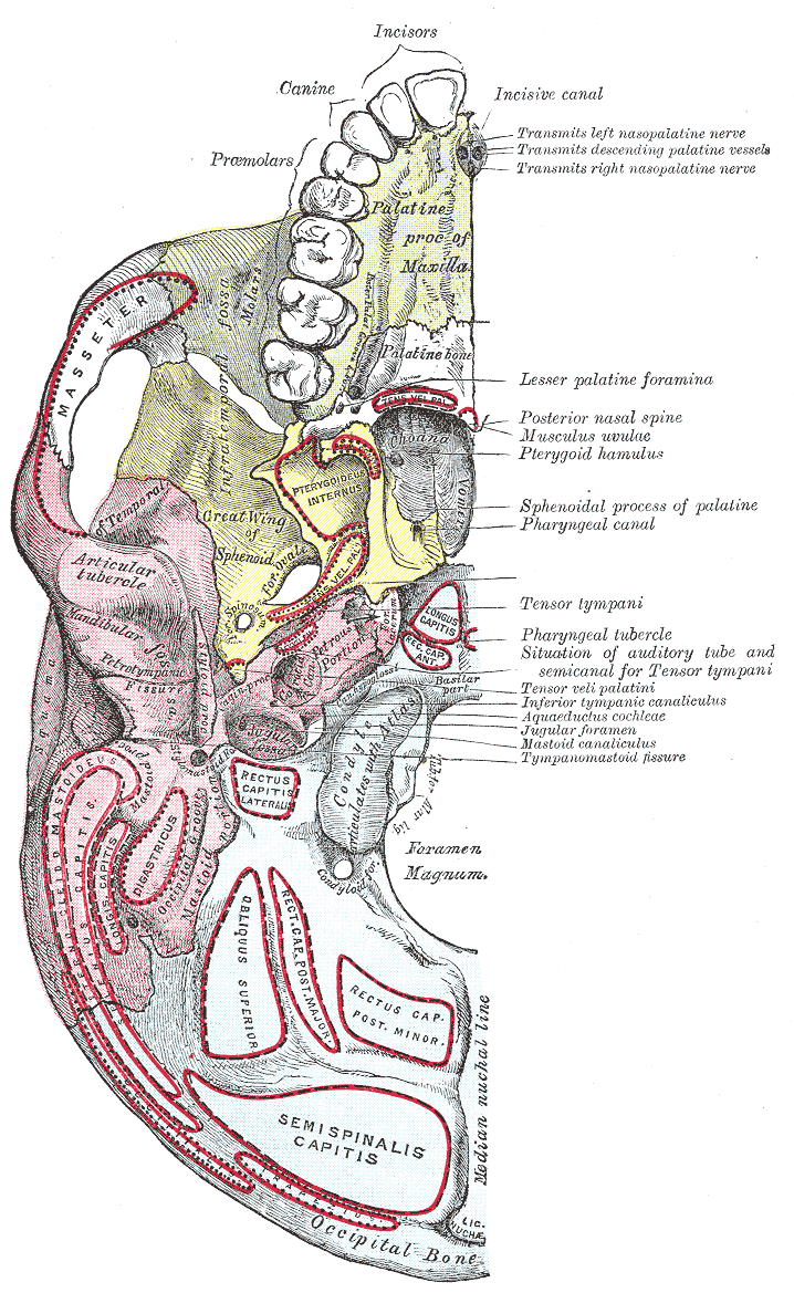
The jugular fossa is a deep depression in the inferior part of the temporal bone at the base of the skull. It lodges the bulb of the internal jugular vein. Structure The jugular fossa is located in the temporal bone, posterior to the carotid canal and the cochlear aqueduct. In the bony ridge dividing the carotid canal from the jugular fossa is the small inferior tympanic canaliculus for the passage of the tympanic branch of the glossopharyngeal nerve. In the lateral part of the jugular fossa is the mastoid canaliculus for the entrance of the auricular branch of the vagus nerve. Behind the jugular fossa is a quadrilateral area, the jugular surface, covered with cartilage in the fresh state, and articulating with the jugular process of the occipital bone. Variation The jugular fossa has variable depth and size in different skulls. Function The jugular fossa lodges the bulb of the internal jugular vein. Clinical significance Abnormally shaped jugular fossae may cause ear ... [...More Info...] [...Related Items...] OR: [Wikipedia] [Google] [Baidu] |
Eardrum
In the anatomy of humans and various other tetrapods, the eardrum, also called the tympanic membrane or myringa, is a thin, cone-shaped membrane that separates the external ear from the middle ear. Its function is to transmit sound from the air to the ossicles inside the middle ear, and then to the oval window in the fluid-filled cochlea. Hence, it ultimately converts and amplifies vibration in the air to vibration in cochlear fluid. The malleus bone bridges the gap between the eardrum and the other ossicles. Rupture or perforation of the eardrum can lead to conductive hearing loss. Collapse or retraction of the eardrum can cause conductive hearing loss or cholesteatoma. Structure Orientation and relations The tympanic membrane is oriented obliquely in the anteroposterior, mediolateral, and superoinferior planes. Consequently, its superoposterior end lies lateral to its anteroinferior end. Anatomically, it relates superiorly to the middle cranial fossa, posteriorly t ... [...More Info...] [...Related Items...] OR: [Wikipedia] [Google] [Baidu] |
Temporal Bone
The temporal bones are situated at the sides and base of the skull, and lateral to the temporal lobes of the cerebral cortex. The temporal bones are overlaid by the sides of the head known as the temples, and house the structures of the ears. The lower seven cranial nerves and the major vessels to and from the brain traverse the temporal bone. Structure The temporal bone consists of four parts— the squamous, mastoid, petrous and tympanic parts. The squamous part is the largest and most superiorly positioned relative to the rest of the bone. The zygomatic process is a long, arched process projecting from the lower region of the squamous part and it articulates with the zygomatic bone. Posteroinferior to the squamous is the mastoid part. Fused with the squamous and mastoid parts and between the sphenoid and occipital bones lies the petrous part, which is shaped like a pyramid. The tympanic part is relatively small and lies inferior to the squamous part, anterior to t ... [...More Info...] [...Related Items...] OR: [Wikipedia] [Google] [Baidu] |
Skull
The skull is a bone protective cavity for the brain. The skull is composed of four types of bone i.e., cranial bones, facial bones, ear ossicles and hyoid bone. However two parts are more prominent: the cranium and the mandible. In humans, these two parts are the neurocranium and the viscerocranium ( facial skeleton) that includes the mandible as its largest bone. The skull forms the anterior-most portion of the skeleton and is a product of cephalisation—housing the brain, and several sensory structures such as the eyes, ears, nose, and mouth. In humans these sensory structures are part of the facial skeleton. Functions of the skull include protection of the brain, fixing the distance between the eyes to allow stereoscopic vision, and fixing the position of the ears to enable sound localisation of the direction and distance of sounds. In some animals, such as horned ungulates (mammals with hooves), the skull also has a defensive function by providing the mount (on the ... [...More Info...] [...Related Items...] OR: [Wikipedia] [Google] [Baidu] |
Base Of The Skull
The base of skull, also known as the cranial base or the cranial floor, is the most inferior area of the skull. It is composed of the endocranium and the lower parts of the calvaria. Structure Structures found at the base of the skull are for example: Bones There are five bones that make up the base of the skull: * Ethmoid bone *Sphenoid bone *Occipital bone *Frontal bone *Temporal bone Sinuses * Occipital sinus * Superior sagittal sinus * Superior petrosal sinus Foramina of the skull * Foramen cecum * Optic foramen * Foramen lacerum * Foramen rotundum *Foramen magnum * Foramen ovale * Jugular foramen * Internal auditory meatus * Mastoid foramen * Sphenoidal emissary foramen * Foramen spinosum Sutures * Frontoethmoidal suture * Sphenofrontal suture * Sphenopetrosal suture *Sphenoethmoidal suture * Petrosquamous suture *Sphenosquamosal suture Other * Sphenoidal lingula * Subarcuate fossa *Dorsum sellae * Jugular process * Petro-occipital fissure * Condylar canal * Jug ... [...More Info...] [...Related Items...] OR: [Wikipedia] [Google] [Baidu] |
Internal Jugular Vein
The internal jugular vein is a paired jugular vein that collects blood from the brain and the superficial parts of the face and neck. This vein runs in the carotid sheath with the common carotid artery and vagus nerve. It begins in the posterior compartment of the jugular foramen, at the base of the skull. It is somewhat dilated at its origin, which is called the ''superior bulb''. This vein also has a common trunk into which drains the anterior branch of the retromandibular vein, the facial vein, and the lingual vein. It runs down the side of the neck in a vertical direction, being at one end lateral to the internal carotid artery, and then lateral to the common carotid artery, and at the root of the neck, it unites with the subclavian vein to form the brachiocephalic vein (innominate vein); a little above its termination is a second dilation, the ''inferior bulb''. Above, it lies upon the rectus capitis lateralis, behind the internal carotid artery and the nerves pas ... [...More Info...] [...Related Items...] OR: [Wikipedia] [Google] [Baidu] |
Carotid Canal
The carotid canal is a passageway in the temporal bone of the skull through which the internal carotid artery enters the middle cranial fossa from the neck. Structure The carotid canal is located within the middle cranial fossa, at the petrous part of the temporal bone. Anteriorly, it is limited by posterior margin of the greater wing of sphenoid bone. Posteromedially, it is limited by basilar part of occipital bone. It is divided in three parts, namely, ascending petrous, transverse petrous, and ascending cavernous parts. The carotid canal has two openings, namely internal and external openings. The internal opening is situated laterally to foramen lacerum. The external opening of the carotid canal is located posterolaterally to the foramen lacerum. Both internal and external openings of the carotid canal lies anterior to the jugular foramen, where the latter is located inside the posterior cranial fossa. The carotid canal is separated from middle ear and inner ear by a thin ... [...More Info...] [...Related Items...] OR: [Wikipedia] [Google] [Baidu] |
Cochlear Aqueduct
Medial to the opening for the carotid canal and close to its posterior border, in front of the jugular fossa, is a triangular depression; at the apex of this is a small opening, the aquaeductus cochleae (or cochlear aqueduct, or aqueduct of cochlea), which lodges a tubular prolongation of the dura mater establishing a communication between the perilymphatic space and the subarachnoid space, and transmits a vein from the cochlea The cochlea is the part of the inner ear involved in hearing. It is a spiral-shaped cavity in the bony labyrinth, in humans making 2.75 turns around its axis, the modiolus. A core component of the cochlea is the Organ of Corti, the sensory or ... to join the internal jugular vein. Additional images File:Gray187.png, Base of skull. Inferior surface. References Bones of the head and neck {{musculoskeletal-stub ... [...More Info...] [...Related Items...] OR: [Wikipedia] [Google] [Baidu] |
Glossopharyngeal Nerve
The glossopharyngeal nerve (), also known as the ninth cranial nerve, cranial nerve IX, or simply CN IX, is a cranial nerve that exits the brainstem from the sides of the upper medulla, just anterior (closer to the nose) to the vagus nerve. Being a mixed nerve (sensorimotor), it carries afferent sensory and efferent motor information. The motor division of the glossopharyngeal nerve is derived from the basal plate of the embryonic medulla oblongata, whereas the sensory division originates from the cranial neural crest. Structure From the anterior portion of the medulla oblongata, the glossopharyngeal nerve passes laterally across or below the flocculus, and leaves the skull through the central part of the jugular foramen. From the superior and inferior ganglia in jugular foramen, it has its own sheath of dura mater. The inferior ganglion on the inferior surface of petrous part of temporal is related with a triangular depression into which the aqueduct of cochlea opens. ... [...More Info...] [...Related Items...] OR: [Wikipedia] [Google] [Baidu] |
Mastoid Canaliculus
In the lateral part of the jugular fossa of the temporal bone is the mastoid canaliculus for the entrance of the auricular branch of the vagus nerve, auricular branch of the vagus nerve. Additional images File:Gray187.png, Base of skull. Inferior surface. References External links * Foramina of the skull {{musculoskeletal-stub ... [...More Info...] [...Related Items...] OR: [Wikipedia] [Google] [Baidu] |
Auricular Branch Of Vagus Nerve
The auricular branch of the vagus nerve is often termed the Alderman's nerve or Arnold's nerve. The latter name is an eponym for Friedrich Arnold. The auricular branch of the vagus nerve supplies sensory innervation to the skin of the ear canal, tragus, and auricle. Path It arises from the superior ganglion of the vagus nerve, and is joined soon after its origin by a filament from the petrous ganglion of the glossopharyngeal; it passes behind the internal jugular vein, and enters the mastoid canaliculus on the lateral wall of the jugular fossa. Traversing the substance of the temporal bone, it crosses the facial canal about above the stylomastoid foramen, and here it gives off an ascending branch which joins the facial nerve. The nerve reaches the surface by passing through the tympanomastoid fissure between the mastoid process and the tympanic part of the temporal bone, and divides into two branches: * one joins the posterior auricular nerve. * the other is distributed t ... [...More Info...] [...Related Items...] OR: [Wikipedia] [Google] [Baidu] |
Vagus Nerve
The vagus nerve, also known as the tenth cranial nerve, cranial nerve X, or simply CN X, is a cranial nerve that interfaces with the parasympathetic control of the heart, lungs, and digestive tract. It comprises two nerves—the left and right vagus nerves—but they are typically referred to collectively as a single subsystem. The vagus is the longest nerve of the autonomic nervous system in the human body and comprises both sensory and motor fibers. The sensory fibers originate from neurons of the nodose ganglion, whereas the motor fibers come from neurons of the dorsal motor nucleus of the vagus and the nucleus ambiguus. The vagus was also historically called the pneumogastric nerve. Structure Upon leaving the medulla oblongata between the olive and the inferior cerebellar peduncle, the vagus nerve extends through the jugular foramen, then passes into the carotid sheath between the internal carotid artery and the internal jugular vein down to the neck, chest, and ... [...More Info...] [...Related Items...] OR: [Wikipedia] [Google] [Baidu] |
Occipital Bone
The occipital bone () is a cranial dermal bone and the main bone of the occiput (back and lower part of the skull). It is trapezoidal in shape and curved on itself like a shallow dish. The occipital bone overlies the occipital lobes of the cerebrum. At the base of skull in the occipital bone, there is a large oval opening called the foramen magnum, which allows the passage of the spinal cord. Like the other cranial bones, it is classed as a flat bone. Due to its many attachments and features, the occipital bone is described in terms of separate parts. From its front to the back is the basilar part, also called the basioccipital, at the sides of the foramen magnum are the lateral parts, also called the exoccipitals, and the back is named as the squamous part. The basilar part is a thick, somewhat quadrilateral piece in front of the foramen magnum and directed towards the pharynx. The squamous part is the curved, expanded plate behind the foramen magnum and is the largest par ... [...More Info...] [...Related Items...] OR: [Wikipedia] [Google] [Baidu] |


