|
Internal Thoracic Artery
The internal thoracic artery (ITA), also known as the internal mammary artery, is an artery that supplies the anterior chest wall and the breasts. It is a paired artery, with one running along each side of the sternum, to continue after its bifurcation as the superior epigastric and musculophrenic arteries. Structure The internal thoracic artery arises from the anterior surface of the subclavian artery near its origin. It has a width of between 1-2 mm. It travels downward on the inside of the rib cage, approximately 1 cm from the sides of the sternum, and thus medial to the nipple. It is accompanied by the internal thoracic vein. It runs deep to the abdominal external oblique muscle, but superficial to the vagus nerve. In adults, the internal thoracic artery lies closest to the sternum at the first intercostal space. The gap between the artery and lateral border of the sternum increases when going downwards, up to 1.1 cm to 1.3 cm at the sixth intercost ... [...More Info...] [...Related Items...] OR: [Wikipedia] [Google] [Baidu] |
Subclavian Artery
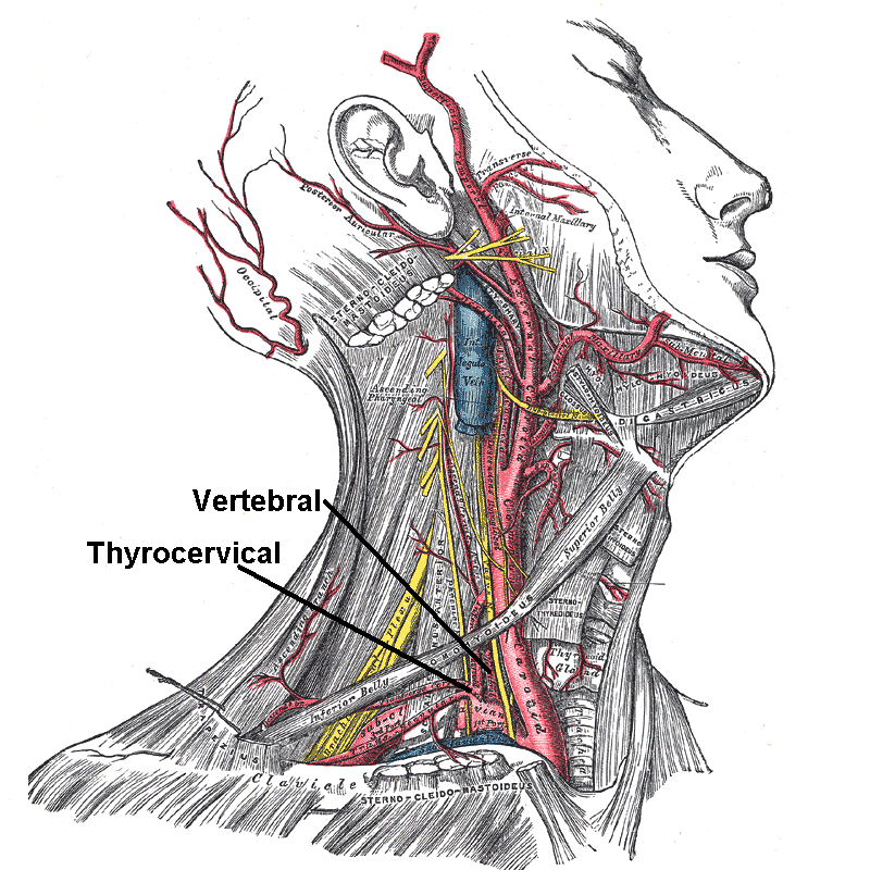
In human anatomy, the subclavian arteries are paired major arteries of the upper thorax, below the clavicle. They receive blood from the aortic arch. The left subclavian artery supplies blood to the left arm and the right subclavian artery supplies blood to the right arm, with some branches supplying the head and thorax. On the left side of the body, the subclavian comes directly off the aortic arch, while on the right side it arises from the relatively short brachiocephalic artery when it bifurcates into the subclavian and the right common carotid artery. The usual branches of the subclavian on both sides of the body are the vertebral artery, the internal thoracic artery, the thyrocervical trunk, the costocervical trunk and the dorsal scapular artery, which may branch off the transverse cervical artery, which is a branch of the thyrocervical trunk. The subclavian becomes the axillary artery at the lateral border of the first rib. Structure From its origin, the subclavian art ... [...More Info...] [...Related Items...] OR: [Wikipedia] [Google] [Baidu] |
Internal Thoracic Vein
In human anatomy, the internal thoracic vein (previously known as the internal mammary vein) is the vein that drains the chest wall and breasts. Structure Bilaterally, the internal thoracic vein arises from the superior epigastric vein, and accompanies the internal thoracic artery along its course. It drains the intercostal veins, although the posterior drainage is often handled by the azygous veins. It terminates in the brachiocephalic vein. It has a width of 2-3 mm. There is either one or two internal thoracic veins accompanying the corresponding artery (internal thoracic artery). If internal thoracic vein is single, it usually runs medial to the artery. If there are double thoracic veins, they run on either side of the internal thoracic artery. Variations Bifurcation of each internal thoracic vein is common. The left internal thoracic vein may bifurcate between ribs 3-4 or remain as a single vein. The right internal thoracic vein may bifurcate between ribs 2-4 or re ... [...More Info...] [...Related Items...] OR: [Wikipedia] [Google] [Baidu] |
Breast
The breasts are two prominences located on the upper ventral region of the torso among humans and other primates. Both sexes develop breasts from the same embryology, embryological tissues. The relative size and development of the breasts is a major secondary sex distinction between females and males. There is also considerable Bra size, variation in size between individuals. Permanent Breast development, breast growth during puberty is caused by estrogens in conjunction with the growth hormone. Female humans are the only mammals that permanently develop breasts at puberty; all other mammals develop their mammary tissue during the latter period of pregnancy. In females, the breast serves as the mammary gland, which produces and secretes milk to feed infants. Subcutaneous fat covers and envelops a network of lactiferous duct, ducts that converge on the nipple, and these tissue (biology), tissues give the breast its distinct size and globular shape. At the ends of the ducts are ... [...More Info...] [...Related Items...] OR: [Wikipedia] [Google] [Baidu] |
Musculophrenic Artery
The intercostal arteries are a group of arteries passing within an intercostal space (the space between two adjacent ribs). There are 9 anterior and 11 posterior intercostal arteries on each side of the body. The anterior intercostal arteries are branches of the internal thoracic artery and its terminal branchthe musculophrenic artery. The posterior intercostal arteries are branches of the supreme intercostal artery and thoracic aorta. Each anterior intercostal artery anastomoses with the corresponding posterior intercostal artery arising from the thoracic aorta. Anterior intercostal arteries Origin The upper six anterior intercostal arteries are branches of the internal thoracic artery (anterior intercostal branches of internal thoracic artery). The internal thoracic artery then divides into its two terminal branches, one of which - the musculophrenic artery - proceeds to issue anterior intercostal arteries to the remaining 7th, 8th, and 9th intercostal spaces; these di ... [...More Info...] [...Related Items...] OR: [Wikipedia] [Google] [Baidu] |
Anastomoses
An anastomosis (, : anastomoses) is a connection or opening between two things (especially cavities or passages) that are normally diverging or branching, such as between blood vessels, leaf#Veins, leaf veins, or streams. Such a connection may be normal (such as the foramen ovale (heart), foramen ovale in a fetus' heart) or abnormal (such as the atrial septal defect#Patent foramen ovale, patent foramen ovale in an adult's heart); it may be acquired (such as an arteriovenous fistula) or innate (such as the arteriovenous shunt of a metarteriole); and it may be natural (such as the aforementioned examples) or artificial (such as a surgical anastomosis). The reestablishment of an anastomosis that had become blocked is called a reanastomosis. Anastomoses that are abnormal, whether congenital disorder, congenital or acquired, are often called fistulas. The term is used in medicine, biology, mycology, geology, and geography. Etymology Anastomosis: medical or Modern Latin, from Greek ἀ ... [...More Info...] [...Related Items...] OR: [Wikipedia] [Google] [Baidu] |
Anterior Intercostal Branches
The intercostal arteries are a group of arteries passing within an intercostal space (the space between two adjacent ribs). There are 9 anterior and 11 posterior intercostal arteries on each side of the body. The anterior intercostal arteries are branches of the internal thoracic artery and its terminal branchthe musculophrenic artery. The posterior intercostal arteries are branches of the supreme intercostal artery and thoracic aorta. Each anterior intercostal artery anastomoses with the corresponding posterior intercostal artery arising from the thoracic aorta. Anterior intercostal arteries Origin The upper six anterior intercostal arteries are branches of the internal thoracic artery (anterior intercostal branches of internal thoracic artery). The internal thoracic artery then divides into its two terminal branches, one of which - the musculophrenic artery - proceeds to issue anterior intercostal arteries to the remaining 7th, 8th, and 9th intercostal spaces; these d ... [...More Info...] [...Related Items...] OR: [Wikipedia] [Google] [Baidu] |
Sternal
The sternum (: sternums or sterna) or breastbone is a long flat bone located in the central part of the chest. It connects to the ribs via cartilage and forms the front of the rib cage, thus helping to protect the heart, lungs, and major blood vessels from injury. Shaped roughly like a necktie, it is one of the largest and longest flat bones of the body. Its three regions are the manubrium, the body, and the xiphoid process. The word ''sternum'' originates from Ancient Greek στέρνον (''stérnon'') 'chest'. Structure The sternum is a narrow, flat bone, forming the middle portion of the front of the chest. The top of the sternum supports the clavicles (collarbones) and its edges join with the costal cartilages of the first two pairs of ribs. The inner surface of the sternum is also the attachment of the sternopericardial ligaments. Its top is also connected to the sternocleidomastoid muscle. The sternum consists of three main parts, listed from the top: * Manubrium * Body ... [...More Info...] [...Related Items...] OR: [Wikipedia] [Google] [Baidu] |
Phrenic Nerve
The phrenic nerve is a mixed nerve that originates from the C3–C5 spinal nerves in the neck. The nerve is important for breathing because it provides exclusive motor control of the diaphragm, the primary muscle of respiration. In humans, the right and left phrenic nerves are primarily supplied by the C4 spinal nerve, but there is also a contribution from the C3 and C5 spinal nerves. From its origin in the neck, the nerve travels downward into the chest to pass between the heart and lungs towards the diaphragm. In addition to motor fibers, the phrenic nerve contains sensory fibers, which receive input from the central tendon of the diaphragm and the mediastinal pleura, as well as some sympathetic nerve fibers. Although the nerve receives contributions from nerve roots of the cervical plexus and the brachial plexus, it is usually considered separate from either plexus. The name of the nerve comes from Ancient Greek ''phren'' 'diaphragm'. Structure The phrenic nerve or ... [...More Info...] [...Related Items...] OR: [Wikipedia] [Google] [Baidu] |
Thymic
The thymus (: thymuses or thymi) is a specialized primary lymphoid organ of the immune system. Within the thymus, T cells mature. T cells are critical to the adaptive immune system, where the body adapts to specific foreign invaders. The thymus is located in the upper front part of the chest, in the anterior superior mediastinum, behind the sternum, and in front of the heart. It is made up of two lobes, each consisting of a central medulla and an outer cortex, surrounded by a capsule. The thymus is made up of immature T cells called thymocytes, as well as lining cells called epithelial cells which help the thymocytes develop. T cells that successfully develop react appropriately with MHC immune receptors of the body (called ''positive selection'') and not against proteins of the body (called ''negative selection''). The thymus is the largest and most active during the neonatal and pre-adolescent periods. By the early teens, the thymus begins to decrease in size and activi ... [...More Info...] [...Related Items...] OR: [Wikipedia] [Google] [Baidu] |
Mediastinal
The mediastinum (from ;: mediastina) is the central compartment of the thoracic cavity. Surrounded by loose connective tissue, it is a region that contains vital organs and structures within the thorax, mainly the heart and its vessels, the esophagus, the trachea, the vagus, phrenic and cardiac nerves, the thoracic duct, the thymus and the lymph nodes of the central chest. Anatomy The mediastinum lies within the thorax and is enclosed on the right and left by pleurae. It is surrounded by the chest wall in front, the lungs to the sides and the spine at the back. It extends from the sternum in front to the vertebral column behind. It contains all the organs of the thorax except the lungs. It is continuous with the loose connective tissue of the neck. The mediastinum can be divided into an upper (or superior) and lower (or inferior) part: * The superior mediastinum starts at the superior thoracic aperture and ends at the thoracic plane. * The inferior mediastinum from this ... [...More Info...] [...Related Items...] OR: [Wikipedia] [Google] [Baidu] |


