|
Hyperpolarization (biology)
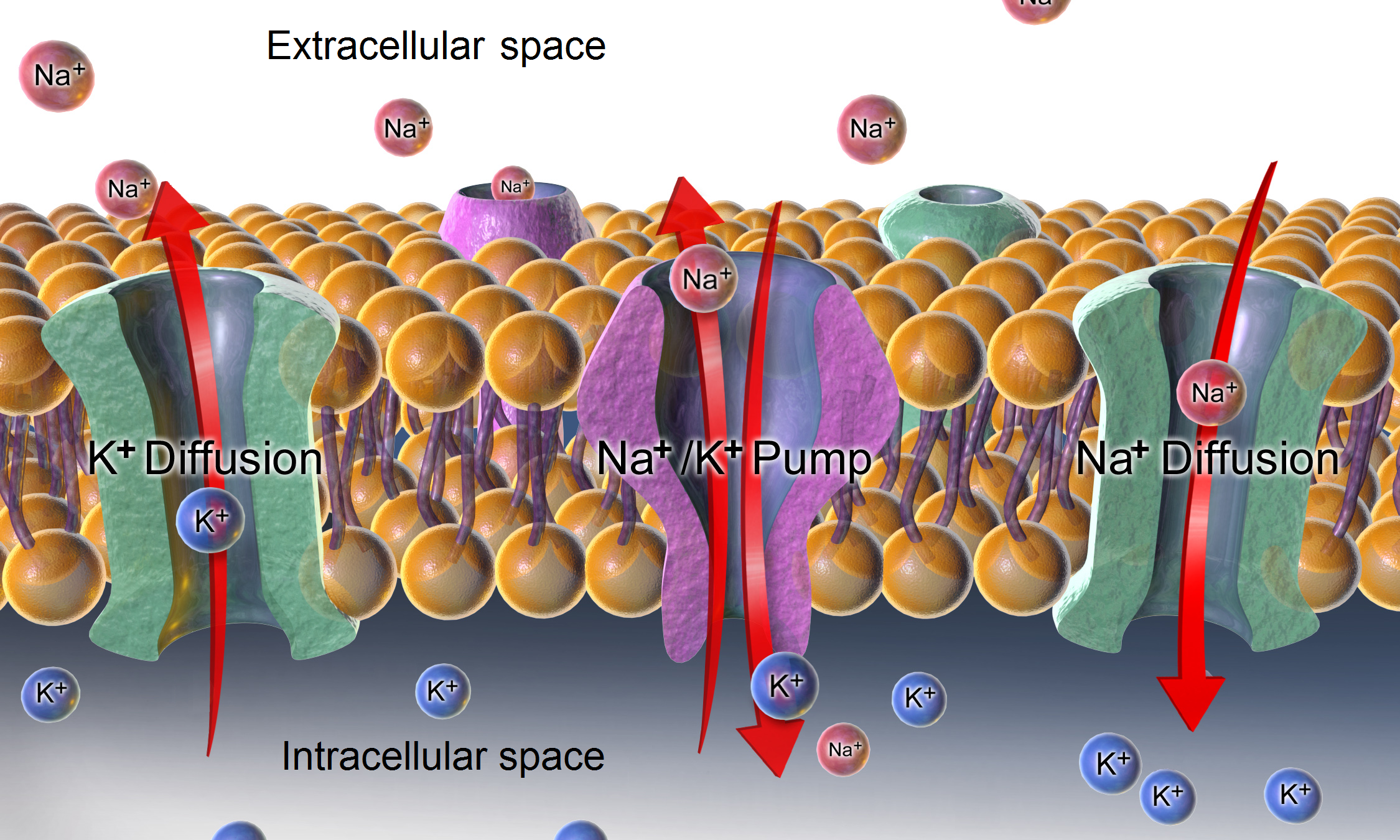
Hyperpolarization is a change in a cell's membrane potential that makes it more negative. Cells typically have a negative resting potential, with neuronal action potentials depolarizing the membrane. When the resting membrane potential is made more negative, it increases the minimum stimulus needed to surpass the needed threshold. Neurons naturally become hyperpolarized at the end of an action potential, which is often referred to as the relative refractory period. Relative refractory periods typically last 2 milliseconds, during which a stronger stimulus is needed to trigger another action potential. Cells can also become hyperpolarized depending on channels and receptors present on the membrane, which can have an inhibitory effect. Hyperpolarization is often caused by efflux of K+ (a cation) through K+ channels, or influx of Cl– (an anion) through Cl– channels. On the other hand, influx of cations, e.g. Na+ through Na+ channels or Ca2+ through Ca2+ cha ... [...More Info...] [...Related Items...] OR: [Wikipedia] [Google] [Baidu] |
Resting Potential
The relatively static membrane potential of quiescent cells is called the resting membrane potential (or resting voltage), as opposed to the specific dynamic electrochemical phenomena called action potential and graded membrane potential. The resting membrane potential has a value of approximately −70 mV or −0.07 V. Apart from the latter two, which occur in excitable cells (neurons, muscles, and some secretory cells in glands), membrane voltage in the majority of non-excitable cells can also undergo changes in response to environmental or intracellular stimuli. The resting potential exists due to the differences in membrane permeabilities for potassium, sodium, calcium, and chloride ions, which in turn result from functional activity of various ion channels, ion transporters, and exchangers. Conventionally, resting membrane potential can be defined as a relatively stable, ground value of transmembrane voltage in animal and plant cells. Because the membrane pe ... [...More Info...] [...Related Items...] OR: [Wikipedia] [Google] [Baidu] |
G Protein-coupled Receptor
G protein-coupled receptors (GPCRs), also known as seven-(pass)-transmembrane domain receptors, 7TM receptors, heptahelical receptors, serpentine receptors, and G protein-linked receptors (GPLR), form a large group of evolutionarily related proteins that are cell surface receptors that detect molecules outside the cell and activate cellular responses. They are coupled with G proteins. They pass through the cell membrane seven times in the form of six loops (three extracellular loops interacting with ligand molecules, three intracellular loops interacting with G proteins, an N-terminal extracellular region and a C-terminal intracellular region) of amino acid residues, which is why they are sometimes referred to as seven-transmembrane receptors. Text was copied from this source, which is available under Attribution 2.5 Generic (CC BY 2.5) licence/ref> Ligands can bind either to the extracellular N-terminus and loops (e.g. glutamate receptors) or to the binding site wi ... [...More Info...] [...Related Items...] OR: [Wikipedia] [Google] [Baidu] |
GABAB Receptor
GABAB receptors (GABABR) are G protein-coupled receptor, G-protein coupled receptors for gamma-aminobutyric acid (GABA), therefore making them metabotropic receptors, that are linked via G-proteins to potassium channels. The changing potassium concentrations hyperpolarize the cell at the end of an action potential. The reversal potential of the GABAB-mediated IPSP (inhibitory postsynaptic potential) is −100 mV, which is much more hyperpolarized than the GABAA receptor, GABAA IPSP. GABAB receptors are found in the central nervous system and the autonomic nervous system, autonomic division of the peripheral nervous system. The receptors were first named in 1981 when their distribution in the CNS was determined, which was determined by Norman Bowery and his team using radioactively labelled baclofen. Functions GABABRs stimulate the opening of Potassium channel, K+ channels, specifically G protein-coupled inwardly-rectifying potassium channel, GIRKs, which brings the neuro ... [...More Info...] [...Related Items...] OR: [Wikipedia] [Google] [Baidu] |
GABAA Receptor
The GABAA receptor (GABAAR) is an ionotropic receptor and ligand-gated ion channel. Its endogenous Ligand (biochemistry), ligand is γ-aminobutyric acid (GABA), the major inhibitory neurotransmitter in the central nervous system. Accurate regulation of GABAergic transmission through appropriate developmental processes, specificity to neural cell types, and responsiveness to activity is crucial for the proper functioning of nearly all aspects of the central nervous system (CNS). Upon opening, the GABAA receptor on the Chemical synapse, postsynaptic cell is selectively permeable to Chloride, chloride ions () and, to a lesser extent, Bicarbonate, bicarbonate ions (). GABAAR are members of the ligand-gated ion channel receptor superfamily, which is a chloride channel family with a dozen or more heterotetrametric subtypes and 19 distinct subunits. These subtypes have distinct brain regional and subcellular localization, age-dependent expression, and the ability to undergo plastic alt ... [...More Info...] [...Related Items...] OR: [Wikipedia] [Google] [Baidu] |
GABA
GABA (gamma-aminobutyric acid, γ-aminobutyric acid) is the chief inhibitory neurotransmitter in the developmentally mature mammalian central nervous system. Its principal role is reducing neuronal excitability throughout the nervous system. GABA is sold as a dietary supplement in many countries. It has been traditionally thought that exogenous GABA (i.e., taken as a supplement) does not cross the blood–brain barrier, but data obtained from more recent research (2010s) in rats describes the notion as being unclear. The carboxylate form of GABA is γ-aminobutyrate. Function Neurotransmitter Two general classes of GABA receptor are known: * GABAA in which the receptor is part of a ligand-gated ion channel complex * GABAB metabotropic receptors, which are G protein-coupled receptors that open or close ion channels via intermediaries (G proteins) Neurons that produce GABA as their output are called GABAergic neurons, and have chiefly inhibitory action at receptors in t ... [...More Info...] [...Related Items...] OR: [Wikipedia] [Google] [Baidu] |
Voltage Clamp
The voltage clamp is an experimental method used by electrophysiologists to measure the ion currents through the membranes of excitable cells, such as neurons, while holding the membrane voltage at a set level. A basic voltage clamp will iteratively measure the membrane potential, and then change the membrane potential (voltage) to a desired value by adding the necessary current. This "clamps" the cell membrane at a desired constant voltage, allowing the voltage clamp to record what currents are delivered. Because the currents applied to the cell must be equal to (and opposite in charge to) the current going across the cell membrane at the set voltage, the recorded currents indicate how the cell reacts to changes in membrane potential. Cell membranes of excitable cells contain many different kinds of ion channels, some of which are voltage-gated. The voltage clamp allows the membrane voltage to be manipulated independently of the ionic currents, allowing the current–voltag ... [...More Info...] [...Related Items...] OR: [Wikipedia] [Google] [Baidu] |
Patch Clamping
The patch clamp technique is a laboratory technique in electrophysiology used to study ionic currents in individual isolated living cells, tissue sections, or patches of cell membrane. The technique is especially useful in the study of excitable cells such as neurons, cardiomyocytes, muscle fibers, and pancreatic beta cells, and can also be applied to the study of bacterial ion channels in specially prepared giant spheroplasts. Patch clamping can be performed using the voltage clamp technique. In this case, the voltage across the cell membrane is controlled by the experimenter and the resulting currents are recorded. Alternatively, the current clamp technique can be used. In this case, the current passing across the membrane is controlled by the experimenter and the resulting changes in voltage are recorded, generally in the form of action potentials. Erwin Neher and Bert Sakmann developed the patch clamp in the late 1970s and early 1980s. This discovery made it possib ... [...More Info...] [...Related Items...] OR: [Wikipedia] [Google] [Baidu] |
Neuroscientist
A neuroscientist (or neurobiologist) is a scientist specializing in neuroscience that deals with the anatomy and function of neurons, Biological neural network, neural circuits, and glia, and their Behavior, behavioral, biological, and psychological roles in health and disease. Neuroscientists generally work as researchers within a college, university, government agency, or private Private industry, industry setting. In research-oriented careers, neuroscientists design and conduct scientific experiments on the nervous system and its functions. They can engage in basic or applied research. Basic research seeks to expand current understanding of the nervous system, whereas applied research seeks to address a specific problem, such as developing a treatment for a neurological disorder. Neuroscientists have numerous career opportunities outside of academic research, including careers in industry, science writing, government program management, science advocacy, and education. A ... [...More Info...] [...Related Items...] OR: [Wikipedia] [Google] [Baidu] |
HCN Channel
Hyperpolarization-activated cyclic nucleotide–gated (HCN) channels are integral membrane proteins that serve as nonselective voltage-gated cation channels in the plasma membranes of heart and brain cells. HCN channels are sometimes referred to as pacemaker channels because they help to generate rhythmic activity within groups of heart and brain cells. HCN channels are activated by membrane hyperpolarization, are permeable to and , and are constitutively open at voltages near the resting membrane potential. HCN channels are encoded by four genes ( HCN1, 2, 3, 4) and are widely expressed throughout the heart and the central nervous system. The current through HCN channels, designated ''I''f or ''I''h, plays a key role in the control of cardiac and neuronal rhythmicity and is called the pacemaker current or "funny" current. Expression of single isoforms in heterologous systems such as human embryonic kidney ( HEK) cells, Chinese hamster ovary ( CHO) cells and ''Xenopus'' ... [...More Info...] [...Related Items...] OR: [Wikipedia] [Google] [Baidu] |


