|
Hypercementosis
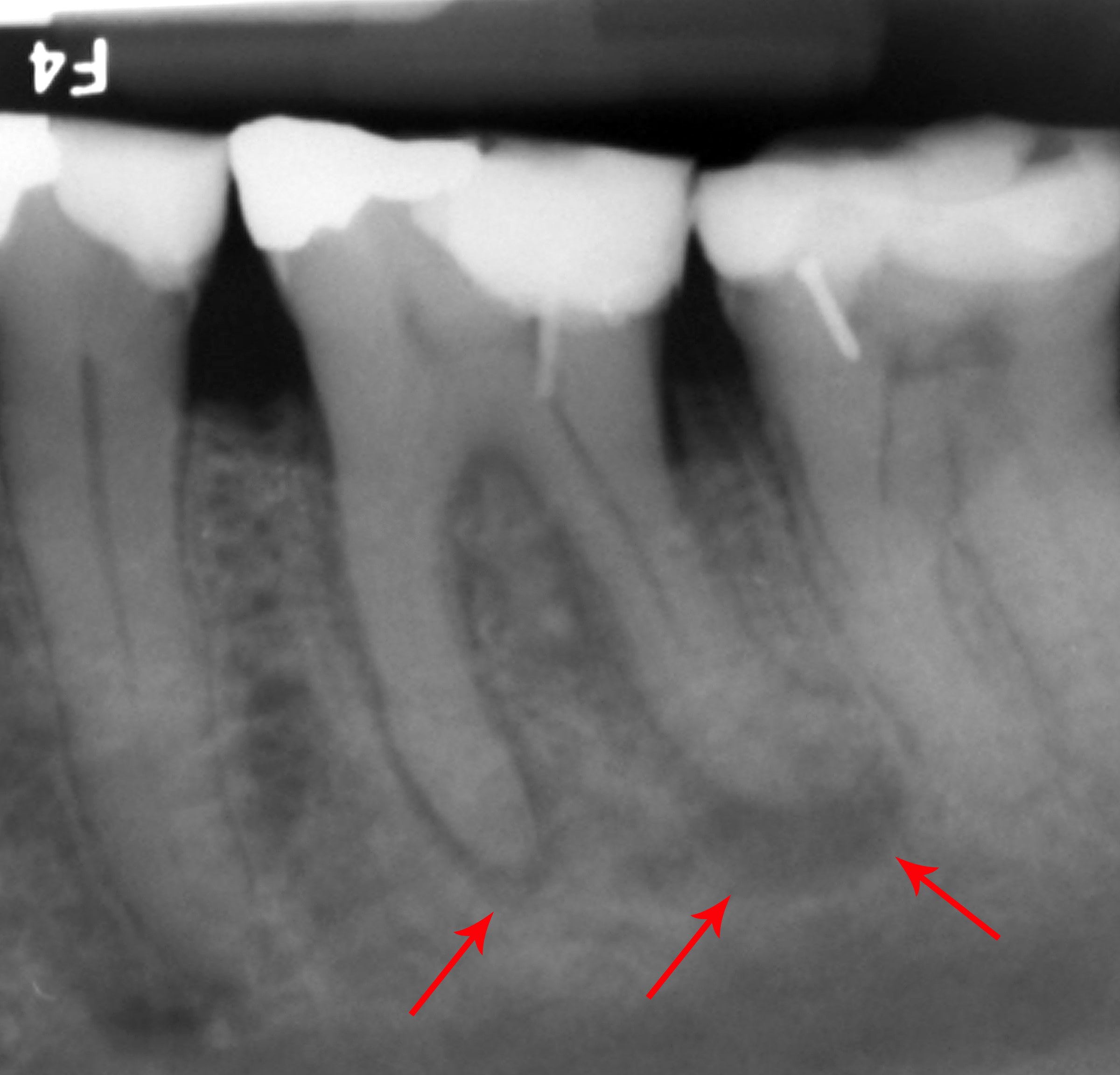
Hypercementosis is an idiopathic, non-Neoplasm, neoplastic condition characterized by the excessive buildup of normal cementum (calcified tissue) on the roots of one or more teeth. A thicker layer of cementum can give the tooth an enlarged appearance, mainly occurring at the Root apex (dental), apex or apices of the tooth. The cellular cementum functions at the bottom half of the tooth roots which contain cementocytes that anchor the tooth into the jaw Dental alveolus, socket, protect the Pulp (tooth), tooth's pulp, and repair external Tooth resorption, root resorption. Signs and symptoms It is experienced as an uncomfortable sensation in the tooth, followed by an aching pain. Excess amounts of cementum may cause pressure on Periodontal fiber, periodontal ligaments and adjacent teeth. The teeth affected may present as asymptomatic. It may be shown on radiographs as a radiopaque (or lighter) mass at each Root apex (dental), root apex to confirm the diagnosis. Cause Trauma and othe ... [...More Info...] [...Related Items...] OR: [Wikipedia] [Google] [Baidu] |
Cementum
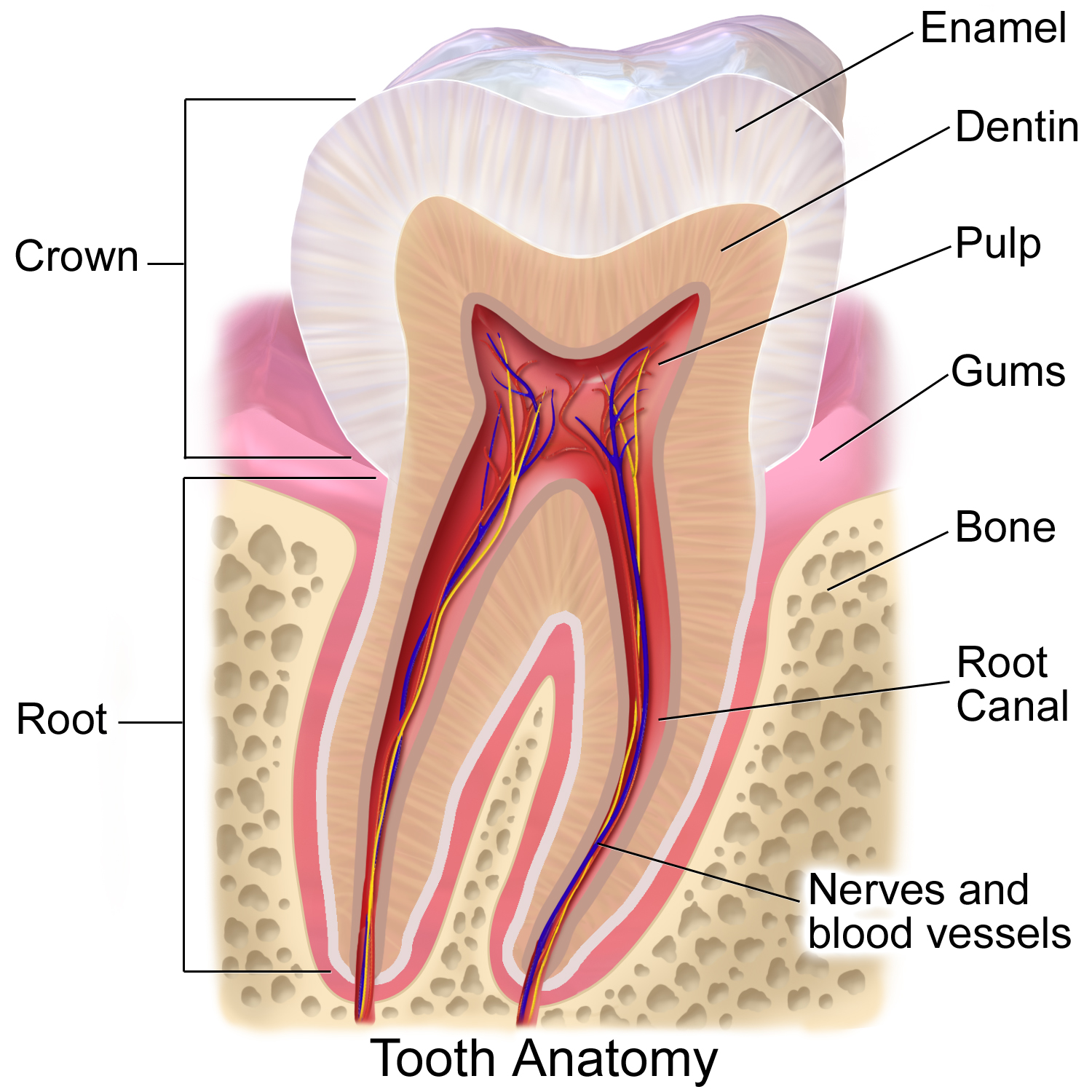
Cementum is a specialized calcified substance covering the root of a tooth. The cementum is the part of the periodontium that attaches the teeth to the alveolar bone by anchoring the periodontal ligament. Structure The cells of cementum are the entrapped cementoblasts, the cementocytes. Each cementocyte lies in its lacuna, similar to the pattern noted in bone. These lacunae also have canaliculi or canals. Unlike those in bone, however, these canals in cementum do not contain nerves, nor do they radiate outward. Instead, the canals are oriented toward the periodontal ligament and contain cementocytic processes that exist to diffuse nutrients from the ligament because it is vascularized. After the apposition of cementum in layers, the cementoblasts that do not become entrapped in cementum line up along the cemental surface along the length of the outer covering of the periodontal ligament. These cementoblasts can form subsequent layers of cementum if the tooth is injured. Sh ... [...More Info...] [...Related Items...] OR: [Wikipedia] [Google] [Baidu] |
Idiopathic
An idiopathic disease is any disease with an unknown cause or mechanism of apparent spontaneous origin. For some medical conditions, one or more causes are somewhat understood, but in a certain percentage of people with the condition, the cause may not be readily apparent or characterized. In these cases, the origin of the condition is said to be idiopathic. With some other medical conditions, the root cause for a large percentage of all cases has not been established—for example, focal segmental glomerulosclerosis or ankylosing spondylitis; the majority of these cases are deemed idiopathic. Certain medical conditions, when idiopathic, notably some forms of epilepsy and stroke, are preferentially described by the synonymous term of cryptogenic. Derivation The term 'idiopathic' derives from Greek ''idios'' "one's own" and ''pathos'' "suffering", so ''idiopathy'' means approximately "a disease of its own kind". Examples Diseases where the cause is seen as wholly or partly ... [...More Info...] [...Related Items...] OR: [Wikipedia] [Google] [Baidu] |
Periapical Granuloma
Periapical granuloma, also sometimes referred to as a radicular granuloma or apical granuloma, is an inflammation at the tip of a dead (nonvital) tooth. It is a lesion or mass that typically starts out as an epithelial lined cyst, and undergoes an inward curvature that results in inflammation of granulation tissue at the root tips of a dead tooth. This is usually due to dental caries or a bacterial infection of the dental pulp. Periapical granuloma is an infrequent disorder that has an occurrence rate between 9.3 and 87.1 percent. Periapical granuloma is not a true granuloma due to the fact that it does not contain granulomatous inflammation; however, ''periapical granuloma'' is a common term used. Symptoms Patients who have a periapical granuloma are usually asymptomatic; however, when there is inflammation, patients could experience temperature sensitivity, pain while chewing solid foods, swelling and sensitivity to a dental percussion test. Generally, periapical granuloma i ... [...More Info...] [...Related Items...] OR: [Wikipedia] [Google] [Baidu] |
Premolar
The premolars, also called premolar Tooth (human), teeth, or bicuspids, are transitional teeth located between the Canine tooth, canine and Molar (tooth), molar teeth. In humans, there are two premolars per dental terminology#Quadrant, quadrant in the permanent teeth, permanent set of teeth, making eight premolars total in the mouth. They have at least two Cusp (dentistry), cusps. Premolars can be considered transitional teeth during chewing, or mastication. They have properties of both the canines, that lie anterior and molars that lie Posterior (anatomy), posterior, and so food can be transferred from the canines to the premolars and finally to the molars for grinding, instead of directly from the canines to the molars. Human anatomy The premolars in humans are the maxillary first premolar, maxillary second premolar, mandibular first premolar, and the mandibular second premolar. Premolar teeth by definition are permanent teeth Anatomical terms of location#Proximal and distal, ... [...More Info...] [...Related Items...] OR: [Wikipedia] [Google] [Baidu] |
Mandibular Molars
The molars or molar teeth are large, flat teeth at the back of the mouth. They are more developed in mammals. They are used primarily to grind food during chewing. The name ''molar'' derives from Latin, ''molaris dens'', meaning "millstone tooth", from ''mola'', millstone and ''dens'', tooth. Molars show a great deal of diversity in size and shape across the mammal groups. The third molar of humans is sometimes vestigial. Human anatomy In humans, the molar teeth have either four or five cusps. Adult humans have 12 molars, in four groups of three at the back of the mouth. The third, rearmost molar in each group is called a wisdom tooth. It is the last tooth to appear, breaking through the front of the gum at about the age of 20, although this varies among individuals and populations, and in many cases the tooth is missing. The human mouth contains upper (maxillary) and lower (mandibular) molars. They are: maxillary first molar, maxillary second molar, maxillary third molar, man ... [...More Info...] [...Related Items...] OR: [Wikipedia] [Google] [Baidu] |
Apical Foramen
In dental anatomy, the apical foramen, literally translated "small opening of the apex," is the tooth's natural opening, found at the root's very tip—that is, the root apex — whereby an artery, vein, and nerve enter the tooth and commingle with the tooth's internal soft tissue, called pulp. Additionally, the apical foramen is the point where the pulp meets the periodontal tissues, the connective tissues that surround and support the tooth. The foramen is located 0.5mm to 1.5mm from the apex of the tooth. Each tooth has an apical foramen. Characteristics The average size of the orifice is 0.3 to 0.4 mm in diameter. There can be two or more foramina separated by a portion of dentin and cementum or by cementum only. If more than one foramen is present on each root, the largest one is designated as the apical foramen and the rest are considered accessory foramina. Apical delta Apical delta refers to the branching pattern of small accessory canals and minor foramina seen ... [...More Info...] [...Related Items...] OR: [Wikipedia] [Google] [Baidu] |
Pulp Necrosis
Pulp necrosis is a clinical diagnostic category indicating the death of cells and tissues in the pulp chamber of a tooth with or without bacterial invasion. It is often the result of many cases of dental trauma, caries and irreversible pulpitis. In the initial stage of the infection, the pulp chamber is partially necrosed for a period of time and if left untreated, the area of cell death expands until the entire pulp necroses. The most common clinical signs present in a tooth with a necrosed pulp would be a grey discoloration of the crown and/or periapical radiolucency. This altered translucency in the tooth is due to disruption and cutting off of the apical neurovascular blood supply. Sequelae of a necrotic pulp include acute apical periodontitis, dental abscess or radicular cyst and discolouration of the tooth. Tests for a necrotic pulp include: vitality testing using a thermal test or an electric pulp tester. Discolouration may be visually obvious, or more subtle. Treat ... [...More Info...] [...Related Items...] OR: [Wikipedia] [Google] [Baidu] |
Concrescence
Concrescence is an uncommon developmental condition of teeth where the cementum overlying the roots of at least two teeth fuse together without the involvement of dentin. Usually, two teeth are involved with the upper second and third molars being most commonly fused together. The prevalence ranges 0.04–0.8% in permanent teeth, with the incidence being highest in the posterior maxilla. Signs and symptoms * Problems with tooth positioning causing cheek biting and traumatic ulcers. * Involved teeth may have difficulty erupting or may not erupt completely. * Possible gum disease (localized periodontal destruction due to aetiological factors, e.g. funnel development leading to plaque accumulation) * Cavities (caries) due to predisposition from crowded teeth and misalignment. * May cause fracture of the tuberosity or floor of the maxillary sinus. Cause The exact cause of concrescence is unknown. However, it may develop during root formation (true/primary concrescence) or after ... [...More Info...] [...Related Items...] OR: [Wikipedia] [Google] [Baidu] |
Dental Extraction
A dental extraction (also referred to as tooth extraction, exodontia, exodontics, or informally, tooth pulling) is the removal of teeth from the dental alveolus (socket) in the alveolar bone. Extractions are performed for a wide variety of reasons, but most commonly to remove teeth which have become unrestorable through tooth decay, periodontal disease, or dental trauma, especially when they are associated with toothache. Sometimes impacted wisdom teeth (wisdom teeth that are stuck and unable to grow normally into the mouth) cause recurrent infections of the gum ( pericoronitis), and may be removed when other conservative treatments have failed (cleaning, antibiotics and operculectomy). In orthodontics, if the teeth are crowded, healthy teeth may be extracted (often bicuspids) to create space so the rest of the teeth can be straightened. Procedure Extractions could be categorized into non-surgical (simple) and surgical, depending on the type of tooth to be removed and o ... [...More Info...] [...Related Items...] OR: [Wikipedia] [Google] [Baidu] |
Hyperplasia
Hyperplasia (from ancient Greek ὑπέρ ''huper'' 'over' + πλάσις ''plasis'' 'formation'), or hypergenesis, is an enlargement of an organ or tissue caused by an increase in the amount of Tissue (biology), organic tissue that results from cell proliferation. It may lead to the Gross anatomy, gross enlargement of an organ, and the term is sometimes confused with benign neoplasia or benign tumor. Hyperplasia is a common preneoplastic response to stimulus. Microscopically, cells resemble normal cells but are increased in numbers. Sometimes cells may also be increased in size (hypertrophy). Hyperplasia is different from hypertrophy in that the Cellular adaptation, adaptive cell change in hypertrophy is an increase in the cell size, ''size'' of cells, whereas hyperplasia involves an increase in the ''number'' of cells. Causes Hyperplasia may be due to any number of causes, including proliferation of basal layer of epidermis to compensate skin loss, Chronic inflammation, chr ... [...More Info...] [...Related Items...] OR: [Wikipedia] [Google] [Baidu] |
Periapical
Dental anatomy is a field of anatomy dedicated to the study of human tooth structures. The development, appearance, and classification of teeth fall within its purview. (The function of teeth as they contact one another falls elsewhere, under dental occlusion.) Tooth formation begins before birth, and the teeth's eventual morphology is dictated during this time. Dental anatomy is also a taxonomical science: it is concerned with the naming of teeth and the structures of which they are made, this information serving a practical purpose in dental treatment. Usually, there are 20 primary ("baby") teeth and 32 permanent teeth, the last four being third molars or "wisdom teeth", each of which may or may not grow in. Among primary teeth, 10 usually are found in the maxilla (upper jaw) and the other 10 in the mandible (lower jaw). Among permanent teeth, 16 are found in the maxilla and the other 16 in the mandible. Each tooth has specific distinguishing features. Growing of tooth ... [...More Info...] [...Related Items...] OR: [Wikipedia] [Google] [Baidu] |
Hydroxyapatite
Hydroxyapatite (International Mineralogical Association, IMA name: hydroxylapatite) (Hap, HAp, or HA) is a naturally occurring mineral form of calcium apatite with the Chemical formula, formula , often written to denote that the Crystal structure, crystal unit cell comprises two entities. It is the Hydroxy group, hydroxyl endmember of the complex apatite, apatite group. The ion can be replaced by fluorine, fluoride or chlorine, chloride, producing fluorapatite or chlorapatite. It crystallizes in the hexagonal (crystal system), hexagonal crystal system. Pure hydroxyapatite powder is white. Naturally occurring apatites can, however, also have brown, yellow, or green colorations, comparable to the discolorations of dental fluorosis. Up to 50% by volume and 70% by weight of human bone is a modified form of hydroxyapatite, known as bone mineral. Carbonated calcium-deficient hydroxyapatite is the main mineral of which dental enamel and dentin are composed. Hydroxyapatite crystals a ... [...More Info...] [...Related Items...] OR: [Wikipedia] [Google] [Baidu] |


