|
Hydrocele Testis
A hydrocele testis is an accumulation of clear fluid within the cavum vaginale, the potential space between the layers of the tunica vaginalis of the testicle. It is the most common form of hydrocele and is often referred to simply as a "hydrocele". A primary hydrocele testis causes a painless enlargement in the scrotum on the affected side and is thought to be due to the defective absorption of fluid secreted between the two layers of the tunica vaginalis (investing membrane). A secondary hydrocele is secondary to either inflammation or a neoplasm in the testis. A hydrocele testis usually occurs on one side, but can also affect both sides. The accumulation can be a marker of physical trauma, infection, tumor or varicocele surgery, but the cause is generally unknown. Indirect inguinal hernia indicates an increased risk of hydrocele testis. Signs and symptoms A hydrocele testis feels like a small fluid-filled balloon inside the scrotum. It is smooth and is mainly in front of the t ... [...More Info...] [...Related Items...] OR: [Wikipedia] [Google] [Baidu] |
Testis
A testicle or testis ( testes) is the gonad in all male bilaterians, including humans, and is Homology (biology), homologous to the ovary in females. Its primary functions are the production of sperm and the secretion of Androgen, androgens, primarily testosterone. The release of testosterone is regulated by luteinizing hormone (LH) from the anterior pituitary gland. Sperm production is controlled by follicle-stimulating hormone (FSH) from the anterior pituitary gland and by testosterone produced within the gonads. Structure Appearance Males have two testicles of similar size contained within the scrotum, which is an extension of the abdominal wall. Scrotal asymmetry, in which one testicle extends farther down into the scrotum than the other, is common. This is because of the differences in the vasculature's anatomy. For 85% of men, the right testis hangs lower than the left one. Measurement and volume The volume of the testicle can be estimated by palpating it and compari ... [...More Info...] [...Related Items...] OR: [Wikipedia] [Google] [Baidu] |
Inguinal Canal
The inguinal canal is a passage in the anterior abdominal wall on each side of the body (one on each side of the midline), which in males, convey the spermatic cords and in females, the round ligament of the uterus. The inguinal canals are larger and more prominent in males. Structure The inguinal canals are situated just above the medial half of the inguinal ligament. The canals are approximately 4 to 6 cm long, angled anteroinferiorly and medially. In males, its diameter is normally 2 cm (±1 cm in standard deviation) at the deep inguinal ring.The diameter has been estimated to be ±2.2cm ±1.08cm in Africans, and 2.1 cm ±0.41cm in Europeans. A first-order approximation is to visualize each canal as a cylinder. Walls To help define the boundaries, these canals are often further approximated as boxes with six sides. Not including the two rings, the remaining four sides are usually called the "anterior wall", "inferior wall ("floor")", "superior wall ("roof")", and "po ... [...More Info...] [...Related Items...] OR: [Wikipedia] [Google] [Baidu] |
Sexual Health
Sexual and reproductive health (SRH) is a field of research, health care, and social activism that explores the health of an individual's reproductive system and sexual well-being during all stages of their life. Sexual and reproductive health is more commonly defined as sexual and reproductive health and rights, to encompass individual agency to make choices about their sexual and reproductive lives. The term can also be further defined more broadly within the framework of the World Health Organization's (WHO) definition of health―as "a state of complete physical, mental and social well-being, and not merely the absence of disease or infirmity"―. WHO has a working definition of sexual health (2006) as '“…''a state of physical, emotional, mental and social well-being in relation to sexuality; it is not merely the absence of disease, dysfunction or infirmity. Sexual health requires a positive and respectful approach to sexuality and sexual relationships, as well as the ... [...More Info...] [...Related Items...] OR: [Wikipedia] [Google] [Baidu] |
Sclerotherapy
Sclerotherapy (the word reflects the Greek ''skleros'', meaning ''hard'') is a procedure used to treat blood vessel malformations ( vascular malformations) and also malformations of the lymphatic system. A medication is injected into the vessels, which makes them shrink. It is used for children and young adults with vascular or lymphatic malformations. In adults, sclerotherapy is often used to treat spider veins, smaller varicose veins, hemorrhoids, and hydroceles. Sclerotherapy is one method for the treatment of spider veins, varicose veins (which are also often treated with surgery, radiofrequency, and laser ablation), and venous malformations. In ultrasound-guided sclerotherapy, ultrasound is used to visualize the underlying vein so the physician can deliver and monitor the injection. Sclerotherapy often takes place under ultrasound guidance after venous abnormalities have been diagnosed with duplex ultrasound. Sclerotherapy under ultrasound guidance and using microfoam s ... [...More Info...] [...Related Items...] OR: [Wikipedia] [Google] [Baidu] |
Needle Aspiration Biopsy
Fine-needle aspiration (FNA) is a diagnostic procedure used to investigate lumps or masses. In this technique, a thin (23–25 gauge (0.52 to 0.64 mm outer diameter)), hollow needle is inserted into the mass for sampling of cells that, after being stained, are examined under a microscope (biopsy). The sampling and biopsy considered together are called fine-needle aspiration biopsy (FNAB) or fine-needle aspiration cytology (FNAC) (the latter to emphasize that any aspiration biopsy involves cytopathology, not histopathology). Fine-needle aspiration biopsies are very safe for minor surgical procedures. Often, a major surgical (excisional or open) biopsy can be avoided by performing a needle aspiration biopsy instead, eliminating the need for hospitalization. In 1981, the first fine-needle aspiration biopsy in the United States was done at Maimonides Medical Center. The modern procedure is widely used to diagnose cancer and inflammatory conditions. Fine needle aspiration i ... [...More Info...] [...Related Items...] OR: [Wikipedia] [Google] [Baidu] |
Urologist
Urology (from Greek οὖρον ''ouron'' "urine" and ''-logia'' "study of"), also known as genitourinary surgery, is the branch of medicine that focuses on surgical and medical diseases of the urinary system and the reproductive organs. Organs under the domain of urology include the kidneys, adrenal glands, ureters, urinary bladder, urethra, and the male reproductive organs (testes, epididymides, vasa deferentia, seminal vesicles, prostate, and penis). The urinary and reproductive tracts are closely linked, and disorders of one often affect the other. Thus a major spectrum of the conditions managed in urology exists under the domain of genitourinary disorders. Urology combines the management of medical (i.e., non-surgical) conditions, such as urinary-tract infections and benign prostatic hyperplasia, with the management of surgical conditions such as bladder or prostate cancer, kidney stones, congenital abnormalities, traumatic injury, and stress incontinence. Urologic ... [...More Info...] [...Related Items...] OR: [Wikipedia] [Google] [Baidu] |
Ernst Von Bergmann
Ernst Gustav Benjamin von Bergmann (16 December 1836 – 25 March 1907) was a Baltic German surgeon. He was the first physician to introduce heat sterilisation of surgical instruments and is known as a pioneer of aseptic surgery. Early life and education Born in Riga, Livonia Governorate (now Latvia), in 1860 he earned his doctorate at the University of Dorpat. He then worked as an assistant at the surgical clinic, and trained for surgery under Georg von Adelmann (his future father-in-law), and Georg von Oettingen. He received his certification in 1864. Career From 1871 to 1878 he was a professor of surgery at Dorpat. In 1878 he became a professor at Würzburg; in 1882 he relocated to the University of Berlin as a successor to Bernhard von Langenbeck. von Bergmann continued as a professor of surgery at Berlin for the remainder of his career. Two of his assistants in Berlin were Curt Schimmelbusch (1860–1895) and Friedrich Gustav von Bramann (1854–1913). [...More Info...] [...Related Items...] OR: [Wikipedia] [Google] [Baidu] |
Aspirator (medical Device)
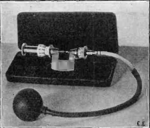
A medical aspirator is a suction machine used to remove mucus, blood, and other bodily fluids from a patient. They can be used during surgical procedures but an operating theater is generally equipped with a central system of vacuum tubes. Most aspirators are therefore portable, for use in ambulances and nursing homes, and can run on mains electricity, AC or electric battery, battery power. They consist of a vacuum pump, a vacuum regulator and gauge, a collection canister, and sometimes a bacterial filter. Plastic tubing is used to continuously draw fluid into the collection canister. In the past manually operated aspirators were used such as ''Potain's aspirator''.p. 97, Minor surgery, Henry R. Wharton, i''System of surgery'' vol. II, Frederic S. Dennis and John S. Billings, eds., Philadelphia: Lea Brothers & Co., 1895. See also * Suction (medicine) References Medical equipment {{medical-equipment-stub ... [...More Info...] [...Related Items...] OR: [Wikipedia] [Google] [Baidu] |
Ultrasound
Ultrasound is sound with frequency, frequencies greater than 20 Hertz, kilohertz. This frequency is the approximate upper audible hearing range, limit of human hearing in healthy young adults. The physical principles of acoustic waves apply to any frequency range, including ultrasound. Ultrasonic devices operate with frequencies from 20 kHz up to several gigahertz. Ultrasound is used in many different fields. Ultrasonic devices are used to detect objects and measure distances. Ultrasound imaging or sonography is often used in medicine. In the nondestructive testing of products and structures, ultrasound is used to detect invisible flaws. Industrially, ultrasound is used for cleaning, mixing, and accelerating chemical processes. Animals such as bats and porpoises use ultrasound for locating prey and obstacles. History Acoustics, the science of sound, starts as far back as Pythagoras in the 6th century BC, who wrote on the mathematical properties of String instrument ... [...More Info...] [...Related Items...] OR: [Wikipedia] [Google] [Baidu] |
Ultrasound Scan ND 0124155309 1600360
Ultrasound is sound with frequencies greater than 20 kilohertz. This frequency is the approximate upper audible limit of human hearing in healthy young adults. The physical principles of acoustic waves apply to any frequency range, including ultrasound. Ultrasonic devices operate with frequencies from 20 kHz up to several gigahertz. Ultrasound is used in many different fields. Ultrasonic devices are used to detect objects and measure distances. Ultrasound imaging or sonography is often used in medicine. In the nondestructive testing of products and structures, ultrasound is used to detect invisible flaws. Industrially, ultrasound is used for cleaning, mixing, and accelerating chemical processes. Animals such as bats and porpoises use ultrasound for locating prey and obstacles. History Acoustics, the science of sound, starts as far back as Pythagoras in the 6th century BC, who wrote on the mathematical properties of stringed instruments. Echolocation in bats was d ... [...More Info...] [...Related Items...] OR: [Wikipedia] [Google] [Baidu] |
Ultrasonography Of Hydrocele
Medical ultrasound includes Medical diagnosis, diagnostic techniques (mainly medical imaging, imaging) using ultrasound, as well as therapeutic ultrasound, therapeutic applications of ultrasound. In diagnosis, it is used to create an image of internal body structures such as tendons, muscles, joints, blood vessels, and internal organs, to measure some characteristics (e.g., distances and velocities) or to generate an informative audible sound. The usage of ultrasound to produce visual images for medicine is called medical ultrasonography or simply sonography, or echography. The practice of examining pregnant women using ultrasound is called obstetric ultrasonography, and was an early development of clinical ultrasonography. The machine used is called an ultrasound machine, a sonograph or an echograph. The visual image formed using this technique is called an ultrasonogram, a sonogram or an echogram. Ultrasound is composed of sound waves with frequency, frequencies greater than ... [...More Info...] [...Related Items...] OR: [Wikipedia] [Google] [Baidu] |
Epididymis
The epididymis (; : epididymides or ) is an elongated tubular genital organ attached to the posterior side of each one of the two male reproductive glands, the testicles. It is a single, narrow, tightly coiled tube in adult humans, in length; uncoiled the tube would be approximately 6 m (20 feet) long. It connects the testicle to the vas deferens in the male reproductive system. The epididymis serves as an interconnection between the multiple efferent ducts at the rear of a testicle (proximally), and the vas deferens (distally). Its primary function is the storage, maturation and transport of sperm cells. Structure The human epididymis is situated posterior and somewhat lateral to the testis. The epididymis is invested completely by the tunica vaginalis (which is continuous with the tunica vaginalis covering the testis). The epididymis can be divided into three main regions: * The head (). The head of the epididymis receives spermatozoa via the efferent ducts of the medi ... [...More Info...] [...Related Items...] OR: [Wikipedia] [Google] [Baidu] |


