|
Greater Trochanter
The greater trochanter of the femur is a large, irregular, quadrilateral eminence and a part of the skeletal system. It is directed lateral and medially and slightly posterior. In the adult it is about 2–4 cm lower than the femoral head.Standring, Susan, editor. ''Gray’s Anatomy: The Anatomical Basis of Clinical Practice''. Forty-First edition, Elsevier Limited, 2016, p. 1327. Because the pelvic outlet in the female is larger than in the male, there is a greater distance between the greater trochanters in the female. It has two surfaces and four borders. It is a traction epiphysis. Surfaces The ''lateral surface'', quadrilateral in form, is broad, rough, convex, and marked by a diagonal impression, which extends from the postero-superior to the antero-inferior angle, and serves for the insertion of the tendon of the gluteus medius. Above the impression is a triangular surface, sometimes rough for part of the tendon of the same muscle, sometimes smooth for the interp ... [...More Info...] [...Related Items...] OR: [Wikipedia] [Google] [Baidu] |
Femur
The femur (; : femurs or femora ), or thigh bone is the only long bone, bone in the thigh — the region of the lower limb between the hip and the knee. In many quadrupeds, four-legged animals the femur is the upper bone of the hindleg. The Femoral head, top of the femur fits into a socket in the pelvis called the hip joint, and the bottom of the femur connects to the shinbone (tibia) and kneecap (patella) to form the knee. In humans the femur is the largest and thickest bone in the body. Structure The femur is the only bone in the upper Human leg, leg. The two femurs converge Anatomical terms of location, medially toward the knees, where they articulate with the Anatomical terms of location, proximal ends of the tibiae. The angle at which the femora converge is an important factor in determining the femoral-tibial angle. In females, thicker pelvic bones cause the femora to converge more than in males. In the condition genu valgum, ''genu valgum'' (knock knee), the femurs conve ... [...More Info...] [...Related Items...] OR: [Wikipedia] [Google] [Baidu] |
Gluteus Maximus
The gluteus maximus is the main extensor muscle of the hip in humans. It is the largest and outermost of the three gluteal muscles and makes up a large part of the shape and appearance of each side of the hips. It is the single largest muscle in the human body. Its thick fleshy mass, in a quadrilateral shape, forms the prominence of the buttocks. The other gluteal muscles are the medius and minimus, and sometimes informally these are collectively referred to as the glutes. Its large size is one of the most characteristic features of the muscular system in humans,Norman Eizenberg et al., ''General Anatomy: Principles and Applications'' (2008), p. 17. connected as it is with the power of maintaining the trunk in the erect posture. Other primates have much flatter hips and cannot sustain standing erectly. The muscle is made up of muscle fascicles lying parallel with one another, and are collected together into larger bundles separated by fibrous septa. Structure The gluteus maxi ... [...More Info...] [...Related Items...] OR: [Wikipedia] [Google] [Baidu] |
Third Trochanter
In human anatomy, the third trochanter is a bony projection occasionally present on the proximal femur near the superior border of the gluteal tuberosity. When present, it is oblong, rounded, or conical in shape and sometimes continuous with the gluteal ridge. It generally occurs bilaterally without significant side to side dimorphism. A structure of minor importance in humans, the incidence of the third trochanter varies from 17 to 72% between ethnic groups and it is frequently reported as more common in females than in males. Structures analogous to the third trochanter are present in other mammals, including some primates, but also in horses. It is called the third trochanter in reference to the greater and lesser trochanters that are always present on the femur. Function Its function is to provide an attachment for the ascending tendon of the gluteus maximus muscle. It may function as (1) a reinforcement mechanism for the proximal femoral diaphysis in response to incre ... [...More Info...] [...Related Items...] OR: [Wikipedia] [Google] [Baidu] |
Vastus Lateralis
The vastus lateralis (), also called the vastus externus, is the largest and most powerful part of the quadriceps femoris, a muscle in the thigh. Together with other muscles of the quadriceps group, it serves to extend the knee joint, moving the lower leg forward. It arises from a series of flat, broad tendons attached to the femur, and attaches to the outer border of the patella. It ultimately joins with the other muscles that make up the quadriceps in the quadriceps tendon, which travels over the knee to connect to the tibia. The vastus lateralis is the recommended site for intramuscular injection in infants less than 7 months old and those unable to walk, with loss of muscular tone.Mann, E. (2016). ''Injection (Intramuscular): Clinician Information.'' The Johanna Briggs Institute. Structure The vastus lateralis muscle arises from several areas of the femur, including the upper part of the intertrochanteric line; the lower, anterior borders of the greater trochanter, to the ... [...More Info...] [...Related Items...] OR: [Wikipedia] [Google] [Baidu] |
Inferior Gemellus Muscle
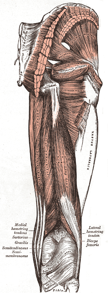
The gemelli muscles are the inferior gemellus muscle and the superior gemellus muscle, two small accessory fasciculi to the tendon of the internal obturator muscle. The gemelli muscles belong to the lateral rotator group of six muscles of the hip that rotate the femur in the hip joint. Superior gemellus muscle The gemelli muscles are two small muscular fasciculi, accessories to the tendon of the internal obturator muscle which is received into a groove between them. The superior gemellus muscle is the higher placed gemellus muscle that arises from the outer (gluteal) surface of the ischial spine, and blends with the upper part of the tendon of the internal obturator. It is smaller than the inferior gemellus. In some people, the fibres of the gemellus superior extend further than average, and are prolonged onto the medial surface of the greater trochanter of the femur. The superior and inferior gemelli are supplied by the inferior gluteal artery. Nerve supply to the superio ... [...More Info...] [...Related Items...] OR: [Wikipedia] [Google] [Baidu] |
Obturator Internus
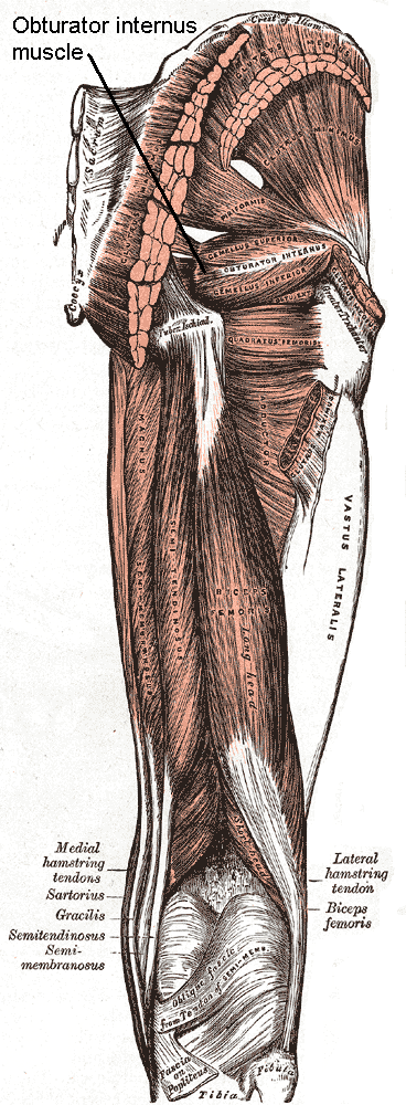
The internal obturator muscle or obturator internus muscle originates on the medial surface of the obturator membrane, the ischium near the membrane, and the rim of the pubis (bone), pubis. It exits the pelvis, pelvic cavity through the lesser sciatic foramen. The internal obturator is situated partly within the lesser pelvis, and partly at the back of the hip-joint. It functions to help laterally rotate femur with hip extension and abduct femur with hip flexion, as well as to steady the femoral head in the acetabulum. Structure Origin The internal obturator muscle arises from the inner surface of the antero-lateral wall of the pelvis. It surrounds the obturator foramen. It is attached to the inferior pubic ramus and ischium, and at the side to the inner surface of the hip bone below and behind the pelvic brim. It reaches from the upper part of the greater sciatic foramen above and behind to the obturator foramen below and in front. It also arises from the pelvic surface of ... [...More Info...] [...Related Items...] OR: [Wikipedia] [Google] [Baidu] |
Obturator Externus
The external obturator muscle or obturator externus muscle (; OE) is a flat, triangular muscle, which covers the outer surface of the anterior wall of the pelvis. It is sometimes considered part of the medial compartment of thigh, and sometimes considered part of the gluteal region. It is also considered to be part of the short external rotators of the hip, along with the gemellus superior and inferior, piriformis, and quadratus femoris. Structure It arises from the margin of bone immediately around the medial side of the obturator membrane and surrounding bone, viz., from the inferior pubic ramus, and the ramus of the ischium; it also arises from the medial two-thirds of the outer surface of the obturator membrane, and from the tendinous arch which completes the canal for the passage of the obturator vessels and nerves. The fibers springing from the pubic arch extend on to the inner surface of the bone, where they obtain a narrow origin between the margin of the for ... [...More Info...] [...Related Items...] OR: [Wikipedia] [Google] [Baidu] |
Trochanteric Fossa
In mammals including humans, the medial surface of the greater trochanter has at its base a deep depression bounded posteriorly by the intertrochanteric crest, called the trochanteric fossa. This fossa is the point of insertion of four muscles. Moving from the inferior-most to the superior-most, they are: the tendon of the obturator externus muscle, the obturator internus, the superior gemellus and inferior gemellus. The width and depth of the trochanteric fossa varies taxonomically.Gray's AnatomyNetter 2003Romer 1956 In reptiliomorphs such as Seymouria or Diadectes and basal reptiles such as Pareiasaurus, the trochanteric fossa (also known as the intertrochanteric fossa) is a very large depression on the ventral/posterior side of the femur. It is bounded medially by the internal trochanter (also known as the lesser trochanter), laterally by the posterior branch of the ventral ridge, and inferiorly by the convergence of the anterior and posterior branches of the ventral ridge a ... [...More Info...] [...Related Items...] OR: [Wikipedia] [Google] [Baidu] |
Bursa (anatomy)
A synovial bursa, usually simply bursa (: bursae or bursas), is a small fluid-filled sac lined by synovial membrane with an inner capillary layer of viscous synovial fluid (similar in consistency to that of a raw egg white). It provides a cushion between bones and tendons and/or muscles around a joint. This helps to reduce friction between the bones and allows free movement. Bursae are found around most major joints of the body. Structure Based on location, there are three types of bursa: subcutaneous, submuscular and subtendinous. A subcutaneous bursa is located between the skin and an underlying bone. It allows skin to move smoothly over the bone. Examples include the prepatellar bursa located over the kneecap and the olecranon bursa at the tip of the elbow. A submuscular bursa is found between a muscle and an underlying bone, or between adjacent muscles. These prevent rubbing of the muscle during movements. A large submuscular bursa, the trochanteric bursa, is found at the ... [...More Info...] [...Related Items...] OR: [Wikipedia] [Google] [Baidu] |
Epiphysis
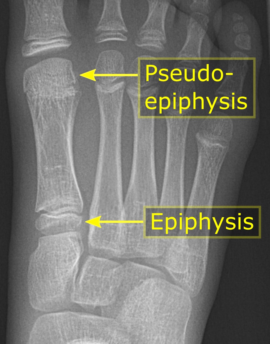
An epiphysis (; : epiphyses) is one of the rounded ends or tips of a long bone that ossify from one or more secondary centers of ossification. Between the epiphysis and diaphysis (the long midsection of the long bone) lies the metaphysis, including the epiphyseal plate (growth plate). During formation of the secondary ossification center, vascular canals (epiphysial canals) stemming from the perichondrium invade the epiphysis, supplying nutrients to the developing secondary centers of ossification. At the joint, the epiphysis is covered with articular cartilage; below that covering is a zone similar to the epiphyseal plate, known as Wikt:subchondral, subchondral bone. The epiphysis is mostly found in mammals but it is also present in some lizards. However, the secondary center of ossification may have evolved multiple times, having been found in the Jurassic sphenodont ''Sapheosaurus'' as well as in the therapsid ''Niassodon, Niassodon mfumukasi.'' The epiphysis is filled wi ... [...More Info...] [...Related Items...] OR: [Wikipedia] [Google] [Baidu] |
Vastus Lateralis
The vastus lateralis (), also called the vastus externus, is the largest and most powerful part of the quadriceps femoris, a muscle in the thigh. Together with other muscles of the quadriceps group, it serves to extend the knee joint, moving the lower leg forward. It arises from a series of flat, broad tendons attached to the femur, and attaches to the outer border of the patella. It ultimately joins with the other muscles that make up the quadriceps in the quadriceps tendon, which travels over the knee to connect to the tibia. The vastus lateralis is the recommended site for intramuscular injection in infants less than 7 months old and those unable to walk, with loss of muscular tone.Mann, E. (2016). ''Injection (Intramuscular): Clinician Information.'' The Johanna Briggs Institute. Structure The vastus lateralis muscle arises from several areas of the femur, including the upper part of the intertrochanteric line; the lower, anterior borders of the greater trochanter, to the ... [...More Info...] [...Related Items...] OR: [Wikipedia] [Google] [Baidu] |


