|
Gonioscopy
In ophthalmology, gonioscopy is a routine procedure that measures the angle between the iris (anatomy), iris and the cornea (the iridocorneal angle), using a goniolens (also known as a gonioscope) together with a slit lamp or operating microscope. Its use is important in diagnosing and monitoring various eye conditions associated with glaucoma. The goniolens or gonioscope The goniolens allows the clinician - usually an ophthalmologist or optometrist - to view the irideocorneal angle through a mirror or prism, without which the angle is masked by total internal reflection from the ocular tissue. The mechanism for this process varies with each type of goniolens. Three examples of goniolenses are the: * Koeppe direct goniolens: this transparent device is placed directly on the cornea along with lubricating fluid (to avoid damaging its surface). The steeper curvature of this goniolens' exterior surface optically eliminates the total internal reflection problem and allows a view o ... [...More Info...] [...Related Items...] OR: [Wikipedia] [Google] [Baidu] |
Sampaolesi Line
Sampaolesi line is a sign which may be observed during a clinical eye examination. During gonioscopy (where the structures of the eye's anterior segment are examined), if an abundance of brown pigment is seen at or anterior to Schwalbe's line, a Sampaolesi line is said to be present. The presence of a Sampaolesi line can signify pigment dispersion syndrome or pseudoexfoliation syndrome. Gonioscopy is performed during eye examinations, which involves placing a mirrored lens on the patient's cornea in order to visualise the angle of the Anterior chamber of eyeball, anterior chamber of the eye. Causes * Idiopathic * Pigment Dispersion Syndrome * Pseudoexfoliation syndrome References Eye diseases {{eye-stub ... [...More Info...] [...Related Items...] OR: [Wikipedia] [Google] [Baidu] |
Total Internal Reflection
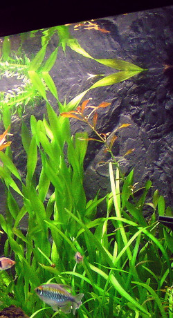
In physics, total internal reflection (TIR) is the phenomenon in which waves arriving at the interface (boundary) from one medium to another (e.g., from water to air) are not refracted into the second ("external") medium, but completely reflected back into the first ("internal") medium. It occurs when the second medium has a higher wave speed (i.e., lower refractive index) than the first, and the waves are incident at a sufficiently oblique angle on the interface. For example, the water-to-air surface in a typical fish tank, when viewed obliquely from below, reflects the underwater scene like a mirror with no loss of brightness (Fig.1). TIR occurs not only with electromagnetic waves such as light and microwaves, but also with other types of waves, including sound and water waves. If the waves are capable of forming a narrow beam (Fig.2), the reflection tends to be described in terms of " rays" rather than waves; in a medium whose properties are independent of direction, such ... [...More Info...] [...Related Items...] OR: [Wikipedia] [Google] [Baidu] |
Slit Lamp
In ophthalmology and optometry, a slit lamp is an instrument consisting of a high-intensity light source that can be focused to shine a thin sheet of light into the eye. It is used in conjunction with a biomicroscope. The lamp facilitates an examination of the anterior segment and posterior segment of the human eye, which includes the eyelid, sclera, conjunctiva, iris (anatomy), iris, natural crystalline lens, and cornea. The binocular slit-lamp examination provides a stereoscopic magnified view of the eye structures in detail, enabling anatomical diagnoses to be made for a variety of eye conditions. A second, hand-held lens is used to examine the retina. History Two conflicting trends emerged in the development of the slit lamp. One trend originated from clinical research and aimed to apply the increasingly complex and advanced technology of the time. [...More Info...] [...Related Items...] OR: [Wikipedia] [Google] [Baidu] |
Glaucoma
Glaucoma is a group of eye diseases that can lead to damage of the optic nerve. The optic nerve transmits visual information from the eye to the brain. Glaucoma may cause vision loss if left untreated. It has been called the "silent thief of sight" because the loss of vision usually occurs slowly over a long period of time. A major risk factor for glaucoma is increased pressure within the eye, known as Intraocular pressure, intraocular pressure (IOP). It is associated with old age, a family history of glaucoma, and certain medical conditions or the use of some medications. The word ''glaucoma'' comes from the Ancient Greek word (), meaning 'gleaming, blue-green, gray'. Of the different types of glaucoma, the most common are called open-angle glaucoma and closed-angle glaucoma. Inside the eye, a liquid called Aqueous humour, aqueous humor helps to maintain shape and provides nutrients. The aqueous humor normally drains through the trabecular meshwork. In open-angle glaucoma, ... [...More Info...] [...Related Items...] OR: [Wikipedia] [Google] [Baidu] |
Pigment Dispersion Syndrome
Pigment dispersion syndrome (PDS) is an eye disorder that can lead to a form of glaucoma known as pigmentary glaucoma. It takes place when pigment cells slough off from the back of the iris and float around in the aqueous humor. Over time, these pigment cells can accumulate in the anterior chamber in such a way that they begin to clog the trabecular meshwork (the major site of aqueous humour drainage), which can in turn prevent the aqueous humour from draining and therefore increases the pressure inside the eye. A common finding in PDS are central, vertical corneal endothelial pigment deposits, known as Krukenberg spindle. With PDS, the intraocular pressure tends to spike at times and then can return to normal. Exercise has been shown to contribute to spikes in pressure as well. When the pressure is great enough to cause damage to the optic nerve, this is called pigmentary glaucoma. As with all types of glaucoma, when damage happens to the optic nerve fibers, the vision loss ... [...More Info...] [...Related Items...] OR: [Wikipedia] [Google] [Baidu] |
Schwalbe's Line
Schwalbe's line is the anatomical line found on the interior surface of the eye's cornea, and delineates the outer limit of the corneal endothelium layer. Specifically, it represents the termination of Descemet's membrane. In many cases it can be seen via gonioscopy In ophthalmology, gonioscopy is a routine procedure that measures the angle between the iris (anatomy), iris and the cornea (the iridocorneal angle), using a goniolens (also known as a gonioscope) together with a slit lamp or operating microscope .... Some evidence suggests that the corneal endothelium actually possesses stem cells that can produce endothelial cells, especially after injury, albeit on a limited scale. References Human eye anatomy {{Eye-stub ... [...More Info...] [...Related Items...] OR: [Wikipedia] [Google] [Baidu] |
Ophthalmology
Ophthalmology (, ) is the branch of medicine that deals with the diagnosis, treatment, and surgery of eye diseases and disorders. An ophthalmologist is a physician who undergoes subspecialty training in medical and surgical eye care. Following a medical degree, a doctor specialising in ophthalmology must pursue additional postgraduate residency training specific to that field. In the United States, following graduation from medical school, one must complete a four-year residency in ophthalmology to become an ophthalmologist. Following residency, additional specialty training (or fellowship) may be sought in a particular aspect of eye pathology. Ophthalmologists prescribe medications to treat ailments, such as eye diseases, implement laser therapy, and perform surgery when needed. Ophthalmologists provide both primary and specialty eye care—medical and surgical. Most ophthalmologists participate in academic research on eye diseases at some point in their training and many inc ... [...More Info...] [...Related Items...] OR: [Wikipedia] [Google] [Baidu] |
Synechia (eye)
Ocular synechia is an eye condition where the iris adheres to either the cornea (i.e. ''anterior synechia'') or lens (i.e. ''posterior synechia'').F. Salmon, J. (2019) Kanski’s Clinical Ophthalmology. 9th Edition, Elsevier. Synechiae can be caused by ocular trauma, iritis or iridocyclitis and may lead to certain types of glaucoma. It is sometimes visible on careful examination but usually more easily through an ophthalmoscope or slit-lamp. Anterior synechia causes closed angle glaucoma, which means that the iris closes the drainage way of aqueous humour which in turn raises the intraocular pressure. Posterior synechia can be observed in cases of anterior uveitis secondary to severe to moderate bacterial keratitis. Posterior synechia also cause glaucoma, but with a different mechanism. In posterior synechia, the iris adheres to the lens, blocking the flow of aqueous humor from the posterior chamber to the anterior chamber. This blocked drainage raises the intraocular pre ... [...More Info...] [...Related Items...] OR: [Wikipedia] [Google] [Baidu] |
Diagnostic Ophthalmology
Diagnosis (: diagnoses) is the identification of the nature and cause of a certain phenomenon. Diagnosis is used in a lot of different disciplines, with variations in the use of logic, analytics, and experience, to determine " cause and effect". In systems engineering and computer science, it is typically used to determine the causes of symptoms, mitigations, and solutions. Computer science and networking * Bayesian network * Complex event processing * Diagnosis (artificial intelligence) * Event correlation * Fault management * Fault tree analysis * Grey problem * RPR problem diagnosis * Remote diagnostics * Root cause analysis * Troubleshooting * Unified Diagnostic Services Mathematics and logic * Bayesian probability * Block Hackam's dictum * Occam's razor * Regression diagnostics * Sutton's law Medicine * Medical diagnosis * Molecular diagnostics Methods * CDR computerized assessment system * Computer-aided diagnosis * Differential diagnosis * Retrospective ... [...More Info...] [...Related Items...] OR: [Wikipedia] [Google] [Baidu] |
Greek Language
Greek (, ; , ) is an Indo-European languages, Indo-European language, constituting an independent Hellenic languages, Hellenic branch within the Indo-European language family. It is native to Greece, Cyprus, Italy (in Calabria and Salento), southern Albania, and other regions of the Balkans, Caucasus, the Black Sea coast, Asia Minor, and the Eastern Mediterranean. It has the list of languages by first written accounts, longest documented history of any Indo-European language, spanning at least 3,400 years of written records. Its writing system is the Greek alphabet, which has been used for approximately 2,800 years; previously, Greek was recorded in writing systems such as Linear B and the Cypriot syllabary. The Greek language holds a very important place in the history of the Western world. Beginning with the epics of Homer, ancient Greek literature includes many works of lasting importance in the European canon. Greek is also the language in which many of the foundational texts ... [...More Info...] [...Related Items...] OR: [Wikipedia] [Google] [Baidu] |
Pseudoexfoliation Syndrome
Pseudoexfoliation syndrome, often abbreviated as PEX and sometimes as PES or PXS, is an aging-related systemic disease manifesting itself primarily in the eyes which is characterized by the accumulation of microscopic granular amyloid-like protein fibers. Its cause is unknown, although there is speculation that there may be a genetics, genetic basis. It is more prevalent in women than men, and in persons past the age of seventy. Its prevalence in different human populations varies; for example, it is prevalent in Scandinavia. The buildup of protein clumps can block normal drainage of the eye fluid called the aqueous humor and can cause, in turn, a buildup of pressure leading to glaucoma and loss of vision (pseudoexfoliation glaucoma, exfoliation glaucoma). As worldwide populations become older because of shifts in demography, PEX may become a matter of greater concern. Signs and symptoms Patients may have no specific symptoms. In some cases, patients may complain of lessened visu ... [...More Info...] [...Related Items...] OR: [Wikipedia] [Google] [Baidu] |
Intraocular Pressure
Intraocular pressure (IOP) is the fluid pressure inside the eye. Tonometry is the method eye care professionals use to determine this. IOP is an important aspect in the evaluation of patients at risk of glaucoma. Most tonometers are calibrated to measure pressure in millimeters of mercury (mmHg). Physiology Intraocular pressure is determined by the production and drainage of aqueous humour by the ciliary body and its drainage via the trabecular meshwork and uveoscleral outflow. The reason for this is because the vitreous humour in the posterior segment has a relatively fixed volume and thus does not affect intraocular pressure regulation. An important quantitative relationship (Goldmann's equation) is as follows: :P_o = \frac + P_v Where: * P_o is the IOP in millimeters of mercury (mmHg) * F the rate of aqueous humour formation in microliters per minute (μL/min) * U the resorption of aqueous humour through the uveoscleral route (μL/min) * C is the facility of outflow in mic ... [...More Info...] [...Related Items...] OR: [Wikipedia] [Google] [Baidu] |


