|
Glycocalix
The glycocalyx, also known as the pericellular matrix, is a glycoprotein and glycolipid covering that surrounds the cell membranes of bacteria, epithelial cells, and other cells. In 1970, Martinez-Palomo discovered the cell coating in animal cells, which is known as the glycocalyx. Animal epithelial cells have a fuzz-like coating on the external surface of their plasma membranes. This viscous coating consists of several carbohydrate moieties of membrane glycolipids and glycoproteins, which serve as backbone molecules for support. Generally, the carbohydrate portion of the glycolipids found on the surface of plasma membranes helps these molecules contribute to cell–cell recognition, communication, and intercellular adhesion. The glycocalyx is a type of identifier that the body uses to distinguish between its own healthy cells and transplanted tissues, diseased cells, or invading organisms. Included in the glycocalyx are cell-adhesion molecules that enable cells to adhere to each ... [...More Info...] [...Related Items...] OR: [Wikipedia] [Google] [Baidu] |
Transmission Electron Microscopy
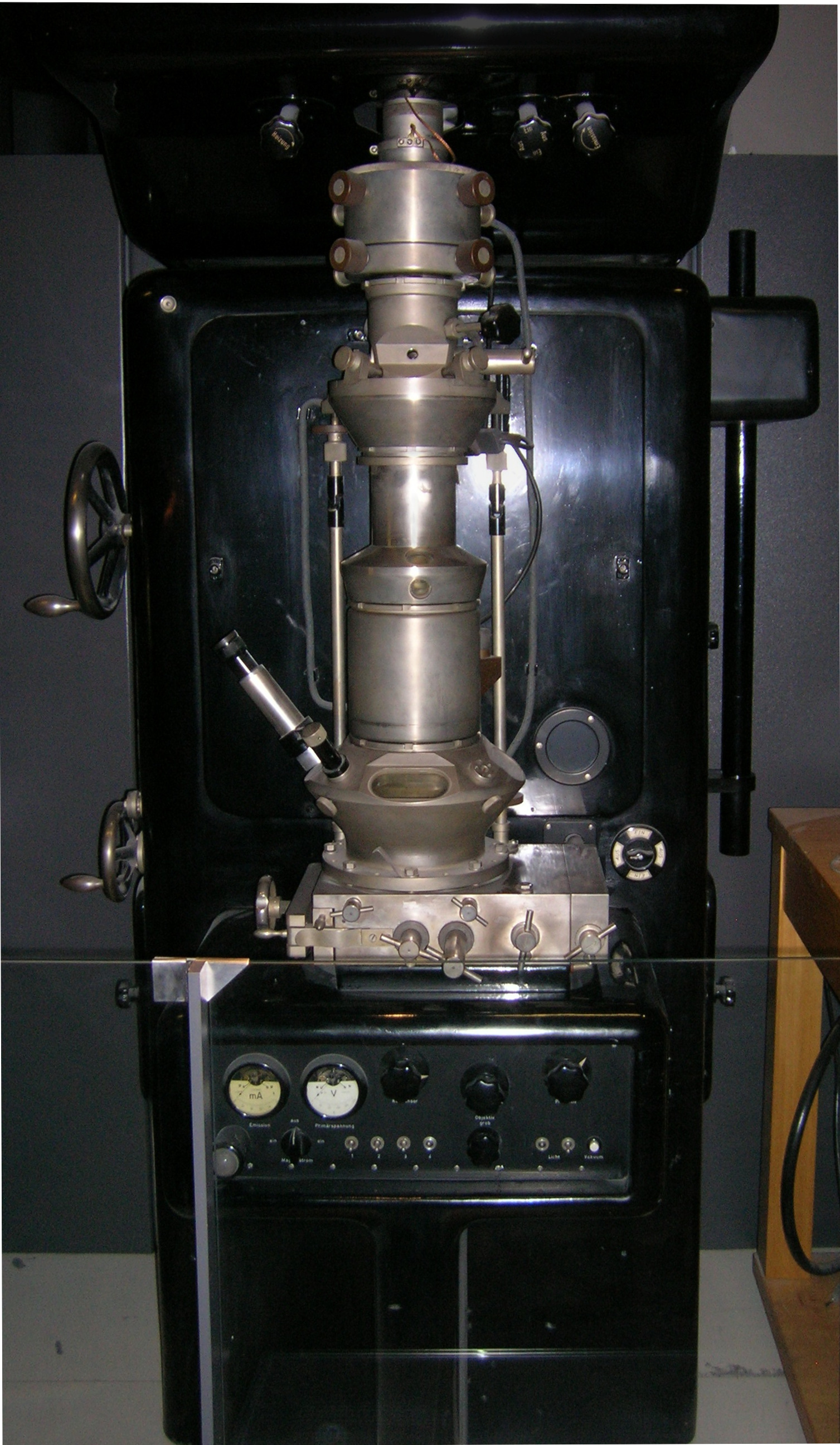
Transmission electron microscopy (TEM) is a microscopy technique in which a beam of electrons is transmitted through a specimen to form an image. The specimen is most often an ultrathin section less than 100 nm thick or a suspension on a grid. An image is formed from the interaction of the electrons with the sample as the beam is transmitted through the specimen. The image is then magnified and focused onto an imaging device, such as a fluorescent screen, a layer of photographic film, or a sensor such as a scintillator attached to a charge-coupled device. Transmission electron microscopes are capable of imaging at a significantly higher resolution than light microscopes, owing to the smaller de Broglie wavelength of electrons. This enables the instrument to capture fine detail—even as small as a single column of atoms, which is thousands of times smaller than a resolvable object seen in a light microscope. Transmission electron microscopy is a major analytical method ... [...More Info...] [...Related Items...] OR: [Wikipedia] [Google] [Baidu] |
Lumen (anatomy)
In biology, a lumen (plural lumina) is the inside space of a tubular structure, such as an artery or intestine. It comes . It can refer to: *The interior of a vessel, such as the central space in an artery, vein or capillary through which blood flows. *The interior of the gastrointestinal tract *The pathways of the bronchi in the lungs *The interior of renal tubules and urinary collecting ducts *The pathways of the female genital tract, starting with a single pathway of the vagina, splitting up in two lumina in the uterus, both of which continue through the Fallopian tubes In cell biology, a lumen is a membrane-defined space that is found inside several organelles, cellular components, or structures: *thylakoid, endoplasmic reticulum, Golgi apparatus, lysosome, mitochondrion, or microtubule Transluminal procedures ''Transluminal procedures'' are procedures occurring through lumina, including: * Natural orifice transluminal endoscopic surgery in the lumina of, for example, ... [...More Info...] [...Related Items...] OR: [Wikipedia] [Google] [Baidu] |
Chemokines
Chemokines (), or chemotactic cytokines, are a family of small cytokines or signaling proteins secreted by cells that induce directional movement of leukocytes, as well as other cell types, including endothelial and epithelial cells. In addition to playing a major role in the activation of host immune responses, chemokines are important for biological processes, including morphogenesis and wound healing, as well as in the pathogenesis of diseases like cancers. Cytokine proteins are classified as chemokines according to behavior and structural characteristics. In addition to being known for mediating chemotaxis, chemokines are all approximately 8-10 kilodaltons in mass and have four cysteine residues in conserved locations that are key to forming their 3-dimensional shape. These proteins have historically been known under several other names including the ''SIS family of cytokines'', ''SIG family of cytokines'', ''SCY family of cytokines'', ''Platelet factor-4 superfamily'' or ... [...More Info...] [...Related Items...] OR: [Wikipedia] [Google] [Baidu] |
Growth Factors
A growth factor is a naturally occurring substance capable of stimulating cell proliferation, wound healing, and occasionally cellular differentiation. Usually it is a secreted protein or a steroid hormone. Growth factors are important for regulating a variety of cellular processes. Growth factors typically act as signaling molecules between cells. Examples are cytokines and hormones that bind to specific receptors on the surface of their target cells. They often promote cell differentiation and maturation, which varies between growth factors. For example, epidermal growth factor (EGF) enhances osteogenic differentiation, while fibroblast growth factors and vascular endothelial growth factors stimulate blood vessel differentiation ( angiogenesis). Comparison to cytokines ''Growth factor'' is sometimes used interchangeably among scientists with the term ''cytokine.'' Historically, cytokines were associated with hematopoietic (blood and lymph forming) cells and immun ... [...More Info...] [...Related Items...] OR: [Wikipedia] [Google] [Baidu] |
Apolipoproteins
Apolipoproteins are proteins that bind lipids (oil-soluble substances such as fats, cholesterol and fat soluble vitamins) to form lipoproteins. They transport lipids in blood, cerebrospinal fluid and lymph. The lipid components of lipoproteins are insoluble in water. However, because of their detergent-like (amphipathic) properties, apolipoproteins and other amphipathic molecules (such as phospholipids) can surround the lipids, creating a lipoprotein particle that is itself water-soluble, and can thus be carried through body fluids (i.e., blood, lymph). In addition to stabilizing lipoprotein structure and solubilizing the lipid component, apolipoproteins interact with lipoprotein receptors and lipid transport proteins, thereby participating in lipoprotein uptake and clearance. They also serve as enzyme cofactors for specific enzymes involved in the metabolism of lipoproteins. Apolipoproteins are also exploited by hepatitis C virus (HCV) to enable virus entry, assembly, and ... [...More Info...] [...Related Items...] OR: [Wikipedia] [Google] [Baidu] |
Lipoprotein Lipase
Lipoprotein lipase (LPL) (EC 3.1.1.34, systematic name triacylglycerol acylhydrolase (lipoprotein-dependent)) is a member of the lipase gene family, which includes pancreatic lipase, hepatic lipase, and endothelial lipase. It is a water-soluble enzyme that hydrolyzes triglycerides in lipoproteins, such as those found in chylomicrons and very low-density lipoproteins (VLDL), into two free fatty acids and one monoacylglycerol molecule: : triacylglycerol + H2O = diacylglycerol + a carboxylate It is also involved in promoting the cellular uptake of chylomicron remnants, cholesterol-rich lipoproteins, and free fatty acids. LPL requires ApoC-II as a cofactor. LPL is attached to the luminal surface of endothelial cells in capillaries by the protein glycosylphosphatidylinositol HDL-binding protein 1 (GPIHBP1) and by heparan sulfated peptidoglycans. It is most widely distributed in adipose, heart, and skeletal muscle tissue, as well as in lactating mammary glands. Synthesis ... [...More Info...] [...Related Items...] OR: [Wikipedia] [Google] [Baidu] |
Antithrombin
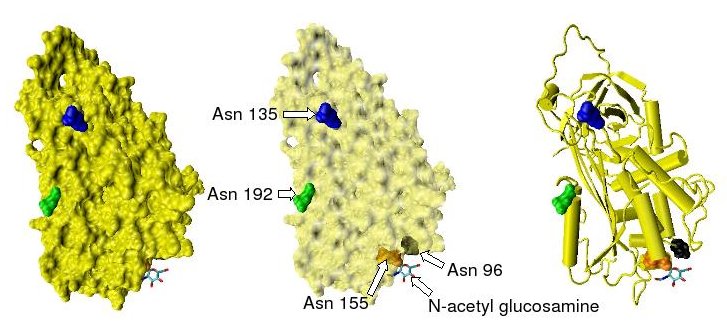
Antithrombin (AT) is a small glycoprotein that inactivates several enzymes of the coagulation system. It is a 432-amino-acid protein produced by the liver. It contains three disulfide bonds and a total of four possible glycosylation sites. α-Antithrombin is the dominant form of antithrombin found in blood plasma and has an oligosaccharide occupying each of its four glycosylation sites. A single glycosylation site remains consistently un-occupied in the minor form of antithrombin, β-antithrombin. Its activity is increased manyfold by the anticoagulant drug heparin, which enhances the binding of antithrombin to factor IIa (prothrombin) and factor Xa. Nomenclature Antithrombin is also termed antithrombin III (AT III). The designations antithrombin I through to antithrombin IV originate in early studies carried out in the 1950s by Seegers, Johnson and Fell. Antithrombin I (AT I) refers to the absorption of thrombin onto fibrin after thrombin has activated fibrinogen. Antithrom ... [...More Info...] [...Related Items...] OR: [Wikipedia] [Google] [Baidu] |
Angiotensin Converting Enzyme
Angiotensin-converting enzyme (), or ACE, is a central component of the renin–angiotensin system (RAS), which controls blood pressure by regulating the volume of fluids in the body. It converts the hormone angiotensin I to the active vasoconstrictor angiotensin II. Therefore, ACE indirectly increases blood pressure by causing blood vessels to constrict. ACE inhibitors are widely used as pharmaceutical drugs for treatment of cardiovascular diseases. Other lesser known functions of ACE are degradation of bradykinin, substance P and amyloid beta-protein. Nomenclature ACE is also known by the following names: * dipeptidyl carboxypeptidase I * peptidase P * dipeptide hydrolase * peptidyl dipeptidase * angiotensin converting enzyme * kininase II * angiotensin I-converting enzyme * carboxycathepsin * dipeptidyl carboxypeptidase * "hypertensin converting enzyme" peptidyl dipeptidase I * peptidyl-dipeptide hydrolase * peptidyldipeptide hydrolase * endothelial cell peptidyl dipeptida ... [...More Info...] [...Related Items...] OR: [Wikipedia] [Google] [Baidu] |
SOD3
Extracellular superoxide dismutase u-Zn'' is an enzyme that in humans is encoded by the ''SOD3'' gene. This gene encodes a member of the superoxide dismutase (SOD) protein family. SODs are antioxidant enzymes that catalyze the dismutation of two superoxide radicals into hydrogen peroxide and oxygen. The product of this gene is thought to protect the brain, lungs, and other tissues from oxidative stress. The protein is secreted into the extracellular space and forms a glycosylated homotetramer that is anchored to the extracellular matrix (ECM) and cell surfaces through an interaction with heparan sulfate proteoglycan and collagen. A fraction of the protein is cleaved near the C-terminus before secretion to generate circulating tetramers that do not interact with the ECM. Among black garden ants (''Lasius niger''), the lifespan of queens Queens is a borough of New York City, coextensive with Queens County, in the U.S. state of New York. Located on Long Island, it is the larg ... [...More Info...] [...Related Items...] OR: [Wikipedia] [Google] [Baidu] |
Endothelial NOS
Endothelial NOS (eNOS), also known as nitric oxide synthase 3 (NOS3) or constitutive NOS (cNOS), is an enzyme that in humans is encoded by the ''NOS3'' gene located in the 7q35-7q36 region of chromosome 7. This enzyme is one of three isoforms that synthesize nitric oxide (NO), a small gaseous and lipophilic molecule that participates in several biological processes. The other isoforms include neuronal nitric oxide synthase (nNOS), which is constitutively expressed in specific neurons of the brain and inducible nitric oxide synthase (iNOS), whose expression is typically induced in inflammatory diseases. eNOS is primarily responsible for the generation of NO in the vascular endothelium, a monolayer of flat cells lining the interior surface of blood vessels, at the interface between circulating blood in the lumen and the remainder of the vessel wall. NO produced by eNOS in the vascular endothelium plays crucial roles in regulating vascular tone, cellular proliferation, leukocyte ad ... [...More Info...] [...Related Items...] OR: [Wikipedia] [Google] [Baidu] |
Thrombocyte
Platelets, also called thrombocytes (from Greek θρόμβος, "clot" and κύτος, "cell"), are a component of blood whose function (along with the coagulation factors) is to react to bleeding from blood vessel injury by clumping, thereby initiating a blood clot. Platelets have no cell nucleus; they are fragments of cytoplasm that are derived from the megakaryocytes of the bone marrow or lung, which then enter the circulation. Platelets are found only in mammals, whereas in other vertebrates (e.g. birds, amphibians), thrombocytes circulate as intact mononuclear cells. One major function of platelets is to contribute to hemostasis: the process of stopping bleeding at the site of interrupted endothelium. They gather at the site and, unless the interruption is physically too large, they plug the hole. First, platelets attach to substances outside the interrupted endothelium: ''adhesion''. Second, they change shape, turn on receptors and secrete chemical messengers: ''acti ... [...More Info...] [...Related Items...] OR: [Wikipedia] [Google] [Baidu] |
Leukocyte
White blood cells, also called leukocytes or leucocytes, are the cells of the immune system that are involved in protecting the body against both infectious disease and foreign invaders. All white blood cells are produced and derived from multipotent cells in the bone marrow known as hematopoietic stem cells. Leukocytes are found throughout the body, including the blood and lymphatic system. All white blood cells have nuclei, which distinguishes them from the other blood cells, the anucleated red blood cells (RBCs) and platelets. The different white blood cells are usually classified by cell lineage ( myeloid cells or lymphoid cells). White blood cells are part of the body's immune system. They help the body fight infection and other diseases. Types of white blood cells are granulocytes (neutrophils, eosinophils, and basophils), and agranulocytes ( monocytes, and lymphocytes (T cells and B cells)). Myeloid cells ( myelocytes) include neutrophils, eosinophils, mast ... [...More Info...] [...Related Items...] OR: [Wikipedia] [Google] [Baidu] |


