|
Gingival Fibers
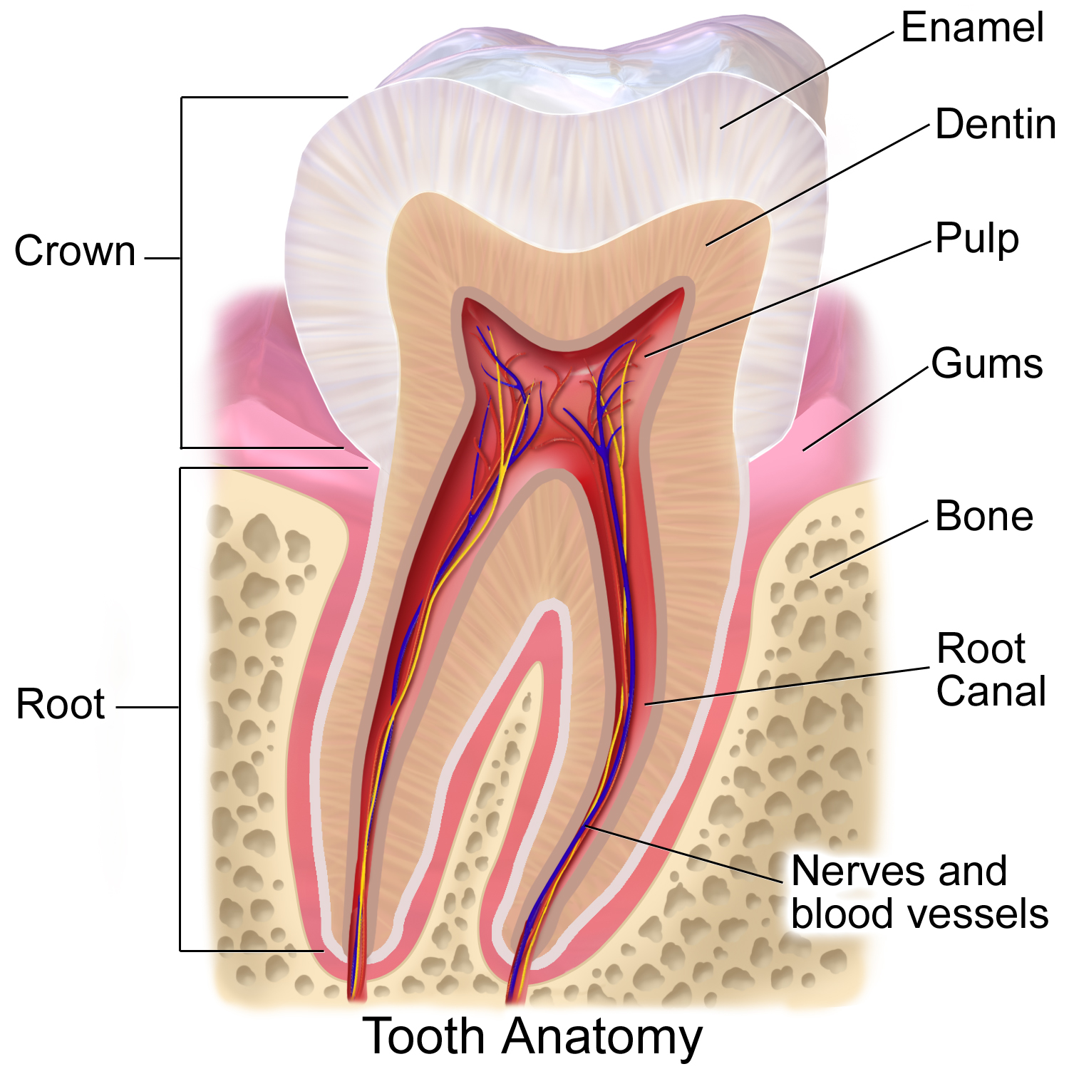
In dental anatomy, the gingival fibers are the connective tissue fibers that inhabit the gingiva, gingival tissue (gums) adjacent to Tooth, teeth and help hold the tissue firmly against the teeth. They are primarily composed of type I collagen, although type III fibers are also involved. These fibers, unlike the fibers of the periodontal ligament, in general, attach the tooth to the gingival tissue, rather than the tooth to the alveolar bone. Functions of the gingival fibers The gingival fibers accomplish the following tasks: *They hold the free gingival margin, marginal gingiva against the tooth *They provide the marginal gingiva with enough rigidity to withstand the forces of mastication without distorting *They serve to stabilize the marginal gingiva by uniting it with both the tissue of the more rigid attached gingiva as well as the cementum layer of the tooth. Gingival fibers and periodontitis In theory, gingival fibers are the protectors against periodontitis, as once they ... [...More Info...] [...Related Items...] OR: [Wikipedia] [Google] [Baidu] |
Dental Anatomy
Dental anatomy is a field of anatomy dedicated to the study of Tooth (human), human tooth structures. The development, appearance, and classification of teeth fall within its purview. (The function of teeth as they contact one another falls elsewhere, under dental occlusion.) Tooth formation begins before birth, and the teeth's eventual Morphology (biology), morphology is dictated during this time. Dental anatomy is also a Taxonomy (general), taxonomical science: it is concerned with the naming of teeth and the structures of which they are made, this information serving a practical purpose in dental treatment. Usually, there are 20 primary ("baby") teeth and 32 permanent teeth, the last four being third molars or "wisdom teeth", each of which may or may not grow in. Among primary teeth, 10 usually are found in the maxilla (upper jaw) and the other 10 in the human mandible, mandible (lower jaw). Among permanent teeth, 16 are found in the maxilla and the other 16 in the mandible. Ea ... [...More Info...] [...Related Items...] OR: [Wikipedia] [Google] [Baidu] |
Periodontitis
Periodontal disease, also known as gum disease, is a set of inflammatory conditions affecting the tissues surrounding the teeth. In its early stage, called gingivitis, the gums become swollen and red and may bleed. It is considered the main cause of tooth loss for adults worldwide. In its more serious form, called periodontitis, the gums can pull away from the tooth, bone can be lost, and the teeth may loosen or fall out. Halitosis (bad breath) may also occur. Periodontal disease typically arises from the development of plaque biofilm, which harbors harmful bacteria such as ''Porphyromonas gingivalis'' and ''Treponema denticola''. These bacteria infect the gum tissue surrounding the teeth, leading to inflammation and, if left untreated, progressive damage to the teeth and gum tissue. Recent meta-analysis have shown that the composition of the oral microbiota and its response to periodontal disease differ between men and women. These differences are particularly notable in t ... [...More Info...] [...Related Items...] OR: [Wikipedia] [Google] [Baidu] |
Anatomical Terms Of Location
Standard anatomical terms of location are used to describe unambiguously the anatomy of humans and other animals. The terms, typically derived from Latin or Greek roots, describe something in its standard anatomical position. This position provides a definition of what is at the front ("anterior"), behind ("posterior") and so on. As part of defining and describing terms, the body is described through the use of anatomical planes and axes. The meaning of terms that are used can change depending on whether a vertebrate is a biped or a quadruped, due to the difference in the neuraxis, or if an invertebrate is a non-bilaterian. A non-bilaterian has no anterior or posterior surface for example but can still have a descriptor used such as proximal or distal in relation to a body part that is nearest to, or furthest from its middle. International organisations have determined vocabularies that are often used as standards for subdisciplines of anatomy. For example, '' Termi ... [...More Info...] [...Related Items...] OR: [Wikipedia] [Google] [Baidu] |
Periodontium
The periodontium () is the specialized tissues that both surround and support the teeth, maintaining them in the maxillary and mandibular bones. Periodontics is the dental specialty that relates specifically to the care and maintenance of these tissues. It provides the support necessary to maintain teeth in function. It consists of four principal components, namely: * Gingiva (the gums) * Periodontal ligament (PDL) * Cementum * Alveolar bone proper Each of these components is distinct in location, architecture, and biochemical properties, which adapt during the life of the structure. For example, as teeth respond to forces or migrate medially, bone resorbs on the pressure side and is added on the tension side. Cementum similarly adapts to wear on the occlusal surfaces of the teeth by apical deposition. The periodontal ligament in itself is an area of high turnover that allows the tooth not only to be suspended in the alveolar bone but also to respond to the forces. Thus, ... [...More Info...] [...Related Items...] OR: [Wikipedia] [Google] [Baidu] |
Junctional Epithelium
In dental anatomy, the junctional epithelium (JE) is that epithelium which lies at, and in health also defines, the base of the gingival sulcus (i.e. where the gums attach to a tooth). The probing depth of the gingival sulcus is measured by a calibrated periodontal probe. In a healthy-case scenario, the probe is gently inserted, slides by the sulcular epithelium (SE), and is stopped by the epithelial attachment (EA). However, the probing depth of the gingival sulcus may be considerably different from the true histological gingival sulcus depth. Location The junctional epithelium, a nonkeratinized stratified squamous epithelium, lies immediately apical to the sulcular epithelium, which lines the gingival sulcus from the base to the free gingival margin, where it interfaces with the epithelium of the oral cavity. The gingival sulcus is bounded by the enamel of the crown of the tooth and the sulcular epithelium. Immediately apical to the base of the pocket, and coronal to the ... [...More Info...] [...Related Items...] OR: [Wikipedia] [Google] [Baidu] |
Sulcular Epithelium
In dental anatomy, the sulcular epithelium is that epithelium Epithelium or epithelial tissue is a thin, continuous, protective layer of cells with little extracellular matrix. An example is the epidermis, the outermost layer of the skin. Epithelial ( mesothelial) tissues line the outer surfaces of man ... which lines the gingival sulcus.Carranza's Clinical Periodontology, W.B. Saunders, 2002, page 23. It is apically bounded by the junctional epithelium and meets the epithelium of the oral cavity at the height of the free gingival margin. The sulcular epithelium is nonkeratinized. References Dental anatomy {{dentistry-stub ... [...More Info...] [...Related Items...] OR: [Wikipedia] [Google] [Baidu] |
Bacteria
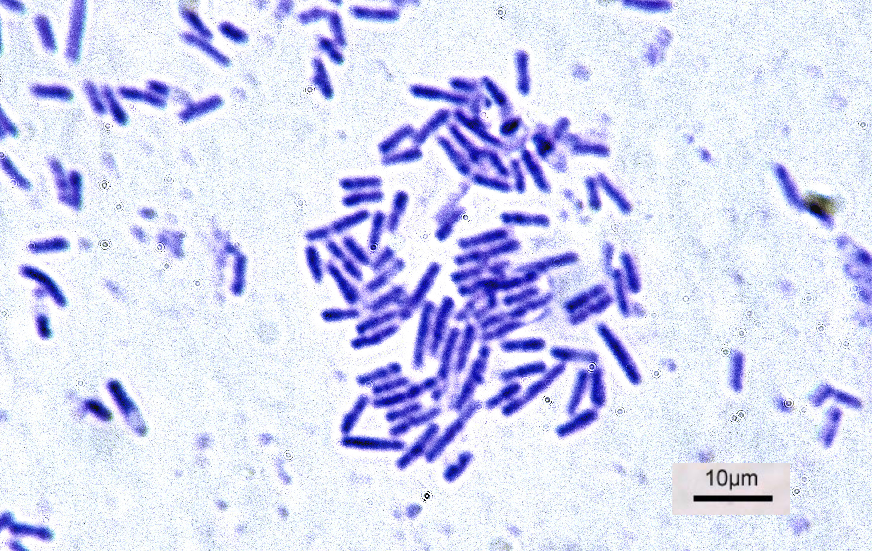
Bacteria (; : bacterium) are ubiquitous, mostly free-living organisms often consisting of one Cell (biology), biological cell. They constitute a large domain (biology), domain of Prokaryote, prokaryotic microorganisms. Typically a few micrometres in length, bacteria were among the first life forms to appear on Earth, and are present in most of its habitats. Bacteria inhabit the air, soil, water, Hot spring, acidic hot springs, radioactive waste, and the deep biosphere of Earth's crust. Bacteria play a vital role in many stages of the nutrient cycle by recycling nutrients and the nitrogen fixation, fixation of nitrogen from the Earth's atmosphere, atmosphere. The nutrient cycle includes the decomposition of cadaver, dead bodies; bacteria are responsible for the putrefaction stage in this process. In the biological communities surrounding hydrothermal vents and cold seeps, extremophile bacteria provide the nutrients needed to sustain life by converting dissolved compounds, suc ... [...More Info...] [...Related Items...] OR: [Wikipedia] [Google] [Baidu] |
Gingival Sulcus
In dental anatomy, the gingival sulcus is an area of potential space between a tooth and the surrounding gingiva, gingival tissue and is lined by sulcular epithelium. The depth of the sulcus (Latin for ''groove'') is bounded by two entities: Commonly used terms of relationship and comparison in dentistry, apically by the gingival fibers of the connective tissue attachment and Commonly used terms of relationship and comparison in dentistry, coronally by the free gingival margin. A healthy sulcular depth is three millimeters or less, which is readily self-cleansable with a properly used toothbrush or the supplemental use of other oral hygiene aids. Anatomy The dentogingival tissues consist of many constituents, such as the enamel or cementum of the tooth and the connective tissue supporting epithelia like the junctional epithelium, the gingival epithelium and the sulcular epithelium. The junctional epithelium is developed during the eruption of teeth when the reduced enamel ... [...More Info...] [...Related Items...] OR: [Wikipedia] [Google] [Baidu] |
Cementum
Cementum is a specialized calcified substance covering the root of a tooth. The cementum is the part of the periodontium that attaches the teeth to the alveolar bone by anchoring the periodontal ligament. Structure The cells of cementum are the entrapped cementoblasts, the cementocytes. Each cementocyte lies in its lacuna, similar to the pattern noted in bone. These lacunae also have canaliculi or canals. Unlike those in bone, however, these canals in cementum do not contain nerves, nor do they radiate outward. Instead, the canals are oriented toward the periodontal ligament and contain cementocytic processes that exist to diffuse nutrients from the ligament because it is vascularized. After the apposition of cementum in layers, the cementoblasts that do not become entrapped in cementum line up along the cemental surface along the length of the outer covering of the periodontal ligament. These cementoblasts can form subsequent layers of cementum if the tooth is injured. Sh ... [...More Info...] [...Related Items...] OR: [Wikipedia] [Google] [Baidu] |
Connective Tissue
Connective tissue is one of the four primary types of animal tissue, a group of cells that are similar in structure, along with epithelial tissue, muscle tissue, and nervous tissue. It develops mostly from the mesenchyme, derived from the mesoderm, the middle embryonic germ layer. Connective tissue is found in between other tissues everywhere in the body, including the nervous system. The three meninges, membranes that envelop the brain and spinal cord, are composed of connective tissue. Most types of connective tissue consists of three main components: elastic and collagen fibers, ground substance, and cells. Blood and lymph are classed as specialized fluid connective tissues that do not contain fiber. All are immersed in the body water. The cells of connective tissue include fibroblasts, adipocytes, macrophages, mast cells and leukocytes. The term "connective tissue" (in German, ) was introduced in 1830 by Johannes Peter Müller. The tissue was already recognized as ... [...More Info...] [...Related Items...] OR: [Wikipedia] [Google] [Baidu] |
Mastication
Chewing or mastication is the process by which food is comminution, crushed and ground by the teeth. It is the first step in the process of digestion, allowing a greater surface area for digestive enzymes to break down the foods. During the mastication process, the food is positioned by the cheek and tongue between the teeth for grinding. The muscles of mastication move the jaws to bring the teeth into intermittent contact, repeatedly occlusion (dentistry), occluding and opening. As chewing continues, the food is made softer and warmer, and the enzymes in saliva begin to break down carbohydrates in the food. After chewing, the food (now called a Bolus (digestion), bolus) is swallowed. It enters the esophagus and via peristalsis continues on to the stomach, where the next step of digestion occurs. Increasing the number of chews per bite stimulates the production of digestive enzymes and peptides and has been shown to increase diet-induced thermogenesis (DIT) by activating the sympa ... [...More Info...] [...Related Items...] OR: [Wikipedia] [Google] [Baidu] |
Free Gingival Margin
In dental anatomy, the free gingival margin is the interface between the sulcular epithelium and the epithelium of the oral cavity. This interface exists at the most coronal point of the gingiva, otherwise known as the crest of the marginal gingiva. Because the short part of gingiva existing above the height of the underlying alveolar process of maxilla, known as the free gingiva, is not bound down to the periosteum that envelops the bone, it is moveable. However, due to the presence of gingival fibers such as the dentogingival and circular fibers, the free gingiva remains pulled up against the surface of the tooth unless being pushed away by, for example, a periodontal probe or the bristles of a toothbrush. Gingival retraction or recession ''Gingival retraction'' or ''gingival recession'' is when there is lateral movement of the gingival margin away from the tooth surface. It is usually termed ''gingival retraction'' as an intentional procedure, and in such cases it is perfor ... [...More Info...] [...Related Items...] OR: [Wikipedia] [Google] [Baidu] |


