|
Gamma Wave
A gamma wave or gamma rhythm is a pattern of neural oscillation in humans with a frequency between 30 and 100 Hz, the 40 Hz point being of particular interest. Gamma waves with frequencies between 30 and 70 hertz may be classified as low gamma, and those between 70 and 150 hertz as high gamma. Gamma rhythms are correlated with large-scale brain network activity and cognitive phenomena such as working memory, attention, and perceptual grouping, and can be increased in amplitude via meditation or neurostimulation. Altered gamma activity has been observed in many mood and cognitive disorders such as Alzheimer's disease, epilepsy, and schizophrenia. Discovery Gamma waves can be detected by electroencephalography or magnetoencephalography. One of the earliest reports of gamma wave activity was recorded from the visual cortex of awake monkeys. Subsequently, significant research activity has concentrated on gamma activity in visual cortex. Gamma activity has also been detect ... [...More Info...] [...Related Items...] OR: [Wikipedia] [Google] [Baidu] |
Eeg Gamma
Electroencephalography (EEG) is a method to record an electrogram of the spontaneous electrical activity of the brain. The bio signals detected by EEG have been shown to represent the postsynaptic potentials of pyramidal neurons in the neocortex and allocortex. It is typically non-invasive, with the EEG electrodes placed along the scalp (commonly called "scalp EEG") using the International 10–20 system, or variations of it. Electrocorticography, involving surgical placement of electrodes, is sometimes called "intracranial EEG". Clinical interpretation of EEG recordings is most often performed by visual inspection of the tracing or quantitative EEG analysis. Voltage fluctuations measured by the EEG bio amplifier and electrodes allow the evaluation of normal brain activity. As the electrical activity monitored by EEG originates in neurons in the underlying brain tissue, the recordings made by the electrodes on the surface of the scalp vary in accordance with their orienta ... [...More Info...] [...Related Items...] OR: [Wikipedia] [Google] [Baidu] |
Magnetoencephalography
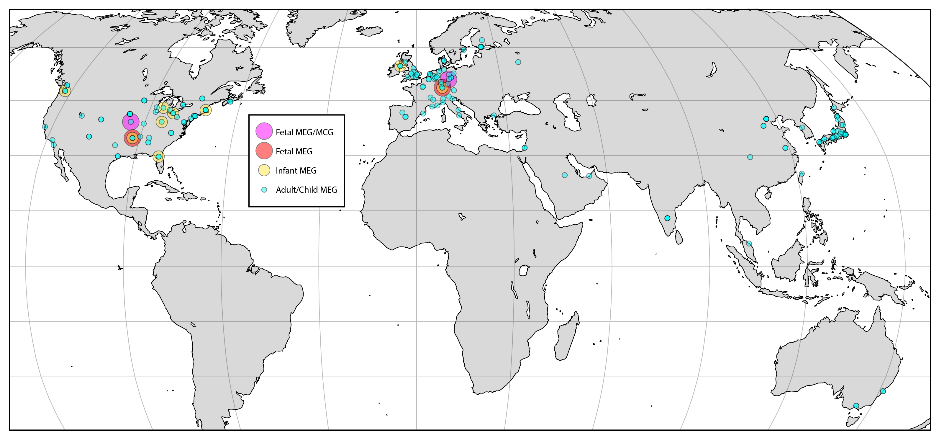
Magnetoencephalography (MEG) is a functional neuroimaging technique for mapping brain activity by recording magnetic fields produced by electric current, electrical currents occurring naturally in the human brain, brain, using very sensitive magnetometers. Arrays of SQUIDs (superconducting quantum interference devices) are currently the most common magnetometer, while the SERF (spin exchange relaxation-free) magnetometer is being investigated for future machines. Applications of MEG include basic research into perceptual and cognitive brain processes, localizing regions affected by pathology before surgical removal, determining the function of various parts of the brain, and neurofeedback. This can be applied in a clinical setting to find locations of abnormalities as well as in an experimental setting to simply measure brain activity. History MEG signals were first measured by University of Illinois physicist David Cohen (physicist), David Cohen in 1968, before the availabili ... [...More Info...] [...Related Items...] OR: [Wikipedia] [Google] [Baidu] |
Independent Component Analysis
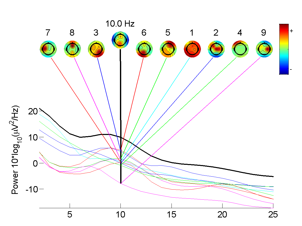
In signal processing, independent component analysis (ICA) is a computational method for separating a multivariate statistics, multivariate signal into additive subcomponents. This is done by assuming that at most one subcomponent is Gaussian and that the subcomponents are Statistical independence, statistically independent from each other. ICA was invented by Jeanny Hérault and Christian Jutten in 1985. ICA is a special case of blind source separation. A common example application of ICA is the "cocktail party problem" of listening in on one person's speech in a noisy room. Introduction Independent component analysis attempts to decompose a multivariate signal into independent non-Gaussian signals. As an example, sound is usually a signal that is composed of the numerical addition, at each time t, of signals from several sources. The question then is whether it is possible to separate these contributing sources from the observed total signal. When the statistical independence ... [...More Info...] [...Related Items...] OR: [Wikipedia] [Google] [Baidu] |
Neuron (journal)
''Neuron'' is a biweekly peer-reviewed scientific journal published by Cell Press, an imprint of Elsevier. Established in 1988, it covers neuroscience and related biological processes. The current editor-in-chief An editor-in-chief (EIC), also known as lead editor or chief editor, is a publication's editorial leader who has final responsibility for its operations and policies. The editor-in-chief heads all departments of the organization and is held accoun ... is Mariela Zirlinger. The founding editors were Lily Jan, A. James Hudspeth, Louis Reichardt, Roger Nicoll, and Zach Hall. A past editor-in-chief was Katja Brose. Transcript and video available. Click on "Transcript" for text. * See alsoA Career in Science Editing: Katja BroseEditor in Chief, Neuron References Neuroscience journals Cell Press academic journals Academic journals established in 1988 English-language journals Biweekly journals {{neuroscience-journal-stub ... [...More Info...] [...Related Items...] OR: [Wikipedia] [Google] [Baidu] |
Saccade
In vision science, a saccade ( ; ; ) is a quick, simultaneous movement of both Eye movement (sensory), eyes between two or more phases of focal points in the same direction. In contrast, in Smooth pursuit, smooth-pursuit movements, the eyes move smoothly instead of in jumps. Controlled cortically by the frontal eye fields (FEF), or subcortically by the superior colliculus, saccades serve as a mechanism for focal points, rapid eye movement, and the fast phase of optokinetic reflex, optokinetic nystagmus. The word appears to have been coined in the 1880s by French ophthalmologist Louis Émile Javal, Émile Javal, who used a mirror on one side of a page to observe eye movement in silent reading, and found that it involves a succession of discontinuous individual movements. Function Humans and many organisms do not look at a scene in steadiness; instead, the eyes move around, locating interesting parts of the scene and building up a three-dimensional 'map' corresponding to the scen ... [...More Info...] [...Related Items...] OR: [Wikipedia] [Google] [Baidu] |
Electromyography
Electromyography (EMG) is a technique for evaluating and recording the electrical activity produced by skeletal muscles. EMG is performed using an instrument called an electromyograph to produce a record called an electromyogram. An electromyograph detects the electric potential generated by muscle cells when these cells are electrically or neurologically activated. The signals can be analyzed to detect abnormalities, activation level, or recruitment order, or to analyze the biomechanics of human or animal movement. Needle EMG is an electrodiagnostic medicine technique commonly used by neurologists. Surface EMG is a non-medical procedure used to assess muscle activation by several professionals, including physiotherapists, kinesiologists and biomedical engineers. In computer science, EMG is also used as middleware in gesture recognition towards allowing the input of physical action to a computer as a form of human-computer interaction. Clinical uses EMG testing has a varie ... [...More Info...] [...Related Items...] OR: [Wikipedia] [Google] [Baidu] |
Scalp
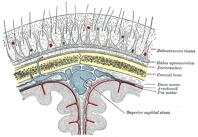
The scalp is the area of the head where head hair grows. It is made up of skin, layers of connective and fibrous tissues, and the membrane of the skull. Anatomically, the scalp is part of the epicranium, a collection of structures covering the cranium. The scalp is bordered by the face at the front, and by the neck at the sides and back. The scientific study of hair and scalp is called trichology. Structure Layers The scalp is usually described as having five layers, which can be remembered using the mnemonic 'SCALP': * S: Skin. The skin of the scalp contains numerous hair follicles and sebaceous glands. * C: Connective tissue. A dense subcutaneous layer of fat and fibrous tissue that lies beneath the skin, containing the nerves and vessels of the scalp. * A: Aponeurosis. The epicranial aponeurosis or galea aponeurotica is a tough layer of dense fibrous tissue which anchors the above layers in place. It runs from the frontalis muscle anteriorly to the occipitalis ... [...More Info...] [...Related Items...] OR: [Wikipedia] [Google] [Baidu] |
Alpha Wave
Alpha waves, or the alpha rhythm, are neural oscillations in the frequency range of 8–12 Hz likely originating from the synchronous and coherent ( in phase or constructive) neocortical neuronal electrical activity possibly involving thalamic pacemaker cells. Historically, they are also called "Berger's waves" after Hans Berger, who first described them when he invented the EEG in 1924. Alpha waves are one type of brain waves detected by electrophysiological methods, e.g., electroencephalography (EEG) or magnetoencephalography (MEG), and can be quantified using power spectra and time-frequency representations of power like quantitative electroencephalography (qEEG). They are predominantly recorded over parieto-occipital brain and were the earliest brain rhythm recorded in humans. Alpha waves can be observed during relaxed wakefulness, especially when there is no mental activity. During the eyes-closed condition, alpha waves are prominent at parietal locations. Atte ... [...More Info...] [...Related Items...] OR: [Wikipedia] [Google] [Baidu] |
Feedforward Neural Network
Feedforward refers to recognition-inference architecture of neural networks. Artificial neural network architectures are based on inputs multiplied by weights to obtain outputs (inputs-to-output): feedforward. Recurrent neural networks, or neural networks with loops allow information from later processing stages to feed back to earlier stages for sequence processing. However, at every stage of inference a feedforward multiplication remains the core, essential for backpropagationRumelhart, David E., Geoffrey E. Hinton, and R. J. Williams.Learning Internal Representations by Error Propagation. David E. Rumelhart, James L. McClelland, and the PDP research group. (editors), Parallel distributed processing: Explorations in the microstructure of cognition, Volume 1: Foundation. MIT Press, 1986. or backpropagation through time. Thus neural networks cannot contain feedback like negative feedback or positive feedback where the outputs feed back to the ''very same'' inputs and modify them, ... [...More Info...] [...Related Items...] OR: [Wikipedia] [Google] [Baidu] |
Cortico-basal Ganglia-thalamo-cortical Loop
The cortico-basal ganglia-thalamo-cortical loop (CBGTC loop) is a system of neural circuits in the brain. The loop involves connections between the cortex, the basal ganglia, the thalamus, and back to the cortex. It is of particular relevance to hyperkinetic and hypokinetic movement disorders, such as Parkinson's disease and Huntington's disease, as well as to mental disorders of control, such as attention deficit hyperactivity disorder (ADHD), obsessive–compulsive disorder (OCD), and Tourette syndrome. The CBGTC loop primarily consists of modulatory dopaminergic projections from the pars compacta of the substantia nigra, and ventral tegmental area as well as excitatory glutamatergic projections from the cortex to the striatum, where these projections form synapses with excitatory and inhibitory pathways that relay back to the cortex. The loop was originally proposed as a part of a model of the basal ganglia called the ''parallel processing model'', which has been criticize ... [...More Info...] [...Related Items...] OR: [Wikipedia] [Google] [Baidu] |
Frontal Lobe
The frontal lobe is the largest of the four major lobes of the brain in mammals, and is located at the front of each cerebral hemisphere (in front of the parietal lobe and the temporal lobe). It is parted from the parietal lobe by a Sulcus (neuroanatomy), groove between tissues called the central sulcus and from the temporal lobe by a deeper groove called the lateral sulcus (Sylvian fissure). The most anterior rounded part of the frontal lobe (though not well-defined) is known as the frontal pole, one of the three Cerebral hemisphere#Poles, poles of the cerebrum. The frontal lobe is covered by the frontal cortex. The frontal cortex includes the premotor cortex and the primary motor cortex – parts of the motor cortex. The front part of the frontal cortex is covered by the prefrontal cortex. The nonprimary motor cortex is a functionally defined portion of the frontal lobe. There are four principal Gyrus, gyri in the frontal lobe. The precentral gyrus is directly anterior to the ... [...More Info...] [...Related Items...] OR: [Wikipedia] [Google] [Baidu] |
Temporal Lobe
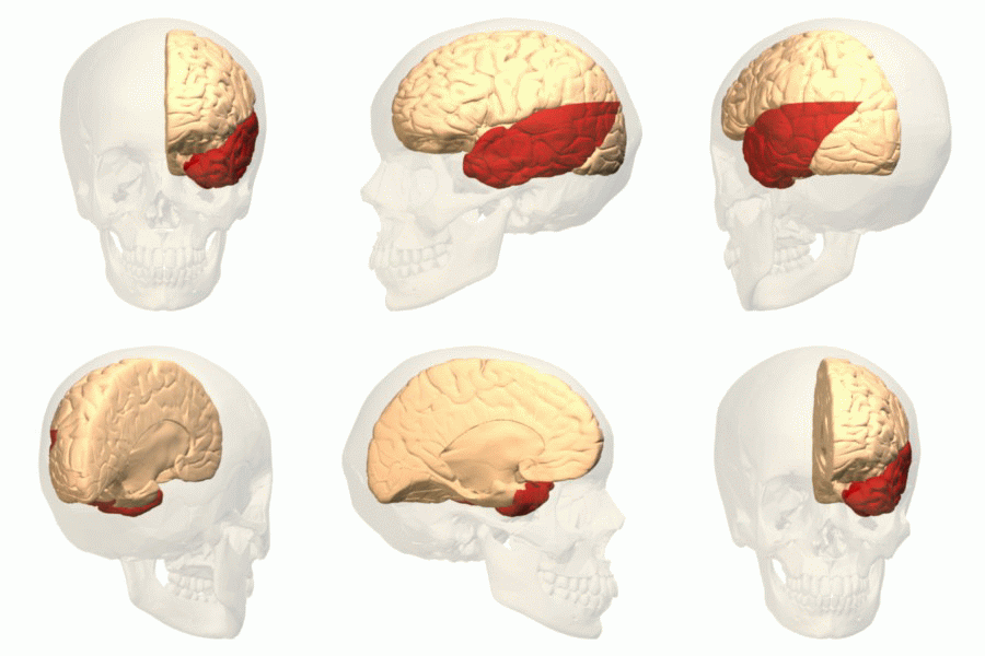
The temporal lobe is one of the four major lobes of the cerebral cortex in the brain of mammals. The temporal lobe is located beneath the lateral fissure on both cerebral hemispheres of the mammalian brain. The temporal lobe is involved in processing sensory input into derived meanings for the appropriate retention of visual memory, language comprehension, and emotion association. ''Temporal'' refers to the head's temples. Structure The temporal lobe consists of structures that are vital for declarative or long-term memory. Declarative (denotative) or explicit memory is conscious memory divided into semantic memory (facts) and episodic memory (events). The medial temporal lobe structures are critical for long-term memory, and include the hippocampal formation, perirhinal cortex, parahippocampal, and entorhinal neocortical regions. The hippocampus is critical for memory formation, and the surrounding medial temporal cortex is currently theorized to be critical f ... [...More Info...] [...Related Items...] OR: [Wikipedia] [Google] [Baidu] |


