|
Flexor Pollicis Longus
The flexor pollicis longus (; FPL, Latin ''flexor'', bender; ''pollicis'', of the thumb; ''longus'', long) is a muscle in the forearm and hand that flexes the thumb. It lies in the same plane as the flexor digitorum profundus. This muscle is unique to humans, being either rudimentary or absent in other primates. A meta-analysis indicated accessory flexor pollicis longus is present in around 48% of the population. Human anatomy Origin and insertion It arises from the grooved anterior (side of palm) surface of the body of the radius, extending from immediately below the radial tuberosity and oblique line to within a short distance of the pronator quadratus muscle.Gray 1918, ''Flexor Pollicis Longus'', paras 20, 25 An occasionally present accessory long head of the flexor pollicis longus muscle is called 'Gantzer's muscle'. It may cause compression of the anterior interosseous nerve. It arises also from the adjacent part of the interosseous membrane of the forearm, and genera ... [...More Info...] [...Related Items...] OR: [Wikipedia] [Google] [Baidu] |
Forearm
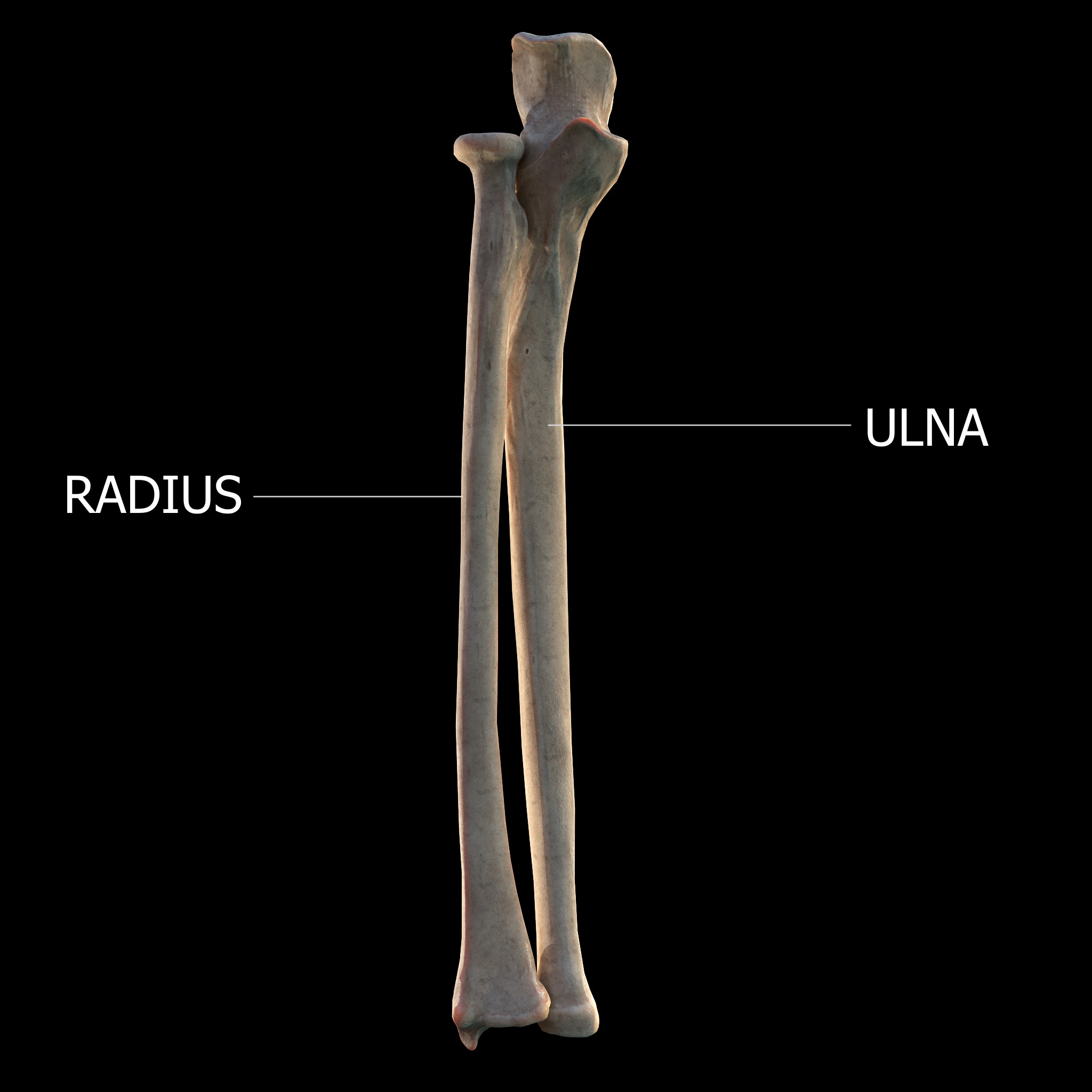
The forearm is the region of the upper limb between the elbow and the wrist. The term forearm is used in anatomy to distinguish it from the arm, a word which is most often used to describe the entire appendage of the upper limb, but which in anatomy, technically, means only the region of the upper arm, whereas the lower "arm" is called the forearm. It is homologous to the region of the leg that lies between the knee and the ankle joints, the crus. The forearm contains two long bones, the radius and the ulna, forming the two radioulnar joints. The interosseous membrane connects these bones. Ultimately, the forearm is covered by skin, the anterior surface usually being less hairy than the posterior surface. The forearm contains many muscles, including the flexors and extensors of the wrist, flexors and extensors of the digits, a flexor of the elbow (brachioradialis), and pronators and supinators that turn the hand to face down or upwards, respectively. In cross-section, ... [...More Info...] [...Related Items...] OR: [Wikipedia] [Google] [Baidu] |
Accessory Muscle
An accessory muscle is a relatively rare anatomical variation where duplication of a muscle may appear anywhere in the muscular system. Treatment is not indicated unless the accessory muscle interferes with normal function. Examples are the sternalis muscle, accessory soleus muscle, extensor digitorum brevis manus and epitrochleoanconeus muscle. An accessory muscle can also refer to a muscle that is not primarily responsible for movement but does provide assistance. Examples of such muscles are the accessory muscles of respiration where the sternocleidomastoid and the scalene muscles (anterior, middle and posterior scalene) are typically considered accessory muscles of respiration.Netter FH. Atlas of Human Anatomy 3rd ed. Icon Learning Systems. Teterboro, New Jersey 2003 - plate 191 See also * Accessory bone An accessory bone or supernumerary bone is a bone that is not normally present in the body, but can be found as a variant in a significant number of people. It poses a ... [...More Info...] [...Related Items...] OR: [Wikipedia] [Google] [Baidu] |
Anterior Interosseous Artery
The anterior interosseous artery (volar interosseous artery) is an artery in the forearm. It is a branch of the common interosseous artery. Course It passes down the forearm on the palmar surface of the interosseous membrane. It is accompanied by the palmar interosseous branch of the median nerve, and overlapped by the contiguous margins of the flexor digitorum profundus and flexor pollicis longus muscles, giving off in this situation muscular branches, and the nutrient arteries of the radius and ulna. At the upper border of the pronator quadratus muscle it pierces the interosseous membrane and reaches the back of the forearm, where it anastomoses with the dorsal interosseous artery. It then descends, in company with the terminal portion of the dorsal interosseous nerve, to the back of the wrist to join the dorsal carpal network. The anterior interosseous artery may give off a slender branch, the median artery, which accompanies the median nerve, and gives offsets to ... [...More Info...] [...Related Items...] OR: [Wikipedia] [Google] [Baidu] |
Aponeurosis
An aponeurosis (; plural: ''aponeuroses'') is a type or a variant of the deep fascia, in the form of a sheet of pearly-white fibrous tissue that attaches sheet-like muscles needing a wide area of attachment. Their primary function is to join muscles and the body parts they act upon, whether bone or other muscles. They have a shiny, whitish-silvery color, are histologically similar to tendons, and are very sparingly supplied with blood vessels and nerves. When dissected, aponeuroses are papery and peel off by sections. The primary regions with thick aponeuroses are in the ventral abdominal region, the dorsal lumbar region, the ventriculus in birds, and the palmar (palms) and plantar (soles) regions. Anatomy Anterior abdominal aponeuroses The anterior abdominal aponeuroses are located just superficial to the rectus abdominis muscle. It has for its borders the external oblique, pectoralis muscles, and the latissimus dorsi. Posterior lumbar aponeuroses The posterior lumbar aponeu ... [...More Info...] [...Related Items...] OR: [Wikipedia] [Google] [Baidu] |
Bone
A bone is a rigid organ that constitutes part of the skeleton in most vertebrate animals. Bones protect the various other organs of the body, produce red and white blood cells, store minerals, provide structure and support for the body, and enable mobility. Bones come in a variety of shapes and sizes and have complex internal and external structures. They are lightweight yet strong and hard and serve multiple functions. Bone tissue (osseous tissue), which is also called bone in the uncountable sense of that word, is hard tissue, a type of specialized connective tissue. It has a honeycomb-like matrix internally, which helps to give the bone rigidity. Bone tissue is made up of different types of bone cells. Osteoblasts and osteocytes are involved in the formation and mineralization of bone; osteoclasts are involved in the resorption of bone tissue. Modified (flattened) osteoblasts become the lining cells that form a protective layer on the bone surface. The mineralize ... [...More Info...] [...Related Items...] OR: [Wikipedia] [Google] [Baidu] |
Adductor Pollicis
In human anatomy, the adductor pollicis muscle is a muscle in the hand that functions to adduct the thumb. It has two heads: transverse and oblique. It is a fleshy, flat, triangular, and fan-shaped muscle deep in the thenar compartment beneath the long flexor tendons and the lumbrical muscles at the center of the palm. It overlies the metacarpal bones and the interosseous muscles. Structure Oblique head The oblique head (Latin: ''adductor obliquus pollicis'') arises by several slips from the capitate bone, the bases of the second and third metacarpals, the intercarpal ligaments, and the sheath of the tendon of the flexor carpi radialis. Gray's Anatomy 1918. (See infobox) From this origin the greater number of fibers pass obliquely downward and converge to a tendon, which, uniting with the tendons of the medial portion of the flexor pollicis brevis and the transverse head of the adductor pollicis, is inserted into the ulnar side of the base of the proximal phalanx of t ... [...More Info...] [...Related Items...] OR: [Wikipedia] [Google] [Baidu] |
Flexor Pollicis Brevis
The flexor pollicis brevis is a muscle in the hand that flexes the thumb. It is one of three thenar muscles. It has both a superficial part and a deep part. Origin and insertion The muscle's superficial head arises from the distal edge of the flexor retinaculum and the tubercle of the trapezium, the most lateral bone in the distal row of carpal bones. It passes along the radial side of the tendon of the flexor pollicis longus. The deeper (and medial) head "varies in size and may be absent." It arises from the trapezoid and capitate bones on the floor of the carpal tunnel, as well as the ligaments of the distal carpal row. Both heads become tendinous and insert together into the radial side of the base of the proximal phalanx of the thumb; at the junction between the tendinous heads there is a sesamoid bone.'' Gray's Anatomy'' 1918, see infobox Innervation The superficial head is usually innervated by the lateral terminal branch of the median nerve. The deep part is often ... [...More Info...] [...Related Items...] OR: [Wikipedia] [Google] [Baidu] |
Carpal Tunnel
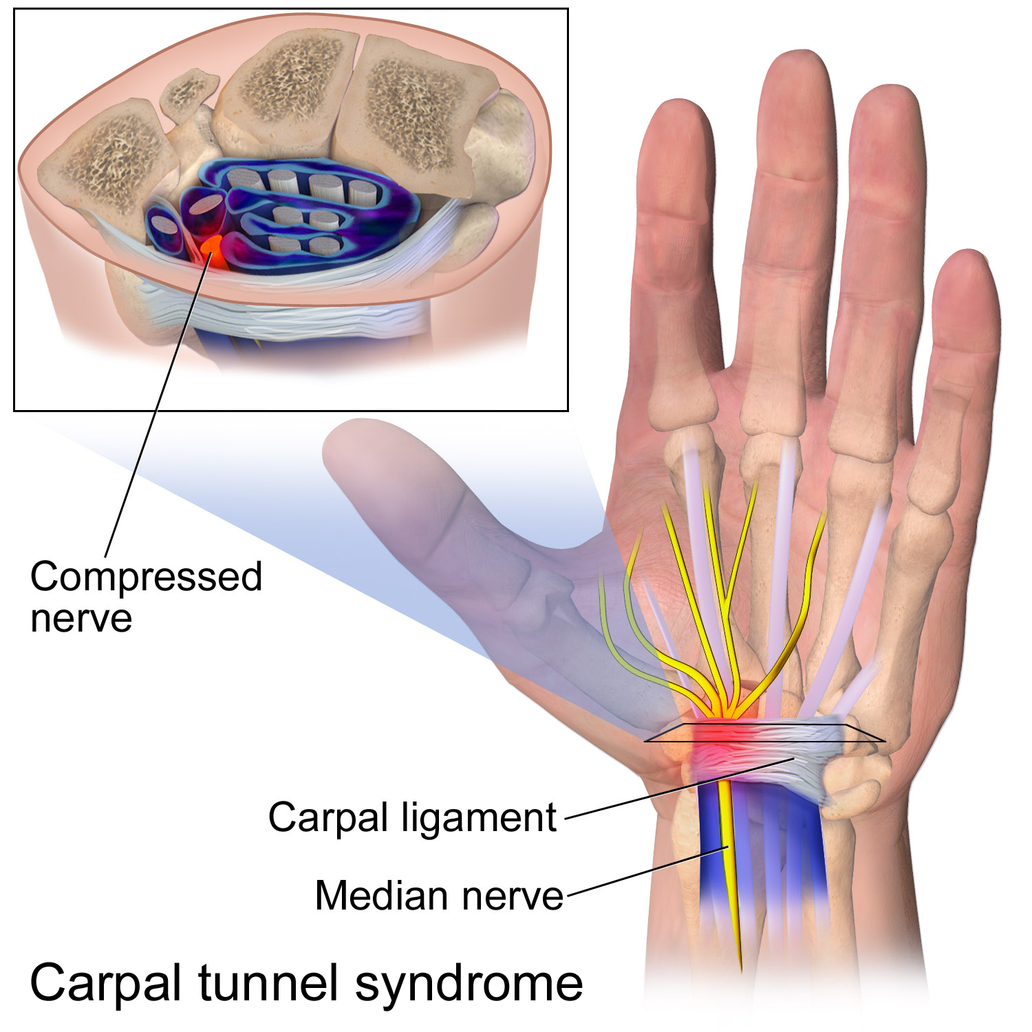
In the human body, the carpal tunnel or carpal canal is the passageway on the palmar side of the wrist that connects the forearm to the hand. The tunnel is bounded by the bones of the wrist and flexor retinaculum from connective tissue. Normally several tendons from the flexor group of forearm muscles and the median nerve pass through it. There are described cases of variable median artery occurrence. When any of the nine long flexor tendons passing through the narrow carpal canal swell or degenerate, the narrowing of the canal may result in the median nerve becoming entrapped or compressed, a common medical condition known as carpal tunnel syndrome (CTS). Structure The carpal bones that make up the wrist form an arch which is convex on the dorsal side of the hand and concave on the palmar side. The groove on the palmar side, the ''sulcus carpi'', is covered by the flexor retinaculum, a sheath of tough connective tissue, thus forming the carpal tunnel. On the side of ... [...More Info...] [...Related Items...] OR: [Wikipedia] [Google] [Baidu] |
Flexor Retinaculum Of The Hand
The flexor retinaculum (transverse carpal ligament, or anterior annular ligament) is a fibrous band on the palmar side of the hand near the wrist. It arches over the carpal bones of the hands, covering them and forming the carpal tunnel. Structure The flexor retinaculum is a strong, fibrous band that covers the carpal bones on the palmar side of the hand near the wrist. It attaches to the bones near the radius and ulna. On the ulnar side, the flexor retinaculum attaches to the pisiform bone and the hook of the hamate bone. On the radial side, it attaches to the tubercle of the scaphoid bone, and to the medial part of the palmar surface and the ridge of the trapezium bone. The flexor retinaculum is continuous with the palmar carpal ligament, and deeper with the palmar aponeurosis. The ulnar artery and ulnar nerve, and the cutaneous branches of the median and ulnar nerves, pass on top of the flexor retinaculum. On the radial side of the retinaculum is the tendon of the flexor ... [...More Info...] [...Related Items...] OR: [Wikipedia] [Google] [Baidu] |
Flexor Digitorum Superficialis Muscle
Flexor digitorum superficialis (''flexor digitorum sublimis'') is an extrinsic flexor muscle of the fingers at the proximal interphalangeal joints. It is in the anterior compartment of the forearm. It is sometimes considered to be the deepest part of the superficial layer of this compartment, and sometimes considered to be a distinct, "intermediate layer" of this compartment. It is relatively common for the Flexor digitorum superficialis to be missing from the little finger, bilaterally and unilaterally, which can cause problems when diagnosing a little finger injury. Structure The muscle has two classically described heads – the humeroulnar and radial – and it is between these heads that the median nerve and ulnar artery pass. The ulnar collateral ligament of elbow joint gives its origin to part of this muscle. Four long tendons come off this muscle near the wrist and travel through the carpal tunnel formed by the flexor retinaculum. These tendons, along with those of f ... [...More Info...] [...Related Items...] OR: [Wikipedia] [Google] [Baidu] |
Tendon
A tendon or sinew is a tough, high-tensile-strength band of dense fibrous connective tissue that connects muscle to bone. It is able to transmit the mechanical forces of muscle contraction to the skeletal system without sacrificing its ability to withstand significant amounts of tension. Tendons are similar to ligaments; both are made of collagen. Ligaments connect one bone to another, while tendons connect muscle to bone. Structure Histologically, tendons consist of dense regular connective tissue. The main cellular component of tendons are specialized fibroblasts called tendon cells (tenocytes). Tenocytes synthesize the extracellular matrix of tendons, abundant in densely packed collagen fibers. The collagen fibers are parallel to each other and organized into tendon fascicles. Individual fascicles are bound by the endotendineum, which is a delicate loose connective tissue containing thin collagen fibrils and elastic fibres. Groups of fascicles are bounded by the epi ... [...More Info...] [...Related Items...] OR: [Wikipedia] [Google] [Baidu] |
Medial Epicondyle Of The Humerus
The medial epicondyle of the humerus is an epicondyle of the humerus bone of the upper arm in humans. It is larger and more prominent than the lateral epicondyle and is directed slightly more posteriorly in the anatomical position. In birds, where the arm is somewhat rotated compared to other tetrapods, it is called the ventral epicondyle of the humerus. In comparative anatomy, the more neutral term entepicondyle is used. The medial epicondyle gives attachment to the ulnar collateral ligament of elbow joint, to the pronator teres, and to a common tendon of origin (the common flexor tendon) of some of the flexor muscles of the forearm: the flexor carpi radialis, the flexor carpi ulnaris, the flexor digitorum superficialis, and the palmaris longus. The medial epicondyle is located on the distal end of the humerus. Additionally, the medial epicondyle is inferior to the medial supracondylar ridge. It is also proximal to the olecranon fossa. The medial epicondyle protects the uln ... [...More Info...] [...Related Items...] OR: [Wikipedia] [Google] [Baidu] |


