|
First Metacarpal Bone
The first metacarpal bone or the metacarpal bone of the thumb is the first bone proximal to the thumb. It is connected to the trapezium of the carpus at the first carpometacarpal joint and to the proximal thumb phalanx at the first metacarpophalangeal joint. Characteristics The first metacarpal bone is short and thick with a shaft thicker and broader than those of the other metacarpal bones. Its narrow shaft connects its widened base and rounded head; the former consisting of a thick cortical bone surrounding the open medullary canal; the latter two consisting of cancellous bone surrounded by a thin cortical shell. Head The head is less rounded and less spherical than those of the other metacarpals, making it better suited for a hinge-like articulation. The distal articular surface is quadrilateral, wide, and flat; thicker and broader transversely and extends much further palmarly than dorsally. On the palmar aspect of the articular surface there is a pair of eminences ... [...More Info...] [...Related Items...] OR: [Wikipedia] [Google] [Baidu] |
Anatomical Terms Of Location
Standard anatomical terms of location are used to unambiguously describe the anatomy of animals, including humans. The terms, typically derived from Latin or Greek roots, describe something in its standard anatomical position. This position provides a definition of what is at the front ("anterior"), behind ("posterior") and so on. As part of defining and describing terms, the body is described through the use of anatomical planes and anatomical axes. The meaning of terms that are used can change depending on whether an organism is bipedal or quadrupedal. Additionally, for some animals such as invertebrates, some terms may not have any meaning at all; for example, an animal that is radially symmetrical will have no anterior surface, but can still have a description that a part is close to the middle ("proximal") or further from the middle ("distal"). International organisations have determined vocabularies that are often used as standard vocabularies for subdisciplines of ana ... [...More Info...] [...Related Items...] OR: [Wikipedia] [Google] [Baidu] |
Opponens Pollicis Muscle
The opponens pollicis is a small, triangular muscle in the hand, which functions to oppose the thumb. It is one of the three thenar muscles. It lies deep to the abductor pollicis brevis and lateral to the flexor pollicis brevis. Structure The opponens pollicis muscle is one of the three thenar muscles. It originates from the flexor retinaculum of the hand and the tubercle of the trapezium. It passes downward and laterally, and is inserted into the whole length of the metacarpal bone of the thumb on its radial side. Innervation Like the other thenar muscles, the opponens pollicis is innervated by the recurrent branch of the median nerve. In 20% of the population, opponens pollicis is innervated by the ulnar nerve. Blood supply The opponens pollicis receives its blood supply from the superficial palmar arch. Function ''Opposition of the thumb'' is a combination of actions that allows the tip of the thumb to touch the tips of other fingers. The part of apposition that this mus ... [...More Info...] [...Related Items...] OR: [Wikipedia] [Google] [Baidu] |
Skeletal System
A skeleton is the structural frame that supports the body of an animal. There are several types of skeletons, including the exoskeleton, which is the stable outer shell of an organism, the endoskeleton, which forms the support structure inside the body, and the hydroskeleton, a flexible internal skeleton supported by fluid pressure. Vertebrates are animals with a vertebral column, and their skeletons are typically composed of bone and cartilage. Invertebrates are animals that lack a vertebral column. The skeletons of invertebrates vary, including hard exoskeleton shells, plated endoskeletons, or spicules. Cartilage is a rigid connective tissue that is found in the skeletal systems of vertebrates and invertebrates. Etymology The term ''skeleton'' comes . ''Sceleton'' is an archaic form of the word. Classification Skeletons can be defined by several attributes. Solid skeletons consist of hard substances, such as bone, cartilage, or cuticle. These can be further divided by locat ... [...More Info...] [...Related Items...] OR: [Wikipedia] [Google] [Baidu] |
Rolando's Fracture
The Rolando fracture is a type of broken finger involving the base of the thumb. It is an intra-articular fracture. It was first described in 1910 by Silvio Rolando.Rolando S. Fracture de la base du premier metacarpien et principalement sur une variete` non encore e`crite. Presse Med 1910;33:303–4 n French It is typically T- or Y-shaped. Treatment There are several proposed methods of treatment. The quality of reduction does not correlate with late symptoms and osteoarthritic changes. Despite this fact, the joint surface should be restored as close to its anatomical position as possible. Some advocate fixation with Kirschner wires, or plate and screw constructions. Another accepted treatment is an external fixator External fixation is a surgical treatment wherein rods are screwed into bone and exit the body to be attached to a stabilizing structure on the outside of the body. It is an alternative to internal fixation, where the components used to provide st ... accompan ... [...More Info...] [...Related Items...] OR: [Wikipedia] [Google] [Baidu] |
Bennett's Fracture
Bennett fracture is a type of partial broken finger involving the base of the thumb, and extends into the carpometacarpal (CMC) joint. Treatment typically requires surgery. This intra-articular fracture is the most common type of fracture of the thumb, and is nearly always accompanied by some degree of subluxation or frank dislocation of the carpometacarpal joint. Symptoms and signs Symptoms of Bennett fracture are instability of the CMC joint of the thumb, accompanied by pain and weakness of the pinch grasp. Characteristic signs include pain, swelling, and ecchymosis around the base of the thumb and thenar eminence, and especially over the CMC joint of the thumb. Physical examination demonstrates instability of the CMC joint of the thumb. The patient will often manifest a weakened ability to grasp objects or perform such tasks as tying shoes and tearing a piece of paper. Other complaints include intense pain experienced upon catching the thumb on an object, such as when ... [...More Info...] [...Related Items...] OR: [Wikipedia] [Google] [Baidu] |
Trapezium (bone)
The trapezium bone (greater multangular bone) is a carpal bone in the hand. It forms the radial border of the carpal tunnel. Structure The trapezium is distinguished by a deep groove on its anterior surface. It is situated at the radial side of the carpus, between the scaphoid and the first metacarpal bone (the metacarpal bone of the thumb). It is homologous with the first distal carpal of reptiles and amphibians. Surfaces The trapezium is an irregular-shaped carpal bone found within the hand. The trapezium is found within the distal row of carpal bones, and is directly adjacent to the metacarpal bone of the thumb. On its ulnar surface are found the trapezoid and scaphoid bones. The '' superior surface'' is directed upward and medialward; medially it is smooth, and articulates with the scaphoid; laterally it is rough and continuous with the lateral surface. The '' inferior surface'' is oval, concave from side to side, convex from before backward, so as to form a saddle-sha ... [...More Info...] [...Related Items...] OR: [Wikipedia] [Google] [Baidu] |
Accessory Bone
An accessory bone or supernumerary bone is a bone that is not normally present in the body, but can be found as a variant in a significant number of people. It poses a risk of being misdiagnosed as bone fractures on radiography. Wrist and hand Os ulnostyloideum The ''os ulnostyloideum'' is an ulnar styloid process that is not fused to the rest of the ulna bone.R. O'Rahilly. ''A survey of carpal and tarsal anomalies.'' J Bone Joint Surg Am. 1953; 35: 626–642 On X-rays, an ''os ulnostyloideum'' is sometimes mistaken for an avulsion fracture of the styloid process. However, the distinction between these is extremely difficult.T.E. Keats, M.W. Anderson. ''Atlas of normal roentgen variants that may simulate disease''. 7th edition, Mosby Inc. 2001 It is alleged that the os ulnostyloideum has a close relationship with or is synonymous with the os triquetrum secundarium. Os centrale The ''os carpi centrale'' (also briefly ''os centrale'') is, where present, located on the ... [...More Info...] [...Related Items...] OR: [Wikipedia] [Google] [Baidu] |
Gray234 - First Metacarpal Bone
Grey (more common in British English) or gray (more common in American English) is an intermediate color between black and white. It is a neutral or achromatic color, meaning literally that it is "without color", because it can be composed of black and white. It is the color of a cloud-covered sky, of ash and of lead. The first recorded use of ''grey'' as a color name in the English language was in 700 CE.Maerz and Paul ''A Dictionary of Color'' New York:1930 McGraw-Hill Page 196 ''Grey'' is the dominant spelling in European and Commonwealth English, while ''gray'' has been the preferred spelling in American English; both spellings are valid in both varieties of English. In Europe and North America, surveys show that grey is the color most commonly associated with neutrality, conformity, boredom, uncertainty, old age, indifference, and modesty. Only one percent of respondents chose it as their favorite color. Etymology ''Grey'' comes from the Middle English ... [...More Info...] [...Related Items...] OR: [Wikipedia] [Google] [Baidu] |
Abductor Pollicis Longus Muscle
In human anatomy, the abductor pollicis longus (APL) is one of the extrinsic muscles of the hand. Its major function is to abduct the thumb at the wrist. Its tendon forms the anterior border of the anatomical snuffbox. Structure The abductor pollicis longus lies immediately below the supinator and is sometimes united with it. It arises from the lateral part of the dorsal surface of the body of the ulna, below the insertion of the anconeus, from the interosseous membrane, and from the middle third of the dorsal surface of the body of the radius.''Gray's Anatomy'' (1918), see infobox Passing obliquely downward and lateralward, it ends in a tendon, which runs through a groove on the lateral side of the lower end of the radius, accompanied by the tendon of the extensor pollicis brevis. The insertion is divided into a distal, superficial part and a proximal, deep part. The superficial part is inserted with one or more tendons into the radial side of the base of the first meta ... [...More Info...] [...Related Items...] OR: [Wikipedia] [Google] [Baidu] |
Dorsal Interossei Of The Hand
In human anatomy, the dorsal interossei (DI) are four muscles in the back of the hand that act to abduct (spread) the index, middle, and ring fingers away from hand's midline (ray of middle finger) and assist in flexion at the metacarpophalangeal joints and extension at the interphalangeal joints of the index, middle and ring fingers. Structure There are four dorsal interossei in each hand. They are specified as 'dorsal' to contrast them with the palmar interossei, which are located on the anterior side of the metacarpals. The dorsal interosseous muscles are bipennate, with each muscle arising by two heads from the adjacent sides of the metacarpal bones, but more extensively from the metacarpal bone of the finger into which the muscle is inserted. They are inserted into the bases of the proximal phalanges and into the extensor expansion of the corresponding extensor digitorum tendon. The middle digit has two dorsal interossei insert onto it while the first digit (thumb) a ... [...More Info...] [...Related Items...] OR: [Wikipedia] [Google] [Baidu] |
Sesamoid Bone
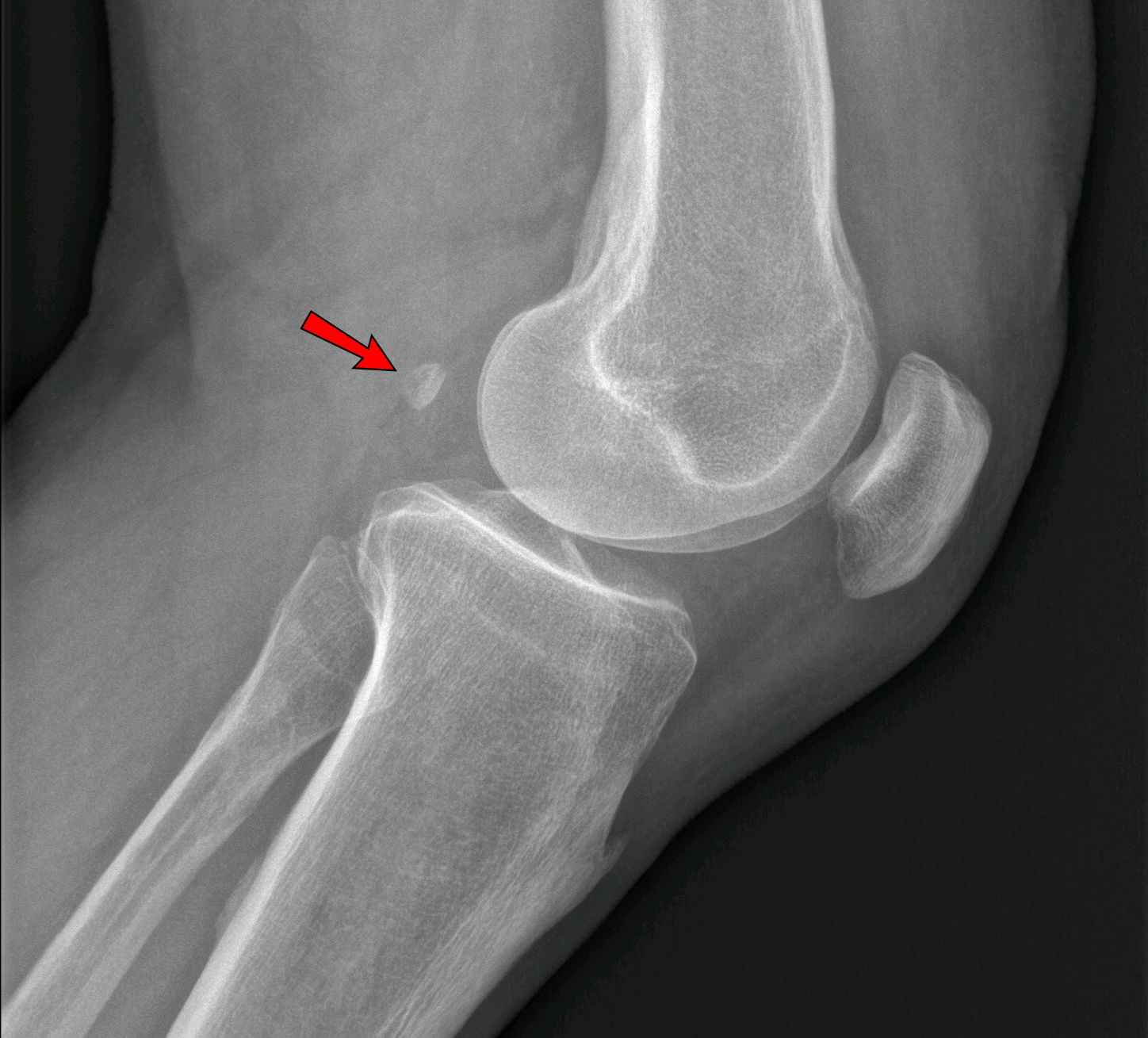
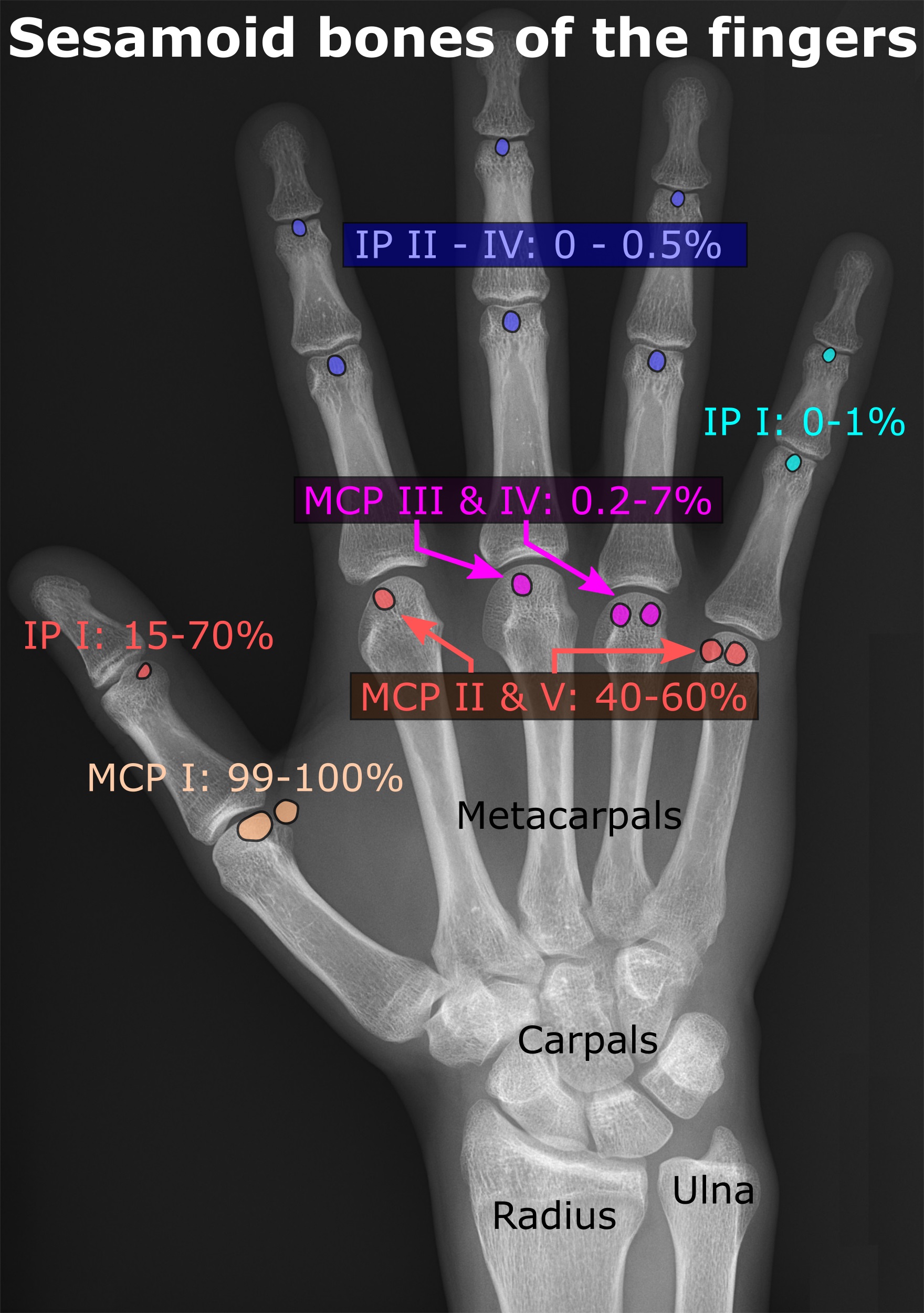
In anatomy, a sesamoid bone () is a bone embedded within a tendon or a muscle. Its name is derived from the Arabic word for 'sesame seed', indicating the small size of most sesamoids. Often, these bones form in response to strain, or can be present as a normal variant. The patella is the largest sesamoid bone in the body. Sesamoids act like pulleys, providing a smooth surface for tendons to slide over, increasing the tendon's ability to transmit muscular forces. Structure Sesamoid bones can be found on joints throughout the body, including: * In the knee—the patella (within the quadriceps tendon). This is the largest sesamoid bone. * In the hand—two sesamoid bones are commonly found in the distal portions of the first metacarpal bone (within the tendons of adductor pollicis and flexor pollicis brevis). There is also commonly a sesamoid bone in distal portions of the second metacarpal bone. * In the wrist—The pisiform of the wrist is a sesamoid bone (within the ten ... [...More Info...] [...Related Items...] OR: [Wikipedia] [Google] [Baidu] |
Metacarpal Bone
In human anatomy, the metacarpal bones or metacarpus form the intermediate part of the skeletal hand located between the phalanges of the fingers and the carpal bones of the wrist, which forms the connection to the forearm. The metacarpal bones are analogous to the metatarsal bones in the foot. Structure The metacarpals form a transverse arch to which the rigid row of distal carpal bones are fixed. The peripheral metacarpals (those of the thumb and little finger) form the sides of the cup of the palmar gutter and as they are brought together they deepen this concavity. The index metacarpal is the most firmly fixed, while the thumb metacarpal articulates with the trapezium and acts independently from the others. The middle metacarpals are tightly united to the carpus by intrinsic interlocking bone elements at their bases. The ring metacarpal is somewhat more mobile while the fifth metacarpal is semi-independent.Tubiana ''et al'' 1998, p 11 Each metacarpal bone consists of a body ... [...More Info...] [...Related Items...] OR: [Wikipedia] [Google] [Baidu] |


_dorsal_view.png)