|
Fibrous Cap
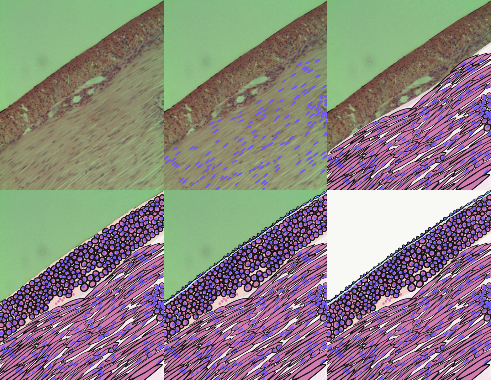
The fibrous cap is a layer of fibrous connective tissue, which is thicker and less cellular than the normal intima, found in atherosclerotic plaques. The fibrous cap contains macrophages and smooth muscle cells. The fibrous cap of an atheroma is composed of bundles of muscle cells, macrophages, foam cells, lymphocytes, collagen Collagen () is the main structural protein in the extracellular matrix found in the body's various connective tissues. As the main component of connective tissue, it is the most abundant protein in mammals, making up from 25% to 35% of the whol ... and elastin. The fibrous cap is prone to rupture and ulceration which can lead to thrombosis. In advanced lesions, further complications may arise including calcification of the fibrous cap. References Angiology {{Pathology-stub ... [...More Info...] [...Related Items...] OR: [Wikipedia] [Google] [Baidu] |
Connective Tissue
Connective tissue is one of the four primary types of animal tissue, along with epithelial tissue, muscle tissue, and nervous tissue. It develops from the mesenchyme derived from the mesoderm the middle embryonic germ layer. Connective tissue is found in between other tissues everywhere in the body, including the nervous system. The three meninges, membranes that envelop the brain and spinal cord are composed of connective tissue. Most types of connective tissue consists of three main components: elastic and collagen fibers, ground substance, and cells. Blood, and lymph are classed as specialized fluid connective tissues that do not contain fiber. All are immersed in the body water. The cells of connective tissue include fibroblasts, adipocytes, macrophages, mast cells and leucocytes. The term "connective tissue" (in German, ''Bindegewebe'') was introduced in 1830 by Johannes Peter Müller. The tissue was already recognized as a distinct class in the 18th century. ... [...More Info...] [...Related Items...] OR: [Wikipedia] [Google] [Baidu] |
Intima
The tunica intima (New Latin "inner coat"), or intima for short, is the innermost tunica (layer) of an artery or vein. It is made up of one layer of endothelial cells and is supported by an internal elastic lamina. The endothelial cells are in direct contact with the blood flow. The three layers of a blood vessel are an inner layer (the tunica intima), a middle layer (the tunica media), and an outer layer (the tunica externa). In dissection, the inner coat (tunica intima) can be separated from the middle (tunica media) by a little maceration, or it may be stripped off in small pieces; but, because of its friability, it cannot be separated as a complete membrane. It is a fine, transparent, colorless structure which is highly elastic, and, after death, is commonly corrugated into longitudinal wrinkles. Structure The structure of the tunica intima depends on the blood vessel type. Elastic arteries – A single layer of Endothelial and a supporting layer of elastin-rich collagen ... [...More Info...] [...Related Items...] OR: [Wikipedia] [Google] [Baidu] |
Atherosclerotic Plaque
An atheroma, or atheromatous plaque, is an abnormal and reversible accumulation of material in the inner layer of an arterial wall. The material consists of mostly macrophage cells, or debris, containing lipids, calcium and a variable amount of fibrous connective tissue. The accumulated material forms a swelling in the artery wall, which may intrude into the lumen of the artery, narrowing it and restricting blood flow. Atheroma is the pathological basis for the disease entity atherosclerosis, a subtype of arteriosclerosis. Signs and symptoms For most people, the first symptoms result from atheroma progression within the heart arteries, most commonly resulting in a heart attack and ensuing debility. The heart arteries are difficult to track because they are small (from about 5 mm down to microscopic), they are hidden deep within the chest and they never stop moving. Additionally, all mass-applied clinical strategies focus on both minimal cost and the overall safety ... [...More Info...] [...Related Items...] OR: [Wikipedia] [Google] [Baidu] |
Macrophages
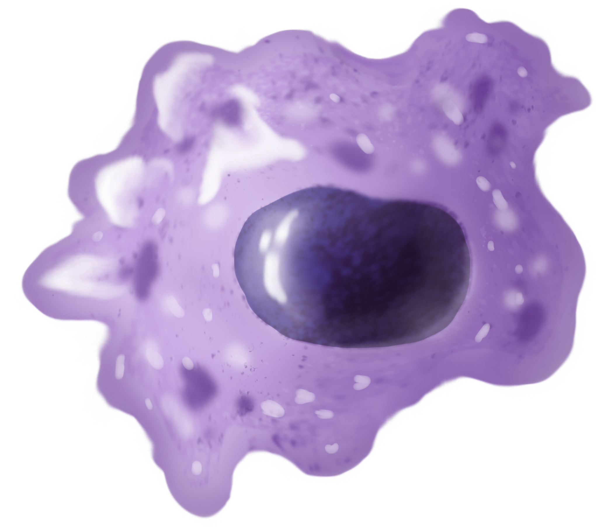
Macrophages (abbreviated as M φ, MΦ or MP) ( el, large eaters, from Greek ''μακρός'' (') = large, ''φαγεῖν'' (') = to eat) are a type of white blood cell of the immune system that engulfs and digests pathogens, such as cancer cells, microbes, cellular debris, and foreign substances, which do not have proteins that are specific to healthy body cells on their surface. The process is called phagocytosis, which acts to defend the host against infection and injury. These large phagocytes are found in essentially all tissues, where they patrol for potential pathogens by amoeboid movement. They take various forms (with various names) throughout the body (e.g., histiocytes, Kupffer cells, alveolar macrophages, microglia, and others), but all are part of the mononuclear phagocyte system. Besides phagocytosis, they play a critical role in nonspecific defense (innate immunity) and also help initiate specific defense mechanisms (adaptive immunity) by recruiting other immune ... [...More Info...] [...Related Items...] OR: [Wikipedia] [Google] [Baidu] |
Smooth Muscle
Smooth muscle is an involuntary non- striated muscle, so-called because it has no sarcomeres and therefore no striations (''bands'' or ''stripes''). It is divided into two subgroups, single-unit and multiunit smooth muscle. Within single-unit muscle, the whole bundle or sheet of smooth muscle cells contracts as a syncytium. Smooth muscle is found in the walls of hollow organs, including the stomach, intestines, bladder and uterus; in the walls of passageways, such as blood, and lymph vessels, and in the tracts of the respiratory, urinary, and reproductive systems. In the eyes, the ciliary muscles, a type of smooth muscle, dilate and contract the iris and alter the shape of the lens. In the skin, smooth muscle cells such as those of the arrector pili cause hair to stand erect in response to cold temperature or fear. Structure Gross anatomy Smooth muscle is grouped into two types: single-unit smooth muscle, also known as visceral smooth muscle, and multiunit ... [...More Info...] [...Related Items...] OR: [Wikipedia] [Google] [Baidu] |
Atheroma
An atheroma, or atheromatous plaque, is an abnormal and reversible accumulation of material in the inner layer of an arterial wall. The material consists of mostly macrophage cells, or debris, containing lipids, calcium and a variable amount of fibrous connective tissue. The accumulated material forms a swelling in the artery wall, which may intrude into the lumen of the artery, narrowing it and restricting blood flow. Atheroma is the pathological basis for the disease entity atherosclerosis, a subtype of arteriosclerosis. Signs and symptoms For most people, the first symptoms result from atheroma progression within the heart arteries, most commonly resulting in a heart attack and ensuing debility. The heart arteries are difficult to track because they are small (from about 5 mm down to microscopic), they are hidden deep within the chest and they never stop moving. Additionally, all mass-applied clinical strategies focus on both minimal cost and the overall safety of ... [...More Info...] [...Related Items...] OR: [Wikipedia] [Google] [Baidu] |
Foam Cells
Foam cells, also called lipid-laden macrophages, are a type of cell that contain cholesterol. These can form a plaque that can lead to atherosclerosis and trigger heart attacks and stroke. Foam cells are fat-laden cells with a M2 macrophage-like phenotype. They contain low density lipoproteins (LDL) and can only be truly detected by examining a fatty plaque under a microscope after it is removed from the body. They are named because the lipoproteins give the cell a foamy appearance. Despite the connection with cardiovascular diseases they might not be inherently dangerous. Some foam cells are derived from smooth muscle cells and present a limited macrophage-like phenotype. Formation Foam cell formation is triggered by a number of factors including the uncontrolled uptake of modified low density lipoproteins (LDL), the upregulation of cholesterol esterification and the impairment of mechanisms associated with cholesterol release. Foam cells are formed when circulating monocy ... [...More Info...] [...Related Items...] OR: [Wikipedia] [Google] [Baidu] |
Collagen
Collagen () is the main structural protein in the extracellular matrix found in the body's various connective tissues. As the main component of connective tissue, it is the most abundant protein in mammals, making up from 25% to 35% of the whole-body protein content. Collagen consists of amino acids bound together to form a triple helix of elongated fibril known as a collagen helix. It is mostly found in connective tissue such as cartilage, bones, tendons, ligaments, and skin. Depending upon the degree of mineralization, collagen tissues may be rigid (bone) or compliant (tendon) or have a gradient from rigid to compliant (cartilage). Collagen is also abundant in corneas, blood vessels, the gut, intervertebral discs, and the dentin in teeth. In muscle tissue, it serves as a major component of the endomysium. Collagen constitutes one to two percent of muscle tissue and accounts for 6% of the weight of the skeletal muscle tissue. The fibroblast is the most common ... [...More Info...] [...Related Items...] OR: [Wikipedia] [Google] [Baidu] |


