|
Eyeball
An eye is a sensory organ that allows an organism to perceive visual information. It detects light and converts it into electro-chemical impulses in neurons (neurones). It is part of an organism's visual system. In higher organisms, the eye is a complex optical Optics is the branch of physics that studies the behaviour and properties of light, including its interactions with matter and the construction of instruments that use or detect it. Optics usually describes the behaviour of visible, ultravio ... system that collects light from the surrounding environment, regulates its intensity through a Iris (anatomy), diaphragm, Focus (optics), focuses it through an adjustable assembly of Lens (anatomy), lenses to form an image, converts this image into a set of electrical signals, and transmits these signals to the brain through neural pathways that connect the eye via the optic nerve to the visual cortex and other areas of the brain. Eyes with resolving power have come in ... [...More Info...] [...Related Items...] OR: [Wikipedia] [Google] [Baidu] |
Human Eye
The human eye is a sensory organ in the visual system that reacts to light, visible light allowing eyesight. Other functions include maintaining the circadian rhythm, and Balance (ability), keeping balance. The eye can be considered as a living optics, optical device. It is approximately spherical in shape, with its outer layers, such as the outermost, white part of the eye (the sclera) and one of its inner layers (the pigmented choroid) keeping the eye essentially stray light, light tight except on the eye's optic axis. In order, along the optic axis, the optical components consist of a first lens (the cornea, cornea—the clear part of the eye) that accounts for most of the optical power of the eye and accomplishes most of the Focus (optics), focusing of light from the outside world; then an aperture (the pupil) in a Diaphragm (optics), diaphragm (the Iris (anatomy), iris—the coloured part of the eye) that controls the amount of light entering the interior of the eye; then an ... [...More Info...] [...Related Items...] OR: [Wikipedia] [Google] [Baidu] |
Optic Nerve
In neuroanatomy, the optic nerve, also known as the second cranial nerve, cranial nerve II, or simply CN II, is a paired cranial nerve that transmits visual system, visual information from the retina to the brain. In humans, the optic nerve is derived from optic stalks during the seventh week of development and is composed of retinal ganglion cell axons and glial cells; it extends from the optic disc to the optic chiasma and continues as the optic tract to the lateral geniculate nucleus, Pretectal area, pretectal nuclei, and superior colliculus. Structure The optic nerve has been classified as the second of twelve paired cranial nerves, but it is technically a myelinated tract of the central nervous system, rather than a classical nerve of the peripheral nervous system because it is derived from an out-pouching of the diencephalon (optic stalks) during embryonic development. As a consequence, the fibers of the optic nerve are covered with myelin produced by oligodendrocytes, r ... [...More Info...] [...Related Items...] OR: [Wikipedia] [Google] [Baidu] |
Lens (anatomy)
The lens, or crystalline lens, is a Transparency and translucency, transparent Biconvex lens, biconvex structure in most land vertebrate eyes. Relatively long, thin fiber cells make up the majority of the lens. These cells vary in architecture and are arranged in concentric layers. New layers of cells are recruited from a thin epithelium at the front of the lens, just below the basement membrane surrounding the lens. As a result the vertebrate lens grows throughout life. The surrounding lens membrane referred to as the lens capsule also grows in a systematic way, ensuring the lens maintains an optically suitable shape in concert with the underlying fiber cells. Thousands of suspensory ligaments are embedded into the capsule at its largest diameter which suspend the lens within the eye. Most of these lens structures are derived from the epithelium of the embryo before birth. Along with the cornea, aqueous humour, aqueous, and vitreous humours, the lens Refraction, refracts light, Fo ... [...More Info...] [...Related Items...] OR: [Wikipedia] [Google] [Baidu] |
Visual System
The visual system is the physiological basis of visual perception (the ability to perception, detect and process light). The system detects, phototransduction, transduces and interprets information concerning light within the visible range to construct an imaging, image and build a mental model of the surrounding environment. The visual system is associated with the eye and functionally divided into the optics, optical system (including cornea and crystalline lens, lens) and the nervous system, neural system (including the retina and visual cortex). The visual system performs a number of complex tasks based on the ''image forming'' functionality of the eye, including the formation of monocular images, the neural mechanisms underlying stereopsis and assessment of distances to (depth perception) and between objects, motion perception, pattern recognition, accurate motor coordination under visual guidance, and colour vision. Together, these facilitate higher order tasks, such as ... [...More Info...] [...Related Items...] OR: [Wikipedia] [Google] [Baidu] |
Sensory Organ
A sense is a biological system used by an organism for sensation, the process of gathering information about the surroundings through the detection of stimuli. Although, in some cultures, five human senses were traditionally identified as such (namely sight, smell, touch, taste, and hearing), many more are now recognized. Senses used by non-human organisms are even greater in variety and number. During sensation, sense organs collect various stimuli (such as a sound or smell) for transduction, meaning transformation into a form that can be understood by the brain. Sensation and perception are fundamental to nearly every aspect of an organism's cognition, behavior and thought. In organisms, a sensory organ consists of a group of interrelated sensory cells that respond to a specific type of physical stimulus. Via cranial and spinal nerves (nerves of the central and peripheral nervous systems that relay sensory information to and from the brain and body), the different ty ... [...More Info...] [...Related Items...] OR: [Wikipedia] [Google] [Baidu] |
Annual Review Of Neuroscience
The ''Annual Review of Neuroscience'' is a peer-reviewed academic journal that publishes review articles relevant to neuroscience. In publication since 1978 by Annual Reviews, founding editor W. Maxwell Cowan led the editorial committee until his death in 2002. Mary E. Hatten and Botond Roska are the current co-editors. As of 2023, it is being published as open access, under the Subscribe to Open model. History In 1975, the nonprofit publisher Annual Reviews had two meetings in New York to coincide with the annual meeting of the Society for Neuroscience. In the meetings, neuroscientists from the US and Canada concurred that it would be useful to establish a journal that published an annual volume of review articles relevant to neuroscience. The board of directors of Annual Reviews gave the final approval for the journal in early 1976, appointing the first editorial committee with W. Maxwell Cowan appointed editor. In April 1976 the editorial committee planned the first ... [...More Info...] [...Related Items...] OR: [Wikipedia] [Google] [Baidu] |
Photosensitive Ganglion Cell
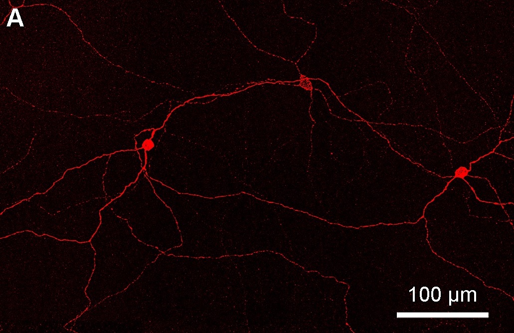
Intrinsically photosensitive retinal ganglion cells (ipRGCs), also called photosensitive retinal ganglion cells (pRGC), or melanopsin-containing retinal ganglion cells (mRGCs), are a type of neuron in the retina of the mammalian eye. The presence of an additional photoreceptor was first suspected in 1927 when mice lacking rod and cone cells still responded to changing light levels through pupil constriction; this suggested that rods and cones are not the only light-sensitive tissue. However, it was unclear whether this light sensitivity arose from an additional retinal photoreceptor or elsewhere in the body. Recent research has shown that these retinal ganglion cells, unlike other retinal ganglion cells, are intrinsically photosensitive due to the presence of melanopsin, a light-sensitive protein. Therefore, they constitute a third class of photoreceptors, in addition to rod and cone cells. Overview Compared to the rods and cones, the ipRGCs respond more sluggishly and ... [...More Info...] [...Related Items...] OR: [Wikipedia] [Google] [Baidu] |
Retinohypothalamic Tract
In neuroanatomy, the retinohypothalamic tract (RHT) is a photic neural input pathway involved in the circadian rhythms of mammals. The origin of the retinohypothalamic tract is the intrinsically photosensitive retinal ganglion cells (ipRGC), which contain the photopigment melanopsin. The axons of the ipRGCs belonging to the retinohypothalamic tract project directly, monosynaptically, to the suprachiasmatic nuclei (SCN) via the optic nerve and the optic chiasm. The suprachiasmatic nuclei receive and interpret information on environmental light, dark and day length, important in the entrainment of the "body clock". They can coordinate peripheral "clocks" and direct the pineal gland to secrete the hormone melatonin. Structure The retinohypothalamic tract consists of retinal ganglion cells. A distinct population of ganglion cells, known as intrinsically photosensitive retinal ganglion cells (ipRGCs), is critically responsible for providing non-image-forming visual signals ... [...More Info...] [...Related Items...] OR: [Wikipedia] [Google] [Baidu] |
Suprachiasmatic Nucleus
The suprachiasmatic nucleus or nuclei (SCN) is a small region of the brain in the hypothalamus, situated directly above the optic chiasm. It is responsible for regulating sleep cycles in animals. Reception of light inputs from photosensitive retinal ganglion cells allow it to coordinate the subordinate cellular clocks of the body and entrain to the environment. The neuronal and hormonal activities it generates regulate many different body functions in an approximately 24-hour cycle. The SCN also interacts with many other regions of the brain. It contains several cell types, neurotransmitters and peptides, including vasopressin and vasoactive intestinal peptide. Disruptions or damage to the SCN has been associated with different mood disorders and sleep disorders, suggesting the significance of the SCN in regulating circadian timing. Neuroanatomy The SCN is situated in the anterior part of the hypothalamus immediately dorsal, or ''superior'' (hence supra) to the optic c ... [...More Info...] [...Related Items...] OR: [Wikipedia] [Google] [Baidu] |
Pupillary Light Reflex
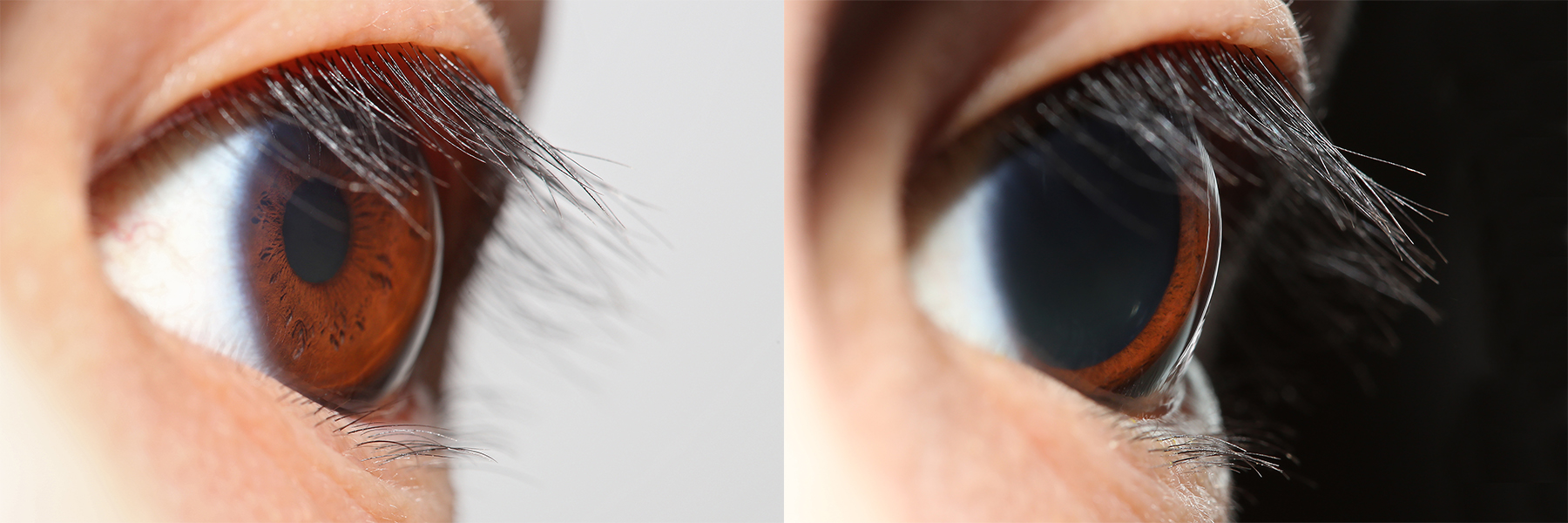
The pupillary light reflex (PLR) or photopupillary reflex is a reflex that controls the diameter of the pupil, in response to the intensity ( luminance) of light that falls on the retinal ganglion cells of the retina in the back of the eye, thereby assisting in adaptation of vision to various levels of lightness/darkness. A greater intensity of light causes the pupil to constrict ( miosis/myosis; thereby allowing less light in), whereas a lower intensity of light causes the pupil to dilate ( mydriasis, expansion; thereby allowing more light in). Thus, the pupillary light reflex regulates the intensity of light entering the eye. Light shone into one eye will cause both pupils to constrict. Terminology The pupil is the dark circular opening in the center of the iris and is where light enters the eye. By analogy with a camera, the pupil is equivalent to aperture, whereas the iris is equivalent to the diaphragm. It may be helpful to consider the ''Pupillary reflex'' as an Iris ... [...More Info...] [...Related Items...] OR: [Wikipedia] [Google] [Baidu] |
Pretectal Area
In neuroanatomy, the pretectal area, or pretectum, is a midbrain structure composed of seven nuclei and comprises part of the subcortical visual system. Through reciprocal bilateral projections from the retina, it is involved primarily in mediating behavioral responses to acute changes in ambient light such as the pupillary light reflex, the optokinetic reflex, and temporary changes to the circadian rhythm. In addition to the pretectum's role in the visual system, the anterior pretectal nucleus has been found to mediate somatosensory and nociceptive information. Location and structure The pretectum is a bilateral group of highly interconnected nuclei located near the junction of the midbrain and forebrain. The pretectum is generally classified as a midbrain structure, although because of its proximity to the forebrain it is sometimes classified as part of the caudal diencephalon (forebrain). Within vertebrates, the pretectum is located directly anterior to the superior col ... [...More Info...] [...Related Items...] OR: [Wikipedia] [Google] [Baidu] |
Insect
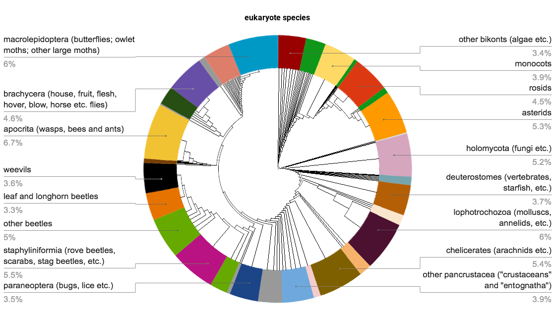
Insects (from Latin ') are Hexapoda, hexapod invertebrates of the class (biology), class Insecta. They are the largest group within the arthropod phylum. Insects have a chitinous exoskeleton, a three-part body (Insect morphology#Head, head, Thorax (insect anatomy), thorax and abdomen (insect anatomy), abdomen), three pairs of jointed Arthropod leg, legs, compound eyes, and a pair of antenna (biology), antennae. Insects are the most diverse group of animals, with more than a million described species; they represent more than half of all animal species. The insect nervous system consists of a insect brain, brain and a ventral nerve cord. Most insects reproduce Oviparous, by laying eggs. Insects Respiratory system of insects, breathe air through a system of Spiracle (arthropods), paired openings along their sides, connected to Trachea#Invertebrates, small tubes that take air directly to the tissues. The blood therefore does not carry oxygen; it is only partly contained in ves ... [...More Info...] [...Related Items...] OR: [Wikipedia] [Google] [Baidu] |


