|
Enamel Cord
The enamel cord, also called enamel septum, is a localization of cells on an enamel organ that appear from the outer enamel epithelium to an enamel knot. The function of the enamel cord and the enamel knot is not known, but they are believed to play a role in the placement of the first cusp developed in a tooth A tooth ( : teeth) is a hard, calcified structure found in the jaws (or mouths) of many vertebrates and used to break down food. Some animals, particularly carnivores and omnivores, also use teeth to help with capturing or wounding prey, .... References * *{{cite book, last=Ten Cate, first=Richard, title=Oral Histology: Development, Structure, and Function, year=1998, edition=5th, publisher=Mosby, location=St. Louis, Mo., isbn=0-8151-2952-1 Parts of tooth ... [...More Info...] [...Related Items...] OR: [Wikipedia] [Google] [Baidu] |
Cell (biology)
The cell is the basic structural and functional unit of life forms. Every cell consists of a cytoplasm enclosed within a membrane, and contains many biomolecules such as proteins, DNA and RNA, as well as many small molecules of nutrients and metabolites.Cell Movements and the Shaping of the Vertebrate Body in Chapter 21 of Molecular Biology of the Cell '' fourth edition, edited by Bruce Alberts (2002) published by Garland Science. The Alberts text discusses how the "cellular building blocks" move to shape developing embryos. It is also common to describe small molecules such ... [...More Info...] [...Related Items...] OR: [Wikipedia] [Google] [Baidu] |
Enamel Organ
The enamel organ, also known as the dental organ, is a cellular aggregation seen in a developing tooth and it lies above the dental papilla. The enamel organ which is differentiated from the primitive oral epithelium lining the stomodeum.The enamel organ is responsible for the formation of enamel, initiation of dentine formation, establishment of the shape of a tooth's crown, and establishment of the dentoenamel junction. The enamel organ has four layers; the inner enamel epithelium, outer enamel epithelium, stratum intermedium, and the stellate reticulum. The dental papilla, the differentiated ectomesenchyme deep to the enamel organ, will produce dentin and the dental pulp. The surrounding ectomesenchyme tissue, the dental follicle, is the primitive cementum, periodontal ligament and alveolar bone beneath the tooth root. The site where the internal enamel epithelium and external enamel epithelium coalesce is the cervical root, important in proliferation of the dental root ... [...More Info...] [...Related Items...] OR: [Wikipedia] [Google] [Baidu] |
Outer Enamel Epithelium
The outer enamel epithelium, also known as the external enamel epithelium, is a layer of cuboidal cells located on the periphery of the enamel organ in a developing tooth. This layer is first seen during the bell stage. The rim of the enamel organ where the outer and inner enamel epithelium join is called the cervical loop The cervical loop is the location on an enamel organ in a developing tooth where the outer enamel epithelium and the inner enamel epithelium join. The cervical loop is a histologic term indicating a specific epithelial structure at the apical s .... References *Cate, A.R. Ten. Oral Histology: development, structure, and function. 5th ed. 1998. . *Ross, Michael H., Gordon I. Kaye, and Wojciech Pawlina. Histology: a text and atlas. 4th edition. 2003. . Tooth development {{dentistry-stub ... [...More Info...] [...Related Items...] OR: [Wikipedia] [Google] [Baidu] |
Enamel Knot
In tooth development, the enamel knot is a localization of cells on an enamel organ that appear thickened in the center of the inner enamel epithelium. The enamel knot is frequently associated with an enamel cord. It is formed in the cap stage and undergoes apoptosis in the bell stage. The enamel knot as signaling center The enamel knot is a signaling center of the tooth that provides positional information for tooth morphogenesis and regulates the growth of tooth cusps. The enamel knot produces a range of molecular signals from all the major signaling families, such as Fibroblast Growth Factors (FGF), Bone morphogenetic proteins (BMP), Hedgehog (Hh) and Wnt signals. These molecular signals direct the growth of the surrounding epithelium and ectomesenchyme Ectomesenchyme has properties similar to mesenchyme. The origin of the ectomesenchyme is disputed. It is either like the mesenchyme, arising from mesodermic cells, or conversely arising from neural crest cells. The neural crest ... [...More Info...] [...Related Items...] OR: [Wikipedia] [Google] [Baidu] |
Cusp (dentistry)
A cusp is a pointed, projecting, or elevated feature. In animals, it is usually used to refer to raised points on the crowns of teeth. The concept is also used with regard to the leaflets of the four heart valves. The mitral valve, which has two cusps, is also known as the bicuspid valve, and the tricuspid valve has three cusps. In humans A cusp is an occlusal or incisal eminence on a tooth. Canine teeth, otherwise known as cuspids, each possess a single cusp, while premolars, otherwise known as bicuspids, possess two each. Molars normally possess either four or five cusps. In certain populations the maxillary molars, especially first molars, will possess a fifth cusp situated on the mesiolingual cusp known as the Cusp of Carabelli. Buccal Cusp- One other variation of the upper first premolar is the 'Uto-Aztecan' upper premolar. It is a bulge on the buccal cusp that is only found in Native American Indians, with highest frequencies of occurrence in Arizona. The nam ... [...More Info...] [...Related Items...] OR: [Wikipedia] [Google] [Baidu] |
Tooth Development
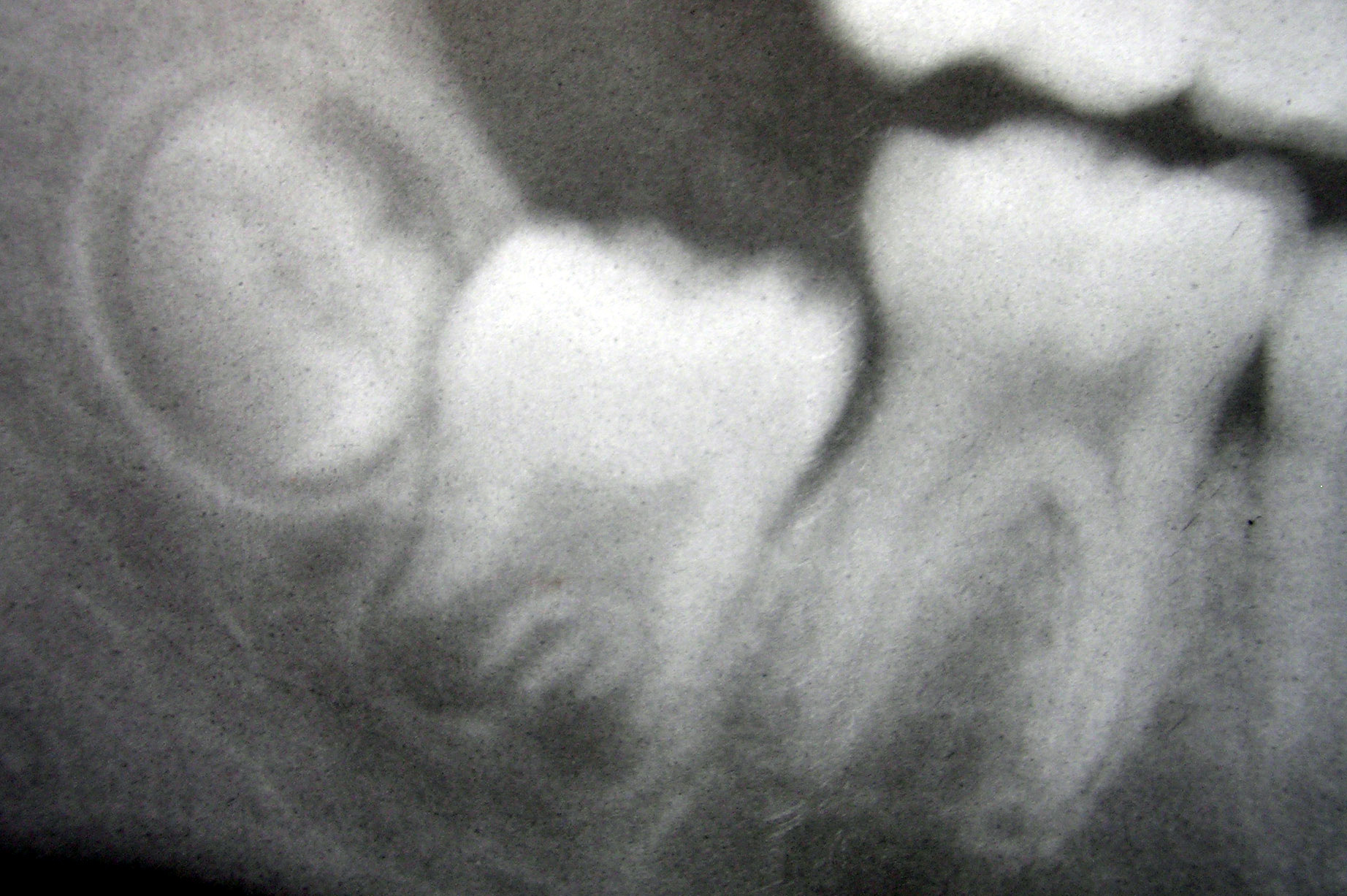
Tooth development or odontogenesis is the complex process by which teeth form from embryonic cells, grow, and erupt into the mouth. For human teeth to have a healthy oral environment, all parts of the tooth must develop during appropriate stages of fetal development. Primary (baby) teeth start to form between the sixth and eighth week of prenatal development, and permanent teeth begin to form in the twentieth week.Ten Cate's Oral Histology, Nanci, Elsevier, 2013, pages 70-94 If teeth do not start to develop at or near these times, they will not develop at all, resulting in hypodontia or anodontia. A significant amount of research has focused on determining the processes that initiate tooth development. It is widely accepted that there is a factor within the tissues of the first pharyngeal arch that is necessary for the development of teeth. Overview The tooth germ is an aggregation of cells that eventually forms a tooth.University of Texas Medical Branch. These cells ar ... [...More Info...] [...Related Items...] OR: [Wikipedia] [Google] [Baidu] |
Tooth
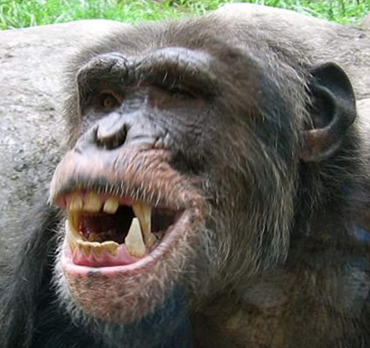
A tooth ( : teeth) is a hard, calcified structure found in the jaws (or mouths) of many vertebrates and used to break down food. Some animals, particularly carnivores and omnivores, also use teeth to help with capturing or wounding prey, tearing food, for defensive purposes, to intimidate other animals often including their own, or to carry prey or their young. The roots of teeth are covered by gums. Teeth are not made of bone, but rather of multiple tissues of varying density and hardness that originate from the embryonic germ layer, the ectoderm. The general structure of teeth is similar across the vertebrates, although there is considerable variation in their form and position. The teeth of mammals have deep roots, and this pattern is also found in some fish, and in crocodilians. In most teleost fish, however, the teeth are attached to the outer surface of the bone, while in lizards they are attached to the inner surface of the jaw by one side. In cartilaginous ... [...More Info...] [...Related Items...] OR: [Wikipedia] [Google] [Baidu] |

