|
Dural Tear
Dural tear is a tear occurring in the dura mater of the brain. It is usually caused as a result of trauma or as a complication following surgery. __TOC__ Diagnosis In case of head injury, a dural tear is likely in case of a depressed skull fracture. A burr hole is made through the normal skull near the fractured portion, and Adson's elevator is introduced. Underlying dura is separated carefully from the overlying depressed bone fragments. The dura that is now visible is carefully examined to exclude any dural tear. Treatment The whole extent of the dural tear is exposed by removing the overlying skull. The ragged edges of the tear are excised. However, care should be taken not to excise too much dura as it may increase the chances for spread of infection into the subarachnoid space. Removing too much dura will also make it difficult to close the tear. If there is not much dural loss, interrupted sutures are made with non-absorbable material. In case of dural loss, the defect is c ... [...More Info...] [...Related Items...] OR: [Wikipedia] [Google] [Baidu] |
Dura Mater
In neuroanatomy, dura mater is a thick membrane made of dense irregular connective tissue that surrounds the brain and spinal cord. It is the outermost of the three layers of membrane called the meninges that protect the central nervous system. The other two meningeal layers are the arachnoid mater and the pia mater. It envelops the arachnoid mater, which is responsible for keeping in the cerebrospinal fluid. It is derived primarily from the neural crest cell population, with postnatal contributions of the paraxial mesoderm. Structure The dura mater has several functions and layers. The dura mater is a membrane that envelops the arachnoid mater. It surrounds and supports the dural sinuses (also called dural venous sinuses, cerebral sinuses, or cranial sinuses) and carries blood from the brain toward the heart. Cranial dura mater has two layers called ''lamellae'', a superficial layer (also called the periosteal layer), which serves as the skull's inner periosteum, called the e ... [...More Info...] [...Related Items...] OR: [Wikipedia] [Google] [Baidu] |
Depressed Skull Fracture
A skull fracture is a break in one or more of the eight bones that form the cranial portion of the skull, usually occurring as a result of blunt force trauma. If the force of the impact is excessive, the bone may fracture at or near the site of the impact and cause damage to the underlying structures within the skull such as the membranes, blood vessels, and brain. While an uncomplicated skull fracture can occur without associated physical or neurological damage and is in itself usually not clinically significant, a fracture in healthy bone indicates that a substantial amount of force has been applied and increases the possibility of associated injury. Any significant blow to the head results in a concussion, with or without loss of consciousness. A fracture in conjunction with an overlying laceration that tears the epidermis and the meninges, or runs through the paranasal sinuses and the middle ear structures, bringing the outside environment into contact with the cranial cav ... [...More Info...] [...Related Items...] OR: [Wikipedia] [Google] [Baidu] |
Burr Hole
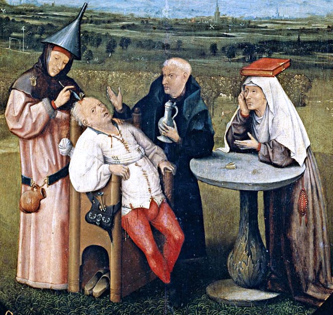
Trepanning, also known as trepanation, trephination, trephining or making a burr hole (the verb ''trepan'' derives from Old French from Medieval Latin from Greek , literally "borer, auger"), is a surgical intervention in which a hole is drilled or scraped into the human skull. The intentional perforation of the cranium exposes the ''dura mater'' to treat health problems related to intracranial diseases or release pressured blood buildup from an injury. It may also refer to any "burr" hole created through other body surfaces, including nail beds. A trephine is an instrument used for cutting out a round piece of skull bone to relieve pressure beneath a surface. In ancient times, holes were drilled into a person who was behaving in what was considered an abnormal way to let out what people believed were evil spirits. Evidence of trepanation has been found in prehistoric human remains from Neolithic times onward. The bone that was trepanned was kept by the prehistoric people and m ... [...More Info...] [...Related Items...] OR: [Wikipedia] [Google] [Baidu] |
Subarachnoid Space
In anatomy, the meninges (, ''singular:'' meninx ( or ), ) are the three membranes that envelop the brain and spinal cord. In mammals, the meninges are the dura mater, the arachnoid mater, and the pia mater. Cerebrospinal fluid is located in the subarachnoid space between the arachnoid mater and the pia mater. The primary function of the meninges is to protect the central nervous system. Structure Dura mater The dura mater ( la, tough mother) (also rarely called ''meninx fibrosa'' or ''pachymeninx'') is a thick, durable membrane, closest to the skull and vertebrae. The dura mater, the outermost part, is a loosely arranged, fibroelastic layer of cells, characterized by multiple interdigitating cell processes, no extracellular collagen, and significant extracellular spaces. The middle region is a mostly fibrous portion. It consists of two layers: the endosteal layer, which lies closest to the skull, and the inner meningeal layer, which lies closer to the brain. It contains la ... [...More Info...] [...Related Items...] OR: [Wikipedia] [Google] [Baidu] |
Diathermy
Diathermy is electrically induced heat or the use of high-frequency electromagnetic currents as a form of physical therapy and in surgical procedures. The earliest observations on the reactions of high-frequency electromagnetic currents upon the human organism were made by Jacques Arsene d'Arsonval. The field was pioneered in 1907 by German physician Karl Franz Nagelschmidt, who coined the term ''diathermy'' from the Greek words ''dia'' and θέρμη ''therma'', literally meaning "heating through" (adj., diather´mal, diather´mic). Diathermy is commonly used for muscle relaxation, and to induce deep heating in tissue for therapeutic purposes in medicine. It is used in physical therapy to deliver moderate heat directly to pathologic lesions in the deeper tissues of the body. Diathermy is produced by three techniques: ultrasound (''ultrasonic diathermy''), short-wave radio frequencies in the range 1–100 MHz (''shortwave diathermy'') or microwaves typically in the 915&nbs ... [...More Info...] [...Related Items...] OR: [Wikipedia] [Google] [Baidu] |
Pericranium
The periosteum is a membrane that covers the outer surface of all bones, except at the articular surfaces (i.e. the parts within a joint space) of long bones. Endosteum lines the inner surface of the medullary cavity of all long bones. Structure The periosteum consists of an outer fibrous layer, and an inner cambium layer (or osteogenic layer). The fibrous layer is of dense irregular connective tissue, containing fibroblasts, while the cambium layer is highly cellular containing progenitor cells that develop into osteoblasts. These osteoblasts are responsible for increasing the width of a long bone and the overall size of the other bone types. After a bone fracture, the progenitor cells develop into osteoblasts and chondroblasts, which are essential to the healing process. The outer fibrous layer and the inner cambium layer is differentiated under electron micrography. As opposed to osseous tissue, the periosteum has nociceptors, sensory neurons that make it very sensitive t ... [...More Info...] [...Related Items...] OR: [Wikipedia] [Google] [Baidu] |
Temporalis
In anatomy, the temporalis muscle, also known as the temporal muscle, is one of the muscles of mastication (chewing). It is a broad, fan-shaped convergent muscle on each side of the head that fills the temporal fossa, superior to the zygomatic arch so it covers much of the temporal bone.Illustrated Anatomy of the Head and Neck, Fehrenbach and Herring, Elsevier, 2012, page 98''Temporal'' refers to the head's temples. Structure In humans, the temporalis muscle arises from the temporal fossa and the deep part of temporal fascia. This is a very broad area of attachment. It passes medial to the zygomatic arch. It forms a tendon which inserts onto the coronoid process of the mandible, with its insertion extending into the retromolar fossa posterior to the most distal mandibular molar.Human Anatomy, Jacobs, Elsevier, 2008, page 194 In other mammals, the muscle usually spans the dorsal part of the skull all the way up to the medial line. There, it may be attached to a sagittal crest, ... [...More Info...] [...Related Items...] OR: [Wikipedia] [Google] [Baidu] |
Brain Disorders
A neurological disorder is any disorder of the nervous system. Structural, biochemical or electrical abnormalities in the brain, spinal cord or other nerves can result in a range of symptoms. Examples of symptoms include paralysis, muscle weakness, poor coordination, loss of sensation, seizures, confusion, pain and altered levels of consciousness. There are many recognized neurological disorders, some relatively common, but many rare. They may be assessed by neurological examination, and studied and treated within the specialities of neurology and clinical neuropsychology. Interventions foneurological disordersinclude preventive measures, lifestyle changes, physiotherapy or other therapy, neurorehabilitation, pain management, medication, operations performed by neurosurgeons or a specific diet. The World Health Organization estimated in 2006 that neurological disorders and their sequelae (direct consequences) affect as many as one billion people worldwide, and ... [...More Info...] [...Related Items...] OR: [Wikipedia] [Google] [Baidu] |
Trauma Surgery
Trauma surgery is a surgical specialty that utilizes both operative and non-operative management to treat traumatic injuries, typically in an acute setting. Trauma surgeons generally complete residency training in general surgery and often fellowship training in trauma or surgical critical care. The trauma surgeon is responsible for initially resuscitating and stabilizing and later evaluating and managing the patient. The attending trauma surgeon also leads the trauma team, which typically includes nurses and support staff as well as resident physicians in teaching hospitals. Training Most United States trauma surgeons practice in larger centers and complete a 1-2 year trauma surgery fellowship, which often includes a surgical critical care fellowship. They may therefore sit for the American Board of Surgery (ABS) certifying examination in Surgical Critical Care. National surgical boards usually supervise European training programs; they also certify for subspecializati ... [...More Info...] [...Related Items...] OR: [Wikipedia] [Google] [Baidu] |


