|
Dictyate
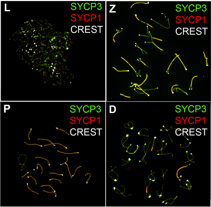
The dictyate or dictyotene is a prolonged resting phase in oogenesis. It occurs in the stage of meiotic prophase I in ootidogenesis. It starts late in fetal life and is terminated shortly before ovulation by the LH surge. Thus, although the majority of oocytes are produced in female fetuses before birth, these pre-eggs remain arrested in the dictyate stage until puberty commences and the cells complete ootidogenesis. In both mouse and human, oocyte DNA of older individuals has substantially more double-strand breaks than that of younger individuals. The dictyate appears to be an adaptation for efficiently removing damages in germ line DNA by homologous recombinational repair. Prophase arrested oocytes have a high capability for efficient repair of DNA damage (naturally occurring), DNA damages. DNA repair capability appears to be a key quality control mechanism in the female germ line and a critical determinant of fertility. Translation halt There are a lot of mRNAs that have been ... [...More Info...] [...Related Items...] OR: [Wikipedia] [Google] [Baidu] |
Oogenesis
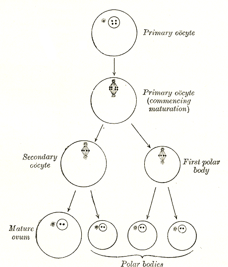
Oogenesis () or ovogenesis is the differentiation of the ovum (egg cell) into a cell competent to further develop when fertilized. It is developed from the primary oocyte by maturation. Oogenesis is initiated before birth during embryonic development. Oogenesis in non-human mammals In mammals, the first part of oogenesis starts in the germinal epithelium, which gives rise to the development of ovarian follicles, the functional unit of the ovary. Oogenesis consists of several sub-processes: oocytogenesis, ootidogenesis, and finally maturation to form an ovum (oogenesis proper). Folliculogenesis is a separate sub-process that accompanies and supports all three oogenetic sub-processes. Oogonium —(Oocytogenesis)—> Primary Oocyte —(Meiosis I)—> First Polar body (Discarded afterward) + Secondary oocyte —(Meiosis II)—> Second Polar Body (Discarded afterward) + Ovum Oocyte meiosis, important to all animal life cycles yet unlike all other instances ... [...More Info...] [...Related Items...] OR: [Wikipedia] [Google] [Baidu] |
Prophase I
Meiosis () is a special type of cell division of germ cells in sexually-reproducing organisms that produces the gametes, the sperm or egg cells. It involves two rounds of division that ultimately result in four cells, each with only one copy of each chromosome (haploid). Additionally, prior to the division, genetic material from the paternal and maternal copies of each chromosome is crossed over, creating new combinations of code on each chromosome. Later on, during fertilisation, the haploid cells produced by meiosis from a male and a female will fuse to create a zygote, a cell with two copies of each chromosome. Errors in meiosis resulting in aneuploidy (an abnormal number of chromosomes) are the leading known cause of miscarriage and the most frequent genetic cause of developmental disabilities. In meiosis, DNA replication is followed by two rounds of cell division to produce four daughter cells, each with half the number of chromosomes as the original parent cell. The tw ... [...More Info...] [...Related Items...] OR: [Wikipedia] [Google] [Baidu] |
Meiotic
Meiosis () is a special type of cell division of germ cells in sexually-reproducing organisms that produces the gametes, the sperm or egg cells. It involves two rounds of division that ultimately result in four cells, each with only one copy of each chromosome (haploid). Additionally, prior to the division, genetic material from the paternal and maternal copies of each chromosome is crossed over, creating new combinations of code on each chromosome. Later on, during fertilisation, the haploid cells produced by meiosis from a male and a female will fuse to create a zygote, a cell with two copies of each chromosome. Errors in meiosis resulting in aneuploidy (an abnormal number of chromosomes) are the leading known cause of miscarriage and the most frequent genetic cause of developmental disabilities. In meiosis, DNA replication is followed by two rounds of cell division to produce four daughter cells, each with half the number of chromosomes as the original parent cell. The t ... [...More Info...] [...Related Items...] OR: [Wikipedia] [Google] [Baidu] |
CPEB
CPEB, or cytoplasmic polyadenylation element binding protein, is a highly conserved RNA-binding protein that promotes the elongation of the polyadenine tail of messenger RNA. CPEB is present at postsynaptic sites and dendrites where it stimulates polyadenylation and translation in response to synaptic activity. CPEB most commonly activates the target RNA for translation, but can also act as a repressor, dependent on its phosphorylation state. As a repressor, CPEB interacts with the deadenylation complex and shortens the polyadenine tail of mRNAs. In animals, CPEB is expressed in several alternative splicing Alternative splicing, alternative RNA splicing, or differential splicing, is an alternative RNA splicing, splicing process during gene expression that allows a single gene to produce different splice variants. For example, some exons of a gene ma ... isoforms that are specific to particular tissues and functions, including the self-cleaving Mammalian CPEB3 ribozyme. CPEB w ... [...More Info...] [...Related Items...] OR: [Wikipedia] [Google] [Baidu] |
Oocytes
An oocyte (, oöcyte, or ovocyte) is a female gametocyte or germ cell involved in reproduction. In other words, it is an immature ovum, or egg cell. An oocyte is produced in a female fetus in the ovary during female gametogenesis. The female germ cells produce a primordial germ cell (PGC), which then undergoes mitosis, forming oogonia. During oogenesis, the oogonia become primary oocytes. An oocyte is a form of genetic material that can be collected for cryoconservation. Formation The formation of an oocyte is called oocytogenesis, which is a part of oogenesis. Oogenesis results in the formation of both primary oocytes during fetal period, and of secondary oocytes after it as part of ovulation. Characteristics Cytoplasm Oocytes are rich in cytoplasm, which contains yolk granules to nourish the cell early in development. Nucleus During the primary oocyte stage of oogenesis, the nucleus is called a germinal vesicle. The only normal human type of secondary oocyte has the ... [...More Info...] [...Related Items...] OR: [Wikipedia] [Google] [Baidu] |
Immature Ovum
An immature ovum is a cell that goes through the process of oogenesis to become an ovum. It can be an oogonium, an oocyte, or an ootid. An oocyte, in turn, can be either primary or secondary, depending on how far it has come in its process of meiosis. Oogonium Oogonia are the cells that turn into primary oocytes in oogenesis. They are diploid. Oogonia are created in early embryonic life. All have turned into primary oocytes at late fetal age. Primary oocyte The primary oocyte is defined by its process of ootidogenesis, which is meiosis. It has duplicated its DNA, so that each chromosome has two chromatids, i.e. 92 chromatids all in all (4C). When meiosis I is completed, one secondary oocyte and one polar body is created. Primary oocytes have been created in late fetal life. This is the stage where immature ova spend most of their lifetime, more specifically in diplotene of prophase I of meiosis. The halt is called dictyate. Most degenerate by atresia, but a few go thro ... [...More Info...] [...Related Items...] OR: [Wikipedia] [Google] [Baidu] |
Embryo
An embryo ( ) is the initial stage of development for a multicellular organism. In organisms that reproduce sexually, embryonic development is the part of the life cycle that begins just after fertilization of the female egg cell by the male sperm cell. The resulting fusion of these two cells produces a single-celled zygote that undergoes many cell divisions that produce cells known as blastomeres. The blastomeres (4-cell stage) are arranged as a solid ball that when reaching a certain size, called a morula, (16-cell stage) takes in fluid to create a cavity called a blastocoel. The structure is then termed a blastula, or a blastocyst in mammals. The mammalian blastocyst hatches before implantating into the endometrial lining of the womb. Once implanted the embryo will continue its development through the next stages of gastrulation, neurulation, and organogenesis. Gastrulation is the formation of the three germ layers that will form all of the different parts of t ... [...More Info...] [...Related Items...] OR: [Wikipedia] [Google] [Baidu] |
EIF-4G
Eukaryotic initiation factors (eIFs) are Protein, proteins or Protein complex, protein complexes involved in the initiation phase of eukaryotic translation. These proteins help stabilize the formation of ribosomal preinitiation complexes around the start codon and are an important input for Post-transcriptional regulation, post-transcription gene regulation. Several initiation factors form a complex with the small 40S ribosomal subunit and Met-tRNAiMet called the 43S preinitiation complex (43S PIC). Additional factors of the eIF4F complex (eIF4A, E, and G) recruit the 43S PIC to the five-prime cap structure of the mRNA, from which the 43S particle scans 5'-->3' along the mRNA to reach an AUG start codon. Recognition of the start codon by the Met-tRNAiMet promotes gated phosphate and eIF1 release to form the 48S preinitiation complex (48S PIC), followed by large 60S ribosomal subunit recruitment to form the Eukaryotic ribosome (80S), 80S ribosome. There exist many more eukaryotic in ... [...More Info...] [...Related Items...] OR: [Wikipedia] [Google] [Baidu] |
EIF-4E
Eukaryotic initiation factors (eIFs) are proteins or protein complexes involved in the initiation phase of eukaryotic translation. These proteins help stabilize the formation of ribosomal preinitiation complexes around the start codon and are an important input for post-transcription gene regulation. Several initiation factors form a complex with the small 40S ribosomal subunit and Met-tRNAiMet called the 43S preinitiation complex (43S PIC). Additional factors of the eIF4F complex (eIF4A, E, and G) recruit the 43S PIC to the five-prime cap structure of the mRNA, from which the 43S particle scans 5'-->3' along the mRNA to reach an AUG start codon. Recognition of the start codon by the Met-tRNAiMet promotes gated phosphate and eIF1 release to form the 48S preinitiation complex (48S PIC), followed by large 60S ribosomal subunit recruitment to form the 80S ribosome. There exist many more eukaryotic initiation factors than prokaryotic initiation factors, reflecting the greater bi ... [...More Info...] [...Related Items...] OR: [Wikipedia] [Google] [Baidu] |
Cytoplasmic Polyadenylation Element
The cytoplasmic polyadenylation element (CPE) is a sequence element found in the 3' untranslated region of messenger RNA. While several sequence elements are known to regulate cytoplasmic polyadenylation, CPE is the best characterized. The most common CPE sequence is UUUUAU, though there are other variations. Binding of CPE binding proteinCPEB to this region promotes the extension of the existing polyadenine tail and, in general, activation of the mRNA for protein translation. This elongation occurs after the mRNA has been exported from the nucleus to the cytoplasm. A longer poly(A) tail attracts more cytoplasmic polyadenine binding proteins (PABPs) which interact with several other cytoplasmic proteins that encourage the mRNA and the ribosome to associate. The lengthening of the poly(A) tail thus has a role in increasing translational efficiency of the mRNA. The polyadenine tails are extended from approximately 40 bases to 150 bases. Cytoplasmic polyadenylation should be disting ... [...More Info...] [...Related Items...] OR: [Wikipedia] [Google] [Baidu] |


