|
Cementum
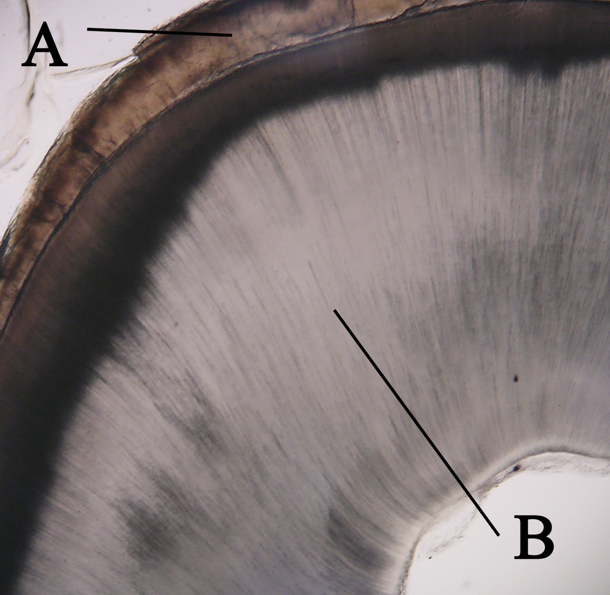
Cementum is a specialized calcified substance covering the root of a tooth. The cementum is the part of the periodontium that attaches the teeth to the alveolar bone by anchoring the periodontal ligament.Illustrated Dental Embryology, Histology, and Anatomy, Bath-Balogh and Fehrenbach, Elsevier, 2011, page 170. Structure The cells of cementum are the entrapped cementoblasts, the cementocytes. Each cementocyte lies in its lacuna, similar to the pattern noted in bone. These lacunae also have canaliculi or canals. Unlike those in bone, however, these canals in cementum do not contain nerves, nor do they radiate outward. Instead, the canals are oriented toward the periodontal ligament and contain cementocytic processes that exist to diffuse nutrients from the ligament because it is vascularized. After the apposition of cementum in layers, the cementoblasts that do not become entrapped in cementum line up along the cemental surface along the length of the outer covering of the perio ... [...More Info...] [...Related Items...] OR: [Wikipedia] [Google] [Baidu] |
Tooth Development
Tooth development or odontogenesis is the complex process by which teeth form from embryonic cells, grow, and erupt into the mouth. For human teeth to have a healthy oral environment, all parts of the tooth must develop during appropriate stages of fetal development. Primary (baby) teeth start to form between the sixth and eighth week of prenatal development, and permanent teeth begin to form in the twentieth week.Ten Cate's Oral Histology, Nanci, Elsevier, 2013, pages 70-94 If teeth do not start to develop at or near these times, they will not develop at all, resulting in hypodontia or anodontia. A significant amount of research has focused on determining the processes that initiate tooth development. It is widely accepted that there is a factor within the tissues of the first pharyngeal arch that is necessary for the development of teeth. Overview The tooth germ is an aggregation of cells that eventually forms a tooth.University of Texas Medical Branch. These cells are de ... [...More Info...] [...Related Items...] OR: [Wikipedia] [Google] [Baidu] |
Periodontal Ligament
The periodontal ligament, commonly abbreviated as the PDL, is a group of specialized connective tissue fibers that essentially attach a tooth to the alveolar bone within which it sits. It inserts into root cementum one side and onto alveolar bone on the other. Structure The PDL consists of principal fibres, loose connective tissue, blast and clast cells, oxytalan fibres and Cell Rest of Malassez. Alveolodental ligament The main principal fiber group is the alveolodental ligament, which consists of five fiber subgroups: alveolar crest, horizontal, oblique, apical, and interradicular on multirooted teeth. Principal fibers other than the alveolodental ligament are the transseptal fibers. All these fibers help the tooth withstand the naturally substantial compressive forces that occur during chewing and remain embedded in the bone. The ends of the principal fibers that are within either cementum or alveolar bone proper are considered Sharpey fibers. * Alveolar crest fibers ('' ... [...More Info...] [...Related Items...] OR: [Wikipedia] [Google] [Baidu] |
Cementicle
A cementicle is a small, spherical or ovoid calcified mass embedded within or attached to the cementum layer on the root surface of a tooth, or lying free within the periodontal ligament. They tend to occur in elderly individuals. There are 3 types: * Free cementicle – not attached to cementum * Attached (sessile) cementicle – attached to the cementum surface (also termed exocementosis) * Embedded (interstitial) cementicle – with advancing age the cementum thickens, and the cementicle may become incorporated into the cementum layer They may be visible on a radiograph (x-ray). They may appear singly or in groups, and are most commonly found at the tip of the root. Their size is variable, but generally they are small (about 0.2 mm – 0.3 mm in diameter). Cementicles are usually acellular, and may contain either fibrillar or afibrillar cementum, or a mixture of both. Cementicles are the result of dystrophic calcification, but the reason why this takes place is unclear. Ceme ... [...More Info...] [...Related Items...] OR: [Wikipedia] [Google] [Baidu] |
Dental Follicle
The dental follicle, also known as dental sac, is made up of mesenchymal cells and fibres surrounding the enamel organ and dental papilla of a developing tooth. It is a vascular fibrous sac containing the developing tooth and its odontogenic organ. The dental follicle (DF) differentiates into the periodontal ligament. In addition, it may be the precursor of other cells of the periodontium, including osteoblasts, cementoblasts and fibroblasts. They develop into the alveolar bone, the cementum with Sharpey's fibers and the periodontal ligament fibers respectively. Similar to dental papilla, the dental follicle provides nutrition to the enamel organ and dental papilla and also have an extremely rich blood supply. Role in tooth eruption The formative role of the dental follicle starts when the crown of the tooth is fully developed and just before tooth eruption into the oral cavity. Although tooth eruption mechanisms have yet to be understood entirely, generally it can be agreed t ... [...More Info...] [...Related Items...] OR: [Wikipedia] [Google] [Baidu] |
Cementoenamel Junction
The cementoenamel junction, frequently abbreviated as the CEJ, is a slightly visible anatomical border identified on a tooth. It is the location where the enamel, which covers the anatomical crown of a tooth, and the cementum, which covers the anatomical root of a tooth, meet. Informally it is known as the neck of the tooth. The border created by these two dental tissues has much significance as it is usually the location where the gingiva attaches to a healthy tooth by fibers called the gingival fibers The gingival fibers are the connective tissue fibers that inhabit the gingival tissue adjacent to teeth and help hold the tissue firmly against the teeth. They are primarily composed of type I collagen, although type III fibers are also involved .... Active recession of the gingiva reveals the cementoenamel junction in the mouth and is usually a sign of an unhealthy condition. There exists a normal variation in the relationship of the cementum and the enamel at the cementoe ... [...More Info...] [...Related Items...] OR: [Wikipedia] [Google] [Baidu] |
Root Resorption
Resorption of the root of the tooth, or root resorption, is the progressive loss of dentin and cementum by the action of odontoclasts. Root resorption is a normal physiological process that occurs in the exfoliation of the primary dentition. However, pathological root resorption occurs in the permanent or secondary dentition and sometimes in the primary dentition. Causes While resorption of bone is a normal physiological response to stimuli throughout the body, root resorption in permanent dentition and sometimes in the primary dentition is pathological. The root is protected internally (endodontium) by pre-dentin and externally on the root surface by cementum and the periodontal ligament. Chronic stimuli that damage these protective layers expose underlying dentin to the action of osteoclasts. Root resorption most commonly occurs due to inflammation caused by: pulp necrosis, trauma, periodontal treatment, orthodontic tooth movement and tooth whitening. Less common causes inc ... [...More Info...] [...Related Items...] OR: [Wikipedia] [Google] [Baidu] |
Tooth
A tooth ( : teeth) is a hard, calcified structure found in the jaws (or mouths) of many vertebrates and used to break down food. Some animals, particularly carnivores and omnivores, also use teeth to help with capturing or wounding prey, tearing food, for defensive purposes, to intimidate other animals often including their own, or to carry prey or their young. The roots of teeth are covered by gums. Teeth are not made of bone, but rather of multiple tissues of varying density and hardness that originate from the embryonic germ layer, the ectoderm. The general structure of teeth is similar across the vertebrates, although there is considerable variation in their form and position. The teeth of mammals have deep roots, and this pattern is also found in some fish, and in crocodilians. In most teleost fish, however, the teeth are attached to the outer surface of the bone, while in lizards they are attached to the inner surface of the jaw by one side. In cartilaginous fish, s ... [...More Info...] [...Related Items...] OR: [Wikipedia] [Google] [Baidu] |
Cementoblasts
A cementoblast is a biological cell that forms from the follicular cells around the root of a tooth, and whose biological function is cementogenesis, which is the formation of cementum (hard tissue that covers the tooth root). The mechanism of differentiation of the cementoblasts is controversial but circumstantial evidence suggests that an epithelium or epithelial component may cause dental sac cells to differentiate into cementoblasts, characterised by an increase in length. Other theories involve Hertwig epithelial root sheath (HERS) being involved. Structure Thus cementoblasts resemble bone-forming osteoblasts but differ functionally and histologically. The cells of cementum are the entrapped cementoblasts, the cementocytes. Each cementocyte lies in its lacuna (plural, lacunae), similar to the pattern noted in bone. These lacunae also have canaliculi or canals. Unlike those in bone, however, these canals in cementum do not contain nerves, nor do they radiate outward. Instead, ... [...More Info...] [...Related Items...] OR: [Wikipedia] [Google] [Baidu] |
Cementoblast
A cementoblast is a biological cell that forms from the follicular cells around the root of a tooth, and whose biological function is cementogenesis, which is the formation of cementum (hard tissue that covers the tooth root). The mechanism of differentiation of the cementoblasts is controversial but circumstantial evidence suggests that an epithelium or epithelial component may cause dental sac cells to differentiate into cementoblasts, characterised by an increase in length. Other theories involve Hertwig epithelial root sheath (HERS) being involved. Structure Thus cementoblasts resemble bone-forming osteoblasts but differ functionally and histologically. The cells of cementum are the entrapped cementoblasts, the cementocytes. Each cementocyte lies in its lacuna (plural, lacunae), similar to the pattern noted in bone. These lacunae also have canaliculi or canals. Unlike those in bone, however, these canals in cementum do not contain nerves, nor do they radiate outward. Instead, ... [...More Info...] [...Related Items...] OR: [Wikipedia] [Google] [Baidu] |
Sharpey's Fibers
Sharpey's fibres (bone fibres, or perforating fibres) are a matrix of connective tissue consisting of bundles of strong predominantly type I collagen fibres connecting periosteum to bone. They are part of the outer fibrous layer of periosteum, entering into the outer circumferential and interstitial lamellae of bone tissue. Sharpey's fibres are also used to attach muscle to the periosteum of bone by merging with the fibrous periosteum and underlying bone as well. A good example is the attachment of the rotator cuff muscles to the blade of the scapula. In the teeth, Sharpey's fibres are the terminal ends of principal fibres (of the periodontal ligament) that insert into the cementum and into the periosteum of the alveolar bone. A study on rats suggests that the three-dimensional structure of Sharpey's fibres intensifies the continuity between the periodontal ligament fibre and the alveolar bone (tooth socket), and acts as a buffer medium against stress. Sharpey's fibres in the pr ... [...More Info...] [...Related Items...] OR: [Wikipedia] [Google] [Baidu] |
Dentin
Dentin () (American English) or dentine ( or ) (British English) ( la, substantia eburnea) is a calcified tissue of the body and, along with enamel, cementum, and pulp, is one of the four major components of teeth. It is usually covered by enamel on the crown and cementum on the root and surrounds the entire pulp. By volume, 45% of dentin consists of the mineral hydroxyapatite, 33% is organic material, and 22% is water. Yellow in appearance, it greatly affects the color of a tooth due to the translucency of enamel. Dentin, which is less mineralized and less brittle than enamel, is necessary for the support of enamel. Dentin rates approximately 3 on the Mohs scale of mineral hardness. There are two main characteristics which distinguish dentin from enamel: firstly, dentin forms throughout life; secondly, dentin is sensitive and can become hypersensitive to changes in temperature due to the sensory function of odontoblasts, especially when enamel recedes and dentin channels becom ... [...More Info...] [...Related Items...] OR: [Wikipedia] [Google] [Baidu] |
Gingival Fibers
The gingival fibers are the connective tissue fibers that inhabit the gingival tissue adjacent to teeth and help hold the tissue firmly against the teeth. They are primarily composed of type I collagen, although type III fibers are also involved. These fibers, unlike the fibers of the periodontal ligament, in general, attach the tooth to the gingival tissue, rather than the tooth to the alveolar bone. Functions of the gingival fibers The gingival fibers accomplish the following tasks: *They hold the marginal gingiva against the tooth *They provide the marginal gingiva with enough rigidity to withstand the forces of mastication without distorting *They serve to stabilize the marginal gingiva by uniting it with both the tissue of the more rigid attached gingiva as well as the cementum layer of the tooth. Gingival fibers and periodontitis In theory, gingival fibers are the protectors against periodontitis, as once they are breached, they cannot be regenerated. When destroyed, the ... [...More Info...] [...Related Items...] OR: [Wikipedia] [Google] [Baidu] |


