|
Cementoblast
A cementoblast is a biological cell that forms from the follicular cells around the root of a tooth, and whose biological function is cementogenesis, which is the formation of cementum (hard tissue that covers the tooth root). The mechanism of differentiation of the cementoblasts is controversial but circumstantial evidence suggests that an epithelium or epithelial component may cause dental sac cells to differentiate into cementoblasts, characterised by an increase in length. Other theories involve Hertwig epithelial root sheath (HERS) being involved. Structure Thus cementoblasts resemble bone-forming osteoblasts but differ functionally and histologically. The cells of cementum are the entrapped cementoblasts, the cementocytes. Each cementocyte lies in its lacuna (plural, lacunae), similar to the pattern noted in bone. These lacunae also have canaliculi or canals. Unlike those in bone, however, these canals in cementum do not contain nerves, nor do they radiate outward. Instead, ... [...More Info...] [...Related Items...] OR: [Wikipedia] [Google] [Baidu] |
Cementoblastoma
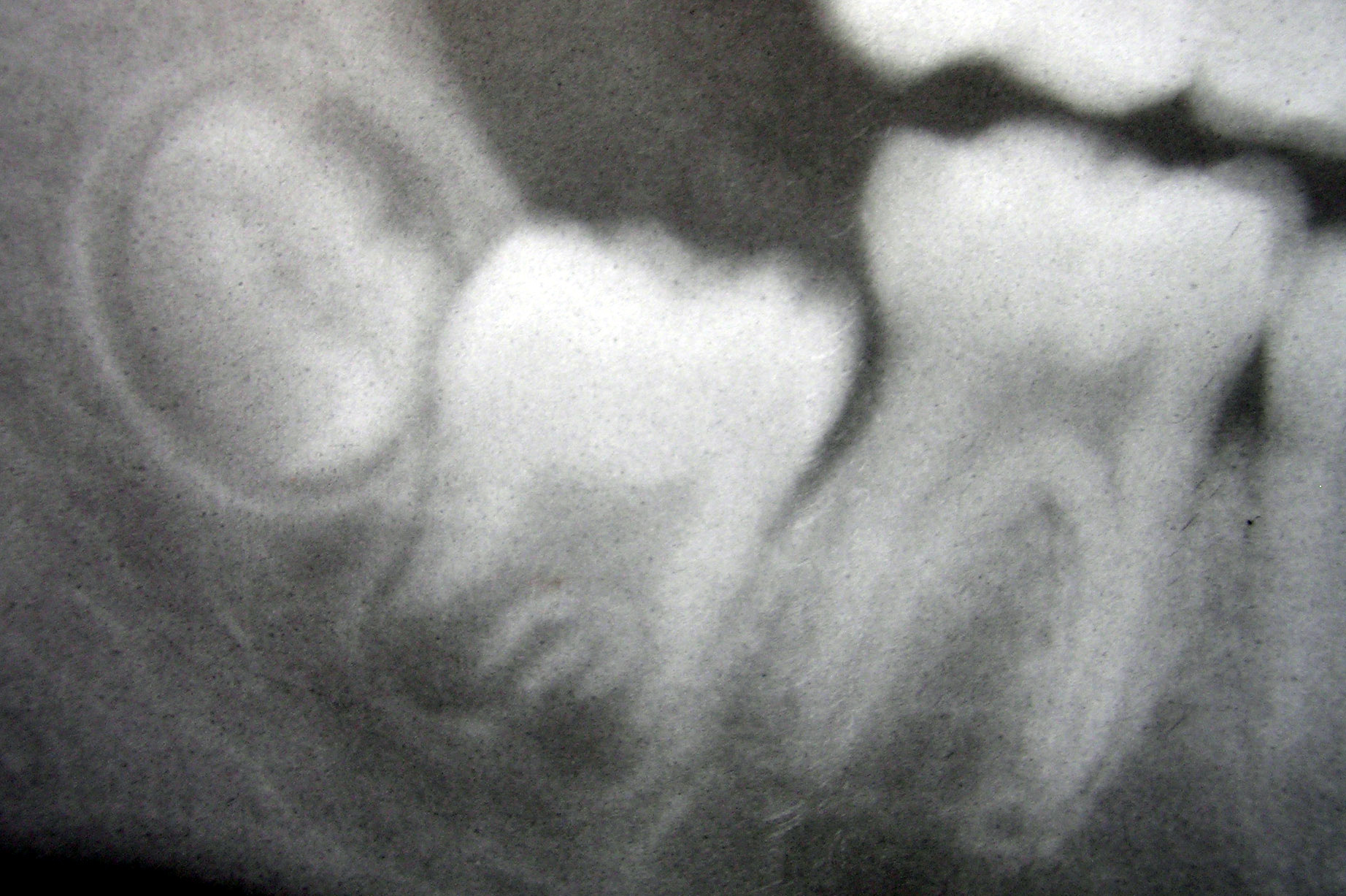
Cementoblastoma, or benign cementoblastoma, is a relatively rare benign neoplasm of the cementum of the teeth. It is derived from ectomesenchyme of odontogenic origin. Cementoblastomas represent less than 0.69–8% of all odontogemic tumors. Signs and symptoms Cementoblastoma usually occurs in people under the age of 25, particularly males. It usually involves the permanent mandibular molars or premolars. The involved tooth usually has a vital pulp. It is attached to the tooth root and may cause its resorption, may involve the pulp canal, grows slowly, tends to expand the overlying cortical plates, and, except for the enlargement produced, is usually asymptomatic. This involves the buccal and lingual aspects of the alveolar ridges. But may be associated with diffuse pain and tooth mobility, but the tooth is still vital. Since a cementoblastoma is a benign neoplasm, it grossly forms a mass of cementum-like tissue as an irregular or round mass attached to the roots of a tooth, us ... [...More Info...] [...Related Items...] OR: [Wikipedia] [Google] [Baidu] |
Cementum
Cementum is a specialized calcified substance covering the root of a tooth. The cementum is the part of the periodontium that attaches the teeth to the alveolar bone by anchoring the periodontal ligament.Illustrated Dental Embryology, Histology, and Anatomy, Bath-Balogh and Fehrenbach, Elsevier, 2011, page 170. Structure The cells of cementum are the entrapped cementoblasts, the cementocytes. Each cementocyte lies in its lacuna, similar to the pattern noted in bone. These lacunae also have canaliculi or canals. Unlike those in bone, however, these canals in cementum do not contain nerves, nor do they radiate outward. Instead, the canals are oriented toward the periodontal ligament and contain cementocytic processes that exist to diffuse nutrients from the ligament because it is vascularized. After the apposition of cementum in layers, the cementoblasts that do not become entrapped in cementum line up along the cemental surface along the length of the outer covering of the per ... [...More Info...] [...Related Items...] OR: [Wikipedia] [Google] [Baidu] |
Tooth Development
Tooth development or odontogenesis is the complex process by which teeth form from embryonic cells, grow, and erupt into the mouth. For human teeth to have a healthy oral environment, all parts of the tooth must develop during appropriate stages of fetal development. Primary (baby) teeth start to form between the sixth and eighth week of prenatal development, and permanent teeth begin to form in the twentieth week.Ten Cate's Oral Histology, Nanci, Elsevier, 2013, pages 70-94 If teeth do not start to develop at or near these times, they will not develop at all, resulting in hypodontia or anodontia. A significant amount of research has focused on determining the processes that initiate tooth development. It is widely accepted that there is a factor within the tissues of the first pharyngeal arch that is necessary for the development of teeth. Overview The tooth germ is an aggregation of cells that eventually forms a tooth.University of Texas Medical Branch. These cells ar ... [...More Info...] [...Related Items...] OR: [Wikipedia] [Google] [Baidu] |
Cementogenesis
Cementogenesis is the formation of cementum, one of the three mineralized substances of a tooth. Cementum covers the roots of teeth and serves to anchor gingival and periodontal fibers of the periodontal ligament by the fibers to the alveolar bone (some types of cementum may also form on the surface of the enamel of the crown at the cementoenamel junction (CEJ)). Process For cementogenesis to begin, Hertwig epithelial root sheath (HERS) must fragment. HERS is a collar of epithelial cells derived from the apical prolongation of the enamel organ. Once the root sheath disintegrates, the newly formed surface of root dentin comes into contact with the undifferentiated cells of the dental sac (dental follicle). This then stimulates the activation of cementoblasts to begin cementogenesis. The external shape of each root is fully determined by the position of the surrounding Hertwig epithelial root sheath. It is believed that either 1) HERS becomes interrupted; 2) infiltrating dental sac ... [...More Info...] [...Related Items...] OR: [Wikipedia] [Google] [Baidu] |
List Of Human Cell Types Derived From The Germ Layers
This is a list of cells in humans derived from the three embryonic germ layers – ectoderm, mesoderm, and endoderm. Cells derived from ectoderm Surface ectoderm Skin * Trichocyte * Keratinocyte Anterior pituitary * Gonadotrope * Corticotrope * Thyrotrope * Somatotrope * Lactotroph Tooth enamel * Ameloblast Neural crest Peripheral nervous system * Neuron * Glia ** Schwann cell ** Satellite glial cell Neuroendocrine system * Chromaffin cell * Glomus cell Skin * Melanocyte ** Nevus cell * Merkel cell Teeth * Odontoblast * Cementoblast Eyes * Corneal keratocyte Neural tube Central nervous system * Neuron * Glia ** Astrocyte ** Ependymocytes ** Muller glia (retina) ** Oligodendrocyte ** Oligodendrocyte progenitor cell ** Pituicyte (posterior pituitary) Pineal gland * Pinealocyte Cells derived from mesoderm Paraxial mesoderm Mesenchymal stem cell =Osteochondroprogenitor cell= * Bone (Osteoblast → Osteocyte) * Cartilage (Chondroblast → Chond ... [...More Info...] [...Related Items...] OR: [Wikipedia] [Google] [Baidu] |
Human Cells
There are many different types of cells in the human body. Cells derived primarily from endoderm Exocrine secretory epithelial cells *Brunner's gland cell in duodenum (enzymes and alkaline mucus) *Insulated goblet cell of respiratory and digestive tracts (mucus secretion) *Stomach ** Foveolar cell (mucus secretion) ** Chief cell ( pepsinogen secretion) ** Parietal cell (hydrochloric acid secretion) * Pancreatic acinar cell (bicarbonate and digestive enzyme secretion) * Paneth cell of small intestine ( lysozyme secretion) *Type II pneumocyte of lung ( surfactant secretion) * Club cell of lung Barrier cells *Type I pneumocyte (lung) * Gall bladder epithelial cell *Centroacinar cell (pancreas) * Intercalated duct cell (pancreas) *Intestinal brush border cell (with microvilli) Hormone-secreting cells * Enteroendocrine cell **K cell (secretes gastric inhibitory peptide) **L cell (secretes glucagon-like peptide-1, peptide YY3-36, oxyntomodulin, and glucagon-like pept ... [...More Info...] [...Related Items...] OR: [Wikipedia] [Google] [Baidu] |
Tooth
A tooth ( : teeth) is a hard, calcified structure found in the jaws (or mouths) of many vertebrates and used to break down food. Some animals, particularly carnivores and omnivores, also use teeth to help with capturing or wounding prey, tearing food, for defensive purposes, to intimidate other animals often including their own, or to carry prey or their young. The roots of teeth are covered by gums. Teeth are not made of bone, but rather of multiple tissues of varying density and hardness that originate from the embryonic germ layer, the ectoderm. The general structure of teeth is similar across the vertebrates, although there is considerable variation in their form and position. The teeth of mammals have deep roots, and this pattern is also found in some fish, and in crocodilians. In most teleost fish, however, the teeth are attached to the outer surface of the bone, while in lizards they are attached to the inner surface of the jaw by one side. In cartilaginous fi ... [...More Info...] [...Related Items...] OR: [Wikipedia] [Google] [Baidu] |
Cell (biology)
The cell is the basic structural and functional unit of life forms. Every cell consists of a cytoplasm enclosed within a membrane, and contains many biomolecules such as proteins, DNA and RNA, as well as many small molecules of nutrients and metabolites.Cell Movements and the Shaping of the Vertebrate Body in Chapter 21 of Molecular Biology of the Cell '' fourth edition, edited by Bruce Alberts (2002) published by Garland Science. The Alberts text discusses how the "cellular building blocks" move to shape developing embryos. It is also common to describe small molecules such as ... [...More Info...] [...Related Items...] OR: [Wikipedia] [Google] [Baidu] |
Follicle (anatomy)
A follicle is a small, spherical or vase-like group of cells enclosing a cavity in which some other structure grows or other material is contained. Thyroid follicles make up the thyroid gland. Follicles are best known as the sockets from which hairs grow in humans and other mammals, but the bristles of annelid The annelids (Annelida , from Latin ', "little ring"), also known as the segmented worms, are a large phylum, with over 22,000 extant species including ragworms, earthworms, and leeches. The species exist in and have adapted to various ecol ... worms also grow from such sockets. References {{reflist Skin anatomy Cells ... [...More Info...] [...Related Items...] OR: [Wikipedia] [Google] [Baidu] |
Epithelial Root Sheath
The Hertwig epithelial root sheath (HERS) or epithelial root sheath is a proliferation of epithelial cells located at the cervical loop of the enamel organ in a developing tooth. Hertwig epithelial root sheath initiates the formation of dentin in the root of a tooth by causing the differentiation of odontoblasts from the dental papilla. The root sheath eventually disintegrates with the periodontal ligament, but residual pieces that do not completely disappear are seen as epithelial cell rests of Malassez (ERM). These rests can become cystic, presenting future periodontal infections. Structure Hertwig epithelial root sheath is derived from the inner and outer enamel epithelium of the enamel organ.Illustrated Dental Embryology, Histology, and Anatomy, Bath-Balogh and Fehrenbach, Elsevier, 2011, p. 66 Function The sheath is also responsible for multiple or accessory roots (medial growth) and lateral or accessory canals in the root (break in epithelium).Ten Cate's Oral Histolog ... [...More Info...] [...Related Items...] OR: [Wikipedia] [Google] [Baidu] |

