|
Calf (leg)
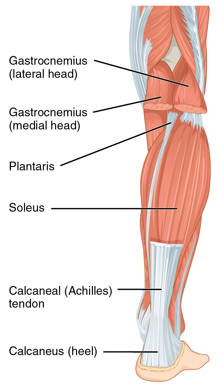
The calf ( : calves; Latin: ''sura'') is the back portion of the lower leg in human anatomy. The muscles within the calf correspond to the posterior compartment of the leg. The two largest muscles within this compartment are known together as the calf muscle and attach to the heel via the Achilles tendon. Several other, smaller muscles attach to the knee, the ankle, and via long tendons to the toes. Etymology From Middle English ''calf'', ''kalf'', from Old Norse ''kalfi'', possibly derived from the same Germanic root as English ''calf'' ("young cow"). Cognate with Icelandic ''kálfi'' ("calf of the leg"). ''Calf'' and ''calf of the leg'' are documented in use in Middle English circa AD 1350 and AD 1425 respectively. Historically, the ''absence of calf'', meaning a lower leg without a prominent calf muscle, was regarded by some authors as a sign of inferiority: ''it is well known that monkeys have no calves, and still less do they exist among the lower orders of mammals''. Str ... [...More Info...] [...Related Items...] OR: [Wikipedia] [Google] [Baidu] |
Anterior Compartment Of The Leg
The anterior compartment of the leg is a fascial compartment of the lower leg. It contains muscles that produce dorsiflexion and participate in inversion and eversion of the foot, as well as vascular and nervous elements, including the anterior tibial artery and veins and the deep fibular nerve. Muscles The muscles of the compartment are: * tibialis anterior * extensor hallucis longus * extensor digitorum longus * fibularis (peroneus) tertius Function The compartment contains muscles that are dorsiflexors and participate in inversion and eversion of the foot. Innervation and blood supply The anterior compartment of the leg is supplied by the deep fibular nerve (deep peroneal nerve), a branch of the common fibular nerve. The nerve contains axons from the L4, L5, and S1 spinal nerves. Blood for the compartment is supplied by the anterior tibial artery, which runs between the tibialis anterior and extensor digitorum longus muscles. When the artery crosses the extensor retin ... [...More Info...] [...Related Items...] OR: [Wikipedia] [Google] [Baidu] |
Posterior Compartment Of The Leg
The posterior compartment of the leg is one of the fascial compartments of the leg and is divided further into deep and superficial compartments. Structure Muscles Superficial posterior compartment Deep posterior compartment Blood supply Posterior tibial artery Innervation The posterior compartment of the leg is supplied by the tibial nerve The tibial nerve is a branch of the sciatic nerve. The tibial nerve passes through the popliteal fossa to pass below the arch of soleus. Structure Popliteal fossa The tibial nerve is the larger terminal branch of the sciatic nerve with root val .... Function * It contains the plantar flexors: Additional images File:From below - Superficial posterior compartment of leg - animation.gif, Superficial posterior compartment. Animation. File:From below - Deep posterior compartment of leg - animation.gif, Deep posterior compartment. Animation. References External links Diagram at patientcareonline.com {{DEFAULTSORT ... [...More Info...] [...Related Items...] OR: [Wikipedia] [Google] [Baidu] |
Leg Cramp
A cramp is a sudden, involuntary, painful skeletal muscle contraction or overshortening associated with electrical activity; while generally temporary and non-damaging, they can cause significant pain and a paralysis-like immobility of the affected muscle. A cramp usually goes away on its own over a period of several seconds, or minutes. Cramps are common and tend to occur at rest, usually at night (nocturnal leg cramps). They are also often associated with pregnancy, physical exercise or overexertion, age (common in older adults), in such cases, cramps are called idiopathic, because there is no underlying pathology. In addition to those benign conditions cramps are also associated with many pathologic conditions. Skeletal muscle cramps may be caused by muscle fatigue or a lack of electrolytes such as sodium (a condition called hyponatremia), potassium (called hypokalemia), or magnesium (called hypomagnesemia). Some skeletal muscle cramps do not have a known cause. Motor ne ... [...More Info...] [...Related Items...] OR: [Wikipedia] [Google] [Baidu] |
Idiopathic
An idiopathic disease is any disease with an unknown cause or mechanism of apparent wikt:spontaneous, spontaneous origin. From Ancient Greek, Greek ἴδιος ''idios'' "one's own" and πάθος ''pathos'' "suffering", ''idiopathy'' means approximately "a disease of its own kind". For some medical conditions, one or more causes are somewhat understood, but in a certain percentage of people with the condition, the cause may not be readily apparent or characterized. In these cases, the origin of the condition is said to be idiopathic. With some other medical conditions, the root cause for a large percentage of all cases have not been established—for example, focal segmental glomerulosclerosis or ankylosing spondylitis; the majority of these cases are deemed idiopathic. Medical advances and this term Advances in medicine, medical science improve the understanding of causes of diseases and the classification of diseases; thus, regarding any particular condition or disease, as more ... [...More Info...] [...Related Items...] OR: [Wikipedia] [Google] [Baidu] |
Varicose Vein
Varicose veins, also known as varicoses, are a medical condition in which superficial veins become enlarged and twisted. These veins typically develop in the legs, just under the skin. Varicose veins usually cause few symptoms. However, some individuals may experience fatigue or pain in the area. Complications can include bleeding or superficial thrombophlebitis. Varices in the scrotum are known as a varicocele, while those around the anus are known as hemorrhoids. Due to the various physical, social, and psychological effects of varicose veins, they can negatively affect one's quality of life. Varicose veins have no specific cause. Risk factors include obesity, lack of exercise, leg trauma, and family history of the condition. They also develop more commonly during pregnancy. Occasionally they result from chronic venous insufficiency. Underlying causes include weak or damaged valves in the veins. They are typically diagnosed by examination, including observation by ultrasound. ... [...More Info...] [...Related Items...] OR: [Wikipedia] [Google] [Baidu] |
Achilles Tendon Rupture
Achilles tendon rupture is when the Achilles tendon, at the back of the ankle, breaks. Symptoms include the sudden onset of sharp pain in the heel. A snapping sound may be heard as the tendon breaks and walking becomes difficult. Rupture typically occurs as a result of a sudden bending up of the foot when the calf muscle is engaged, direct trauma, or long-standing tendonitis. Other risk factors include the use of fluoroquinolones, a significant change in exercise, rheumatoid arthritis, gout, or corticosteroid use. Diagnosis is typically based on symptoms and examination and supported by medical imaging. Prevention may include stretching before activity and gradual progression of exercise intensity. Treatment may consist of surgical repair or conservative management. Quick return to weight bearing (within 4 weeks) appears okay and is often recommended. While surgery traditionally results in a small decrease in the risk of re-rupture, the risk of other complications is greater. ... [...More Info...] [...Related Items...] OR: [Wikipedia] [Google] [Baidu] |
Compartment Syndrome
Compartment syndrome is a condition in which increased pressure within one of the body's anatomical compartments results in insufficient blood supply to tissue within that space. There are two main types: acute and chronic. Compartments of the leg or arm are most commonly involved. Symptoms of acute compartment syndrome (ACS) can include severe pain, poor pulses, decreased ability to move, numbness, or a pale color of the affected limb. It is most commonly due to physical trauma such as a bone fracture (up to 75% of cases) or crush injury, but it can also be caused by acute exertion during sport. It can also occur after blood flow returns following a period of poor blood flow. Diagnosis is generally based upon a person's symptoms and may be supported by measurement of intracompartmental pressure before, during, and after activity. Normal compartment pressure should be within 12-18 mmHg; anything greater than that is considered abnormal and would need treatment. Treatment i ... [...More Info...] [...Related Items...] OR: [Wikipedia] [Google] [Baidu] |
Deep Vein Thrombosis
Deep vein thrombosis (DVT) is a type of venous thrombosis involving the formation of a blood clot in a deep vein, most commonly in the legs or pelvis. A minority of DVTs occur in the arms. Symptoms can include pain, swelling, redness, and enlarged veins in the affected area, but some DVTs have no symptoms. The most common life-threatening concern with DVT is the potential for a clot to embolize (detach from the veins), travel as an embolus through the right side of the heart, and become lodged in a pulmonary artery that supplies blood to the lungs. This is called a pulmonary embolism (PE). DVT and PE comprise the cardiovascular disease of venous thromboembolism (VTE). About two-thirds of VTE manifests as DVT only, with one-third manifesting as PE with or without DVT. The most frequent long-term DVT complication is post-thrombotic syndrome, which can cause pain, swelling, a sensation of heaviness, itching, and in severe cases, ulcers. Recurrent VTE occurs in about 30% of those i ... [...More Info...] [...Related Items...] OR: [Wikipedia] [Google] [Baidu] |
Symptom
Signs and symptoms are the observed or detectable signs, and experienced symptoms of an illness, injury, or condition. A sign for example may be a higher or lower temperature than normal, raised or lowered blood pressure or an abnormality showing on a medical scan. A symptom is something out of the ordinary that is experienced by an individual such as feeling feverish, a headache or other pain or pains in the body. Signs and symptoms Signs A medical sign is an objective observable indication of a disease, injury, or abnormal physiological state that may be detected during a physical examination, examining the patient history, or diagnostic procedure. These signs are visible or otherwise detectable such as a rash or bruise. Medical signs, along with symptoms, assist in formulating diagnostic hypothesis. Examples of signs include elevated blood pressure, nail clubbing of the fingernails or toenails, staggering gait, and arcus senilis and arcus juvenilis of the eyes. Indicati ... [...More Info...] [...Related Items...] OR: [Wikipedia] [Google] [Baidu] |
Medical Condition
A disease is a particular abnormal condition that negatively affects the structure or function (biology), function of all or part of an organism, and that is not immediately due to any external injury. Diseases are often known to be medical conditions that are associated with specific signs and symptoms. A disease may be caused by external factors such as pathogens or by internal dysfunctions. For example, internal dysfunctions of the immune system can produce a variety of different diseases, including various forms of immunodeficiency, hypersensitivity, allergy, allergies and autoimmune disorders. In humans, ''disease'' is often used more broadly to refer to any condition that causes pain, Abnormality (behavior), dysfunction, distress (medicine), distress, social problems, or death to the person affected, or similar problems for those in contact with the person. In this broader sense, it sometimes includes injury, injuries, disability, disabilities, #Disorder, disorders, s ... [...More Info...] [...Related Items...] OR: [Wikipedia] [Google] [Baidu] |
Sural Nerve
The sural nerve ''(L4-S1)'' is generally considered a pure cutaneous nerve of the posterolateral leg to the lateral ankle. The sural nerve originates from a combination of either the sural communicating branch and medial sural cutaneous nerve, or the lateral sural cutaneous nerve. This group of nerves is termed the sural nerve complex. There are eight documented variations of the sural nerve complex. Once formed the sural nerve takes its course midline posterior to posterolateral around the lateral malleolus. The sural nerve terminates as the lateral dorsal cutaneous nerve. Anatomy The sural nerve ''(L4-S1)'' is a cutaneous sensory nerve of the posterolateral calf with cutaneous innervation to the distal one-third of the lower leg. Formation of the ''sural nerve'' is the result of either anastomosis of the medial sural cutaneous nerve and the sural communicating nerve, or it may be found as a continuation of the lateral sural cutaneous nerve traveling parallel to the medial ... [...More Info...] [...Related Items...] OR: [Wikipedia] [Google] [Baidu] |
Tibialis Posterior
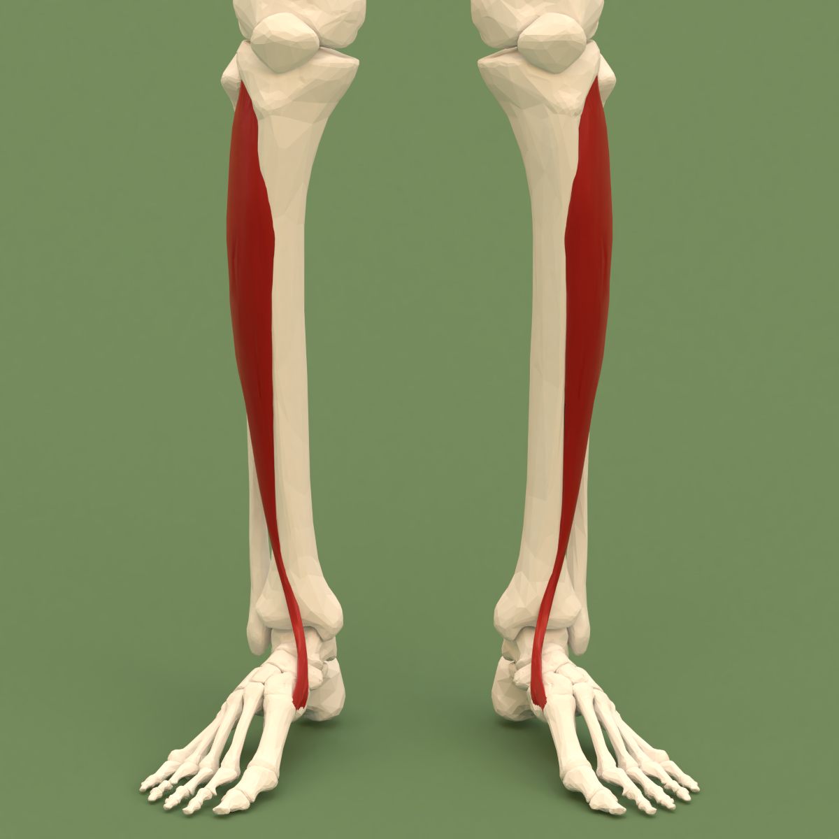
The tibialis posterior muscle is the most central of all the leg muscles, and is located in the deep posterior compartment of the leg. It is the key stabilizing muscle of the lower leg. Structure The tibialis posterior muscle originates on the inner posterior border of the fibula laterally. It is also attached to the interosseous membrane medially, which attaches to the tibia and fibula. The tendon of the tibialis posterior muscle (sometimes called the posterior tibial tendon) descends posterior to the medial malleolus. It terminates by dividing into plantar, main, and recurrent components. The main portion inserts into the tuberosity of the navicular bone. The smaller portion inserts into the plantar surface of the medial cuneiform. The plantar portion inserts into the bases of the second, third and fourth metatarsals, the intermediate and lateral cuneiforms and the cuboid. The recurrent portion inserts into the sustentaculum tali of the calcaneus. Blood is supplied to the m ... [...More Info...] [...Related Items...] OR: [Wikipedia] [Google] [Baidu] |


