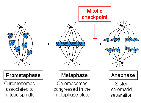
|
Cleavage Furrow
In cell biology, the cleavage furrow is the indentation of the cell's surface that begins the progression of cleavage, by which animal and some algal cells undergo cytokinesis, the final splitting of the membrane, in the process of cell division. The same proteins responsible for muscle contraction, actin and myosin, begin the process of forming the cleavage furrow, creating an actomyosin ring. Other cytoskeletal proteins and actin binding proteins are involved in the procedure. Mechanism Plant cells do not perform cytokinesis through this exact method but the two procedures are not totally different. Animal cells form an actin-myosin contractile ring within the equatorial region of the cell membrane that constricts to form the cleavage furrow. In plant cells, Golgi vesicle secretions form a cell plate or septum on the equatorial plane of the cell wall by the action of microtubules of the phragmoplast. The cleavage furrow in animal cells and the phragmoplast in plant cell ... [...More Info...] [...Related Items...] OR: [Wikipedia] [Google] [Baidu] |
Cell Cycle
The cell cycle, or cell-division cycle, is the sequential series of events that take place in a cell (biology), cell that causes it to divide into two daughter cells. These events include the growth of the cell, duplication of its DNA (DNA replication) and some of its organelles, and subsequently the partitioning of its cytoplasm, chromosomes and other components into two daughter cells in a process called cell division. In eukaryotic cells (having a cell nucleus) including animal, plant, fungal, and protist cells, the cell cycle is divided into two main stages: interphase, and the M phase that includes mitosis and cytokinesis. During interphase, the cell grows, accumulating nutrients needed for mitosis, and replicates its DNA and some of its organelles. During the M phase, the replicated Chromosome, chromosomes, organelles, and cytoplasm separate into two new daughter cells. To ensure the proper replication of cellular components and division, there are control mechanisms kno ... [...More Info...] [...Related Items...] OR: [Wikipedia] [Google] [Baidu] |
Exocytosis
Exocytosis is a term for the active transport process that transports large molecules from cell to the extracellular area. Hormones, proteins and neurotransmitters are examples of large molecules that can be transported out of the cell. Exocytosis is a crucial transport mechanism that enables polar molecules to flow through the cell membranes’ hydrophobic lipid bilayer. The transport process is essential to hormone secretion, immune response and neurotransmission. Both prokaryotes and eukaryotes undergo exocytosis. Prokaryotes secrete molecules and cellular waste through translocons that are localized to the cell membrane. In addition, they secrete molecules to other cells through specialized organs. Eukaryotes rely on multiple cellular processes to perform the exocytosis process. Eukaryotes have several organelles and a nucleus in the cytoplasm that are connected through multiple transport routes, that is formally known as the secretory pathway. This is a complex pathway with ... [...More Info...] [...Related Items...] OR: [Wikipedia] [Google] [Baidu] |
Phosphatidylethanolamine
Phosphatidylethanolamine (PE) is a class of phospholipids found in biological membranes. They are synthesized by the addition of cytidine diphosphate-ethanolamine to diglycerides, releasing cytidine monophosphate. S-Adenosyl methionine, ''S''-Adenosyl methionine can subsequently Methylation, methylate the amine of phosphatidylethanolamines to yield phosphatidylcholines. Function In cells Phosphatidylethanolamines are found in all living cells, composing 25% of all phospholipids. In human physiology, they are found particularly in nervous tissue such as the white matter of brain, nerves, neural tissue, and in spinal cord, where they make up 45% of all phospholipids. Phosphatidylethanolamines play a role in membrane fusion and in disassembly of the contractile ring during cytokinesis in cell division. Additionally, it is thought that phosphatidylethanolamine regulates membrane curvature. Phosphatidylethanolamine is an important precursor, substrate (biochemistry), substrate, or d ... [...More Info...] [...Related Items...] OR: [Wikipedia] [Google] [Baidu] |
Cytoplasm
The cytoplasm describes all the material within a eukaryotic or prokaryotic cell, enclosed by the cell membrane, including the organelles and excluding the nucleus in eukaryotic cells. The material inside the nucleus of a eukaryotic cell and contained within the nuclear membrane is termed the nucleoplasm. The main components of the cytoplasm are the cytosol (a gel-like substance), the cell's internal sub-structures, and various cytoplasmic inclusions. In eukaryotes the cytoplasm also includes the nucleus, and other membrane-bound organelles.The cytoplasm is about 80% water and is usually colorless. The submicroscopic ground cell substance, or cytoplasmic matrix, that remains after the exclusion of the cell organelles and particles is groundplasm. It is the hyaloplasm of light microscopy, a highly complex, polyphasic system in which all resolvable cytoplasmic elements are suspended, including the larger organelles such as the ribosomes, mitochondria, plant plasti ... [...More Info...] [...Related Items...] OR: [Wikipedia] [Google] [Baidu] |
Motor Proteins
Motor proteins are a class of molecular motors that can move along the cytoskeleton of cells. They do this by converting chemical energy into mechanical work by the hydrolysis of ATP. Cellular functions Motor proteins are the driving force behind most active transport of proteins and vesicles in the cytoplasm. Kinesins and cytoplasmic dyneins play essential roles in intracellular transport such as axonal transport and in the formation of the spindle apparatus and the separation of the chromosomes during mitosis and meiosis. Axonemal dynein, found in cilia and flagella, is crucial to cell motility, for example in spermatozoa, and fluid transport, for example in trachea. The muscle protein myosin "motors" the contraction of muscle fibers in animals. Diseases associated with motor protein defects The importance of motor proteins in cells becomes evident when they fail to fulfill their function. For example, kinesin deficiencies have been identified as the cause for Ch ... [...More Info...] [...Related Items...] OR: [Wikipedia] [Google] [Baidu] |
Chromosomes
A chromosome is a package of DNA containing part or all of the genetic material of an organism. In most chromosomes, the very long thin DNA fibers are coated with nucleosome-forming packaging proteins; in eukaryotic cells, the most important of these proteins are the histones. Aided by chaperone proteins, the histones bind to and condense the DNA molecule to maintain its integrity. These eukaryotic chromosomes display a complex three-dimensional structure that has a significant role in transcriptional regulation. Normally, chromosomes are visible under a light microscope only during the metaphase of cell division, where all chromosomes are aligned in the center of the cell in their condensed form. Before this stage occurs, each chromosome is duplicated ( S phase), and the two copies are joined by a centromere—resulting in either an X-shaped structure if the centromere is located equatorially, or a two-armed structure if the centromere is located distally; the joined ... [...More Info...] [...Related Items...] OR: [Wikipedia] [Google] [Baidu] |
Spindle Fibers
In cell biology, the spindle apparatus is the cytoskeletal structure of eukaryotic cells that forms during cell division to separate sister chromatids between daughter cells. It is referred to as the mitotic spindle during mitosis, a process that produces genetically identical daughter cells, or the meiotic spindle during meiosis, a process that produces gametes with half the number of chromosomes of the parent cell. Besides chromosomes, the spindle apparatus is composed of hundreds of proteins. Microtubules comprise the most abundant components of the machinery. Spindle structure Attachment of microtubules to chromosomes is mediated by kinetochores, which actively monitor spindle formation and prevent premature anaphase onset. Microtubule polymerization and depolymerization dynamic drive chromosome congression. Depolymerization of microtubules generates tension at kinetochores; bipolar attachment of sister kinetochores to microtubules emanating from opposite cell poles coupl ... [...More Info...] [...Related Items...] OR: [Wikipedia] [Google] [Baidu] |
Telophase
Telophase () is the final stage in both meiosis and mitosis in a eukaryotic cell. During telophase, the effects of prophase and prometaphase (the nucleolus and nuclear membrane disintegrating) are reversed. As chromosomes reach the cell poles, a nuclear envelope is re-assembled around each set of chromatids, the nucleoli reappear, and chromosomes begin to decondense back into the expanded chromatin that is present during interphase. The mitotic spindle is disassembled and remaining spindle microtubules are depolymerized. Telophase accounts for approximately 2% of the cell cycle's duration. Cytokinesis typically begins before late telophase and, when complete, segregates the two daughter nuclei between a pair of separate daughter cells. Telophase is primarily driven by the dephosphorylation of mitotic cyclin-dependent kinase (Cdk) substrates. Dephosphorylation of Cdk substrates The phosphorylation of the protein targets of M-Cdks (Mitotic Cyclin-dependent Kinases) drives sp ... [...More Info...] [...Related Items...] OR: [Wikipedia] [Google] [Baidu] |
Anaphase
Anaphase () is the stage of mitosis after the process of metaphase, when replicated chromosomes are split and the newly-copied chromosomes (daughter chromatids) are moved to opposite poles of the cell. Chromosomes also reach their overall maximum condensation in late anaphase, to help chromosome segregation and the re-formation of the nucleus. Anaphase starts when the anaphase promoting complex marks an Chaperone (protein), inhibitory chaperone called securin for destruction by Ubiquitin, ubiquitylating it. Securin is a protein which inhibits a protease known as separase. The destruction of securin unleashes separase which then breaks down cohesin, a protein responsible for holding sister chromatids together. At this point, three subclasses of microtubule unique to mitosis are involved in creating the forces necessary to separate the chromatids: kinetochore microtubules, interpolar microtubules, and Aster_(cell_biology)#Astral_microtubules, astral microtubules. The centromere ... [...More Info...] [...Related Items...] OR: [Wikipedia] [Google] [Baidu] |
Metaphase
Metaphase ( and ) is a stage of mitosis in the eukaryotic cell cycle in which chromosomes are at their second-most condensed and coiled stage (they are at their most condensed in anaphase). These chromosomes, carrying genetic information, align in the equator of the cell between the spindle poles at the metaphase plate, before being separated into each of the two daughter nuclei. This alignment marks the beginning of metaphase. Metaphase accounts for approximately 4% of the cell cycle's duration. In metaphase, microtubules from both duplicated centrosomes on opposite poles of the cell have completed attachment to kinetochores on condensed chromosomes. The centromeres of the chromosomes convene themselves on the metaphase plate, an imaginary line that is equidistant from the two spindle poles. This even alignment is due to the counterbalance of the pulling powers generated by the opposing kinetochore microtubules, analogous to a tug-of-war between two people of equal strength, ... [...More Info...] [...Related Items...] OR: [Wikipedia] [Google] [Baidu] |
Prophase
Prophase () is the first stage of cell division in both mitosis and meiosis. Beginning after interphase, DNA has already been replicated when the cell enters prophase. The main occurrences in prophase are the condensation of the chromatin reticulum and the disappearance of the nucleolus. Staining and microscopy Microscopy can be used to visualize condensed chromosomes as they move through meiosis and mitosis. Various DNA stains are used to treat cells such that condensing chromosomes can be visualized as the move through prophase. The giemsa G-banding technique is commonly used to identify mammalian chromosomes, but utilizing the technology on plant cells was originally difficult due to the high degree of chromosome compaction in plant cells. G-banding was fully realized for plant chromosomes in 1990. During both meiotic and mitotic prophase, giemsa staining can be applied to cells to elicit G-banding in chromosomes. Silver staining, a more modern technology, in conj ... [...More Info...] [...Related Items...] OR: [Wikipedia] [Google] [Baidu] |


