|
Central Canal
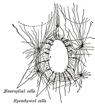
The central canal (also known as spinal foramen or ependymal canal) is the cerebrospinal fluid-filled space that runs through the spinal cord. The central canal lies below and is connected to the ventricular system of the brain, from which it receives cerebrospinal fluid, and shares the same ependymal lining. The central canal helps to transport nutrients to the spinal cord as well as protect it by cushioning the impact of a force when the spine is affected. The central canal represents the adult remainder of the central cavity of the neural tube. It generally occludes (closes off) with age. Structure The central canal below at the ventricular system of the brain, beginning at a region called the obex where the fourth ventricle, a cavity present in the brainstem, narrows. The central canal is located in the third of the spinal cord in the cervical vertebrae, cervical and thoracic spine, thoracic regions. In the lumbar spine it enlarges and is located more centrally. At the c ... [...More Info...] [...Related Items...] OR: [Wikipedia] [Google] [Baidu] |
Spinal Cord
The spinal cord is a long, thin, tubular structure made up of nervous tissue that extends from the medulla oblongata in the lower brainstem to the lumbar region of the vertebral column (backbone) of vertebrate animals. The center of the spinal cord is hollow and contains a structure called the central canal, which contains cerebrospinal fluid. The spinal cord is also covered by meninges and enclosed by the neural arches. Together, the brain and spinal cord make up the central nervous system. In humans, the spinal cord is a continuation of the brainstem and anatomically begins at the occipital bone, passing out of the foramen magnum and then enters the spinal canal at the beginning of the cervical vertebrae. The spinal cord extends down to between the first and second lumbar vertebrae, where it tapers to become the cauda equina. The enclosing bony vertebral column protects the relatively shorter spinal cord. It is around long in adult men and around long in adult women. The diam ... [...More Info...] [...Related Items...] OR: [Wikipedia] [Google] [Baidu] |
Columnar Epithelium
Epithelium or epithelial tissue is a thin, continuous, protective layer of cells with little extracellular matrix. An example is the epidermis, the outermost layer of the skin. Epithelial ( mesothelial) tissues line the outer surfaces of many internal organs, the corresponding inner surfaces of body cavities, and the inner surfaces of blood vessels. Epithelial tissue is one of the four basic types of animal tissue, along with connective tissue, muscle tissue and nervous tissue. These tissues also lack blood or lymph supply. The tissue is supplied by nerves. There are three principal shapes of epithelial cell: squamous (scaly), columnar, and cuboidal. These can be arranged in a singular layer of cells as simple epithelium, either simple squamous, simple columnar, or simple cuboidal, or in layers of two or more cells deep as stratified (layered), or ''compound'', either squamous, columnar or cuboidal. In some tissues, a layer of columnar cells may appear to be stratified due to ... [...More Info...] [...Related Items...] OR: [Wikipedia] [Google] [Baidu] |
Tethered Cord
Tethered cord syndrome (TCS) refers to a group of neurological disorders that relate to malformations of the spinal cord.Tethered Spinal Cord , Columbia University Department of Neurosurgery. Various forms include tight , lipomeningomyelocele, split cord malformations ( diastematomyelia), occult, dermal sinus tracts, and [...More Info...] [...Related Items...] OR: [Wikipedia] [Google] [Baidu] |
Syringohydromyelia
Syringomyelia is a generic term referring to a disorder in which a cyst or cavity forms within the spinal cord. Often, syringomyelia is used as a generic term before an etiology is determined. This cyst, called a Syrinx (medicine), syrinx, can expand and elongate over time, destroying the spinal cord. The damage may result in loss of feeling, paralysis, weakness, and stiffness in the back, shoulders, and extremities. Syringomyelia may also cause a loss of the ability to feel extremes of hot or cold, especially in the hands. It may also lead to a cape-like bilateral loss of pain and temperature sensation along the upper chest and arms. The combination of symptoms varies from one patient to another depending on the location of the syrinx within the spinal cord, as well as its extent. Syringomyelia has a prevalence estimated at 8.4 cases per 100,000 people, with symptoms usually beginning in young adulthood. Signs of the disorder tend to develop slowly, although sudden onset may oc ... [...More Info...] [...Related Items...] OR: [Wikipedia] [Google] [Baidu] |
Myelomeningocele
Spina bifida (SB; ; Latin for 'split spine') is a birth defect in which there is incomplete closing of the spine and the membranes around the spinal cord during early development in pregnancy. There are three main types: spina bifida occulta, meningocele and myelomeningocele. Meningocele and myelomeningocele may be grouped as spina bifida cystica. The most common location is the lower back, but in rare cases it may be in the middle back or neck. Occulta has no or only mild signs, which may include a hairy patch, dimple, dark spot or swelling on the back at the site of the gap in the spine. Meningocele typically causes mild problems, with a sac of fluid present at the gap in the spine. Myelomeningocele, also known as open spina bifida, is the most severe form. Problems associated with this form include poor ability to walk, impaired bladder or bowel control, accumulation of fluid in the brain, a tethered spinal cord and latex allergy. Some experts believe such an allergy ca ... [...More Info...] [...Related Items...] OR: [Wikipedia] [Google] [Baidu] |
Spinal Tumor
Spinal tumors are neoplasms located in either the vertebral column or the spinal cord. There are three main types of spinal tumors classified based on their location: extradural and intradural (intradural-intramedullary and intradural-extramedullary). Extradural tumors are located outside the dura mater lining and are most commonly metastatic. Intradural tumors are located inside the dura mater lining and are further subdivided into intramedullary and extramedullary tumors. Intradural-intramedullary tumors are located within the dura and spinal cord parenchyma, while intradural-extramedullary tumors are located within the dura but outside the spinal cord parenchyma. The most common presenting symptom of spinal tumors is nocturnal back pain. Other common symptoms include muscle weakness, sensory loss, and difficulty walking. Loss of bowel and bladder control may occur during the later stages of the disease. The cause of spinal tumors is unknown. Most extradural tumors are metastati ... [...More Info...] [...Related Items...] OR: [Wikipedia] [Google] [Baidu] |
Arnold-Chiari Syndrome
In neurology, the Chiari malformation ( ; CM) is a structural defect in the cerebellum, characterized by a downward displacement of one or both cerebellar tonsils through the foramen magnum (the opening at the base of the skull). CMs can cause headaches, difficulty swallowing, vomiting, dizziness, neck pain, unsteady gait, poor hand coordination, numbness and tingling of the hands and feet, and speech problems. Less often, people may experience ringing or buzzing in the ears, weakness, slow heart rhythm, fast heart rhythm, curvature of the spine (scoliosis) related to spinal cord impairment, abnormal breathing such as in central sleep apnea, and, in severe cases, paralysis. CM can sometimes lead to non-communicating hydrocephalus as a result of obstruction of cerebrospinal fluid (CSF) outflow. The CSF outflow is caused by phase difference in outflow and influx of blood in the vasculature of the brain. The malformation is named after the Austrian pathologist Hans Chiari. ... [...More Info...] [...Related Items...] OR: [Wikipedia] [Google] [Baidu] |
Spina Bifida
Spina bifida (SB; ; Latin for 'split spine') is a birth defect in which there is incomplete closing of the vertebral column, spine and the meninges, membranes around the spinal cord during embryonic development, early development in pregnancy. There are three main types: spina bifida occulta, meningocele and myelomeningocele. Meningocele and myelomeningocele may be grouped as spina bifida cystica. The most common location is the Lumbar vertebrae, lower back, but in rare cases it may be in the Thoracic vertebrae, middle back or Cervical vertebrae, neck. Occulta has no or only mild signs, which may include a hairy patch, dimple, dark spot or swelling on the back at the site of the gap in the spine. Meningocele typically causes mild problems, with a sac of fluid present at the gap in the spine. Myelomeningocele, also known as open spina bifida, is the most severe form. Problems associated with this form include poor ability to walk, impaired Neurogenic bladder dysfunction, bladder o ... [...More Info...] [...Related Items...] OR: [Wikipedia] [Google] [Baidu] |
Anterior White Commissure
The anterior white commissure (ventral white commissure) is a bundle of nerve fibers which cross the midline of the spinal cord just anterior (in front of) to the gray commissure ( Rexed lamina X). A delta fibers (Aδ fibers) and C fibers carrying pain sensation in the spinothalamic tract contribute to this commissure, as do fibers of the anterior corticospinal tract, which carry motor signals from the primary motor cortex. Two of the five sensory modalities, pain and temperature, cross sides at the anterior white commissure, reaching the contralateral side about two vertebral levels rostral to their origin. The spinothalamic tract thus decussates very soon after entering the spinal cord, ascending in the spinal cord, contralateral to the side from where it provides (pain and temperature) sensory information. Therefore, a lesion that is caudal to the sensory decussation where the remaining three-fifths modalities decussate (at the superior medulla) will result in contralat ... [...More Info...] [...Related Items...] OR: [Wikipedia] [Google] [Baidu] |
White Matter
White matter refers to areas of the central nervous system that are mainly made up of myelinated axons, also called Nerve tract, tracts. Long thought to be passive tissue, white matter affects learning and brain functions, modulating the distribution of action potentials, acting as a relay and coordinating communication between different brain regions. White matter is named for its relatively light appearance resulting from the lipid content of myelin. Its white color in prepared specimens is due to its usual preservation in formaldehyde. It appears pinkish-white to the naked eye otherwise, because myelin is composed largely of lipid tissue veined with capillaries. Structure White matter White matter is composed of bundles, which connect various grey matter areas (the locations of nerve cell bodies) of the brain to each other, and carry nerve impulses between neurons. Myelin acts as an insulator, which allows saltatory conduction, electrical signals to jump, rather than cour ... [...More Info...] [...Related Items...] OR: [Wikipedia] [Google] [Baidu] |
Syringomyelia
Syringomyelia is a generic term referring to a disorder in which a cyst or cavity forms within the spinal cord. Often, syringomyelia is used as a generic term before an etiology is determined. This cyst, called a syrinx, can expand and elongate over time, destroying the spinal cord. The damage may result in loss of feeling, paralysis, weakness, and stiffness in the back, shoulders, and extremities. Syringomyelia may also cause a loss of the ability to feel extremes of hot or cold, especially in the hands. It may also lead to a cape-like bilateral loss of pain and temperature sensation along the upper chest and arms. The combination of symptoms varies from one patient to another depending on the location of the syrinx within the spinal cord, as well as its extent. Syringomyelia has a prevalence estimated at 8.4 cases per 100,000 people, with symptoms usually beginning in young adulthood. Signs of the disorder tend to develop slowly, although sudden onset may occur with coughing, ... [...More Info...] [...Related Items...] OR: [Wikipedia] [Google] [Baidu] |


