|
Cementogenesis
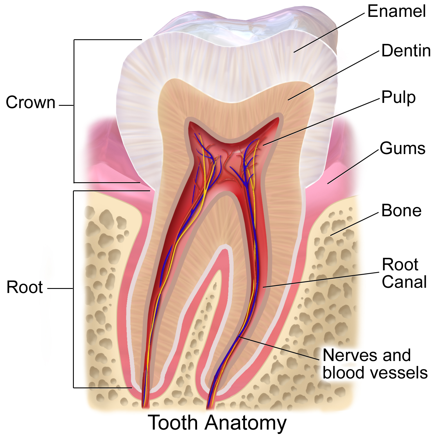
In animal tooth development, cementogenesis is the formation of cementum, one of the three mineralized substances of a tooth. Cementum covers the roots of teeth and serves to anchor gingival and periodontal fibers of the periodontal ligament by the fibers to the alveolar bone (some types of cementum may also form on the surface of the enamel of the crown at the cementoenamel junction (CEJ)). Process For cementogenesis to begin, Hertwig epithelial root sheath (HERS) must fragment. HERS is a collar of epithelial cells derived from the apical prolongation of the enamel organ. Once the root sheath disintegrates, the newly formed surface of root dentin comes into contact with the undifferentiated cells of the dental sac ( dental follicle). This then stimulates the activation of cementoblasts to begin cementogenesis. The external shape of each root is fully determined by the position of the surrounding Hertwig epithelial root sheath. It is believed that either 1) HERS becomes inte ... [...More Info...] [...Related Items...] OR: [Wikipedia] [Google] [Baidu] |
Cementoblast
A cementoblast is a biological cell that forms from the follicular cells around the root of a tooth, and whose biological function is cementogenesis, which is the formation of cementum (hard tissue that covers the tooth root). The mechanism of differentiation of the cementoblasts is controversial but circumstantial evidence suggests that an epithelium or epithelial component may cause dental sac cells to differentiate into cementoblasts, characterised by an increase in length. Other theories involve Hertwig epithelial root sheath (HERS) being involved. Martha Somerman and her laboratory played a key role in identifying and characterizing cementoblasts, the cells responsible for forming cementum, a vital mineralized tissue covering tooth roots. Structure Thus cementoblasts resemble bone-forming osteoblasts but differ functionally and histologically. The cells of cementum are the entrapped cementoblasts, the cementocytes. Each cementocyte lies in its lacuna (plural, lacunae), si ... [...More Info...] [...Related Items...] OR: [Wikipedia] [Google] [Baidu] |
Cementoblasts
A cementoblast is a biological cell that forms from the follicular cells around the root of a tooth, and whose biological function is cementogenesis, which is the formation of cementum (hard tissue that covers the tooth root). The mechanism of differentiation of the cementoblasts is controversial but circumstantial evidence suggests that an epithelium or epithelial component may cause dental sac cells to differentiate into cementoblasts, characterised by an increase in length. Other theories involve Hertwig epithelial root sheath (HERS) being involved. Martha Somerman and her laboratory played a key role in identifying and characterizing cementoblasts, the cells responsible for forming cementum, a vital mineralized tissue covering tooth roots. Structure Thus cementoblasts resemble bone-forming osteoblasts but differ functionally and histologically. The cells of cementum are the entrapped cementoblasts, the cementocytes. Each cementocyte lies in its lacuna (plural, lacunae), simi ... [...More Info...] [...Related Items...] OR: [Wikipedia] [Google] [Baidu] |
Cementum
Cementum is a specialized calcified substance covering the root of a tooth. The cementum is the part of the periodontium that attaches the teeth to the alveolar bone by anchoring the periodontal ligament. Structure The cells of cementum are the entrapped cementoblasts, the cementocytes. Each cementocyte lies in its lacuna, similar to the pattern noted in bone. These lacunae also have canaliculi or canals. Unlike those in bone, however, these canals in cementum do not contain nerves, nor do they radiate outward. Instead, the canals are oriented toward the periodontal ligament and contain cementocytic processes that exist to diffuse nutrients from the ligament because it is vascularized. After the apposition of cementum in layers, the cementoblasts that do not become entrapped in cementum line up along the cemental surface along the length of the outer covering of the periodontal ligament. These cementoblasts can form subsequent layers of cementum if the tooth is injured. Sh ... [...More Info...] [...Related Items...] OR: [Wikipedia] [Google] [Baidu] |
Animal Tooth Development
Tooth development or odontogenesis is the process in which teeth develop and grow into the mouth. Tooth development varies among species. Tooth development in vertebrates Fish In fish, Hox gene expression regulates mechanisms for teeth, tooth initiation. However, sharks continuously produce new teeth throughout their lives via a drastically different mechanism. Shark teeth form from modified scale (zoology), scales near the tongue and move outward on the jaw in rows until they are eventually dislodged. Their scales, called dermal denticles, and teeth are homologous organs. Mammals Generally, tooth development in non-human mammals is similar to human tooth development. The variations usually lie in the morphology, Dentition#Dental formula, number, development timeline, and types of teeth. However, some mammals' teeth do develop differently than humans'. In mouse, mice, Wnt signaling pathway, WNT signals are required for the initiation of teeth, tooth development. Rodents' teet ... [...More Info...] [...Related Items...] OR: [Wikipedia] [Google] [Baidu] |
Tooth
A tooth (: teeth) is a hard, calcified structure found in the jaws (or mouths) of many vertebrates and used to break down food. Some animals, particularly carnivores and omnivores, also use teeth to help with capturing or wounding prey, tearing food, for defensive purposes, to intimidate other animals often including their own, or to carry prey or their young. The roots of teeth are covered by gums. Teeth are not made of bone, but rather of multiple tissues of varying density and hardness that originate from the outermost embryonic germ layer, the ectoderm. The general structure of teeth is similar across the vertebrates, although there is considerable variation in their form and position. The teeth of mammals have deep roots, and this pattern is also found in some fish, and in crocodilians. In most teleost fish, however, the teeth are attached to the outer surface of the bone, while in lizards they are attached to the inner surface of the jaw by one side. In cartila ... [...More Info...] [...Related Items...] OR: [Wikipedia] [Google] [Baidu] |
Gums
The gums or gingiva (: gingivae) consist of the mucosal tissue that lies over the mandible and maxilla inside the mouth. Gum health and disease can have an effect on general health. Structure The gums are part of the soft tissue lining of the mouth. They surround the teeth and provide a seal around them. Unlike the soft tissue linings of the lips and cheeks, most of the gums are tightly bound to the underlying bone which helps resist the friction of food passing over them. Thus when healthy, it presents an effective barrier to the barrage of periodontal insults to deeper tissue. Healthy gums are usually coral pink in light skinned people, and may be naturally darker with melanin pigmentation. Changes in color, particularly increased redness, together with swelling and an increased tendency to bleed, suggest an inflammation that is possibly due to the accumulation of bacterial plaque. Overall, the clinical appearance of the tissue reflects the underlying histology, both in hea ... [...More Info...] [...Related Items...] OR: [Wikipedia] [Google] [Baidu] |
Periodontium
The periodontium () is the specialized tissues that both surround and support the teeth, maintaining them in the maxillary and mandibular bones. Periodontics is the dental specialty that relates specifically to the care and maintenance of these tissues. It provides the support necessary to maintain teeth in function. It consists of four principal components, namely: * Gingiva (the gums) * Periodontal ligament (PDL) * Cementum * Alveolar bone proper Each of these components is distinct in location, architecture, and biochemical properties, which adapt during the life of the structure. For example, as teeth respond to forces or migrate medially, bone resorbs on the pressure side and is added on the tension side. Cementum similarly adapts to wear on the occlusal surfaces of the teeth by apical deposition. The periodontal ligament in itself is an area of high turnover that allows the tooth not only to be suspended in the alveolar bone but also to respond to the forces. Thus, ... [...More Info...] [...Related Items...] OR: [Wikipedia] [Google] [Baidu] |
Periodontal Fiber
The periodontal ligament, commonly abbreviated as the PDL, are a group of specialized connective tissue fibers that essentially attach a tooth to the alveolar bone within which they sit. It inserts into root cementum on one side and onto alveolar bone on the other. Structure The PDL consists of principal fibers, loose connective tissue, blast and clast cells, oxytalan fibers and cell rest of Malassez. Alveolodental ligament The main principal fiber group is the alveolodental ligament, which consists of five fiber subgroups: alveolar crest, horizontal, oblique, apical, and interradicular on multirooted teeth. Principal fibers other than the alveolodental ligament are the transseptal fibers. All these fibers help the tooth withstand the naturally substantial compressive forces that occur during chewing and remain embedded in the bone. The ends of the principal fibers that are within either cementum or alveolar bone proper are considered Sharpey fibers. * Alveolar crest ... [...More Info...] [...Related Items...] OR: [Wikipedia] [Google] [Baidu] |
Alveolar Bone
The alveolar process () is the portion of bone containing the tooth sockets on the jaw bones (in humans, the maxilla and the mandible). The alveolar process is covered by gums within the mouth, terminating roughly along the line of the mandibular canal. Partially comprising compact bone, it is penetrated by many small openings for blood vessels and connective fibres. The bone is of clinical, phonetic and forensic significance. Terminology The term ''alveolar'' () ('hollow') refers to the cavities of the tooth sockets, known as dental alveoli. The alveolar process is also called the ''alveolar bone'' or ''alveolar ridge''. In phonetics, the term refers more specifically to the ridges on the inside of the mouth which can be felt with the tongue, either on roof of the mouth between the upper teeth and the hard palate or on the bottom of the mouth behind the lower teeth. The curved portion of the process is referred to as the alveolar arch. The alveolar bone proper, also ca ... [...More Info...] [...Related Items...] OR: [Wikipedia] [Google] [Baidu] |
Tooth Enamel
Tooth enamel is one of the four major Tissue (biology), tissues that make up the tooth in humans and many animals, including some species of fish. It makes up the normally visible part of the tooth, covering the Crown (tooth), crown. The other major tissues are dentin, cementum, and Pulp (tooth), dental pulp. It is a very hard, white to off-white, highly mineralised substance that acts as a barrier to protect the tooth but can become susceptible to degradation, especially by acids from food and drink. In rare circumstances enamel fails to form, leaving the underlying dentin exposed on the surface. Features Enamel is the hardest substance in the human body and contains the highest percentage of minerals (at 96%),Ross ''et al.'', p. 485 with water and organic material composing the rest.Ten Cate's Oral Histology, Nancy, Elsevier, pp. 70–94 The primary mineral is hydroxyapatite, which is a crystalline calcium phosphate. Enamel is formed on the tooth while the tooth develops wit ... [...More Info...] [...Related Items...] OR: [Wikipedia] [Google] [Baidu] |
Crown (tooth)
In dentistry, the crown is the visible part of the tooth above the gingival margin and is an essential component of dental anatomy. Covered by Tooth enamel, enamel, the crown plays a crucial role in cutting, tearing, and grinding food. Its shape and structure vary depending on the type and function of the tooth (incisors, Canine tooth, canines, premolars, or Molar (tooth), molars), and differ between Deciduous teeth, primary dentition and Permanent teeth, permanent dentition. The crown also contributes to facial aesthetics, speech, and oral health. Anatomical crown vs clinical crown The anatomical crown refers to the portion of the tooth covered by enamel, regardless of whether it is visible. The clinical crown is the part of the tooth that is visible in the mouth. In a healthy young adult, the gums typically follow the contour where enamel meets the root, so the clinical and anatomical crowns are similar in size. However, with age or periodontal disease, this may change. Te ... [...More Info...] [...Related Items...] OR: [Wikipedia] [Google] [Baidu] |


