|
Catenin
Catenins are a family of proteins found in complexes with cadherin cell adhesion molecules of animal cells. The first two catenins that were identified became known as α-catenin and β-catenin. α-Catenin can bind to β-catenin and can also bind filamentous actin (F-actin). β-Catenin binds directly to the cytoplasmic tail of classical cadherins. Additional catenins such as γ-catenin and δ-catenin have been identified. The name "catenin" was originally selected ('catena' means 'chain' in Latin) because it was suspected that catenins might link cadherins to the cytoskeleton. Types * α-catenin * β-catenin * γ-catenin * δ-catenin All but α-catenin contain armadillo repeats. They exhibit a high degree of protein dynamics, alone or in complex. Function Several types of catenins work with N-cadherins to play an important role in learning and memory. Cell-cell adhesion complexes are required for simple epithelia in higher organisms to maintain structure, function an ... [...More Info...] [...Related Items...] OR: [Wikipedia] [Google] [Baidu] |
Beta-catenin
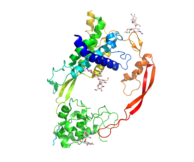
Catenin beta-1, also known as β-catenin (''beta''-catenin), is a protein that in humans is encoded by the ''CTNNB1'' gene. β-Catenin is a dual function protein, involved in regulation and coordination of cell–cell adhesion and gene transcription. In humans, the CTNNB1 protein is encoded by the ''CTNNB1'' gene. In ''Drosophila'', the homologous protein is called ''armadillo''. β-catenin is a subunit of the cadherin protein complex and acts as an intracellular signal transducer in the Wnt signaling pathway. It is a member of the catenin protein family and homologous to Plakoglobin, γ-catenin, also known as plakoglobin. β-Catenin is widely expressed in many tissues. In cardiac muscle, β-catenin localizes to adherens junctions in intercalated disc structures, which are critical for electrical and mechanical coupling between adjacent cardiomyocytes. Mutations and overexpression of β-catenin are associated with many cancers, including hepatocellular carcinoma, colorectal cance ... [...More Info...] [...Related Items...] OR: [Wikipedia] [Google] [Baidu] |
Wnt Signaling Pathway
In cellular biology, the Wnt signaling pathways are a group of signal transduction pathways which begin with proteins that pass signals into a cell through cell surface receptors. The name Wnt, pronounced "wint", is a portmanteau created from the names Wingless and Int-1. Wnt signaling pathways use either nearby cell-cell communication (paracrine) or same-cell communication (autocrine). They are highly evolutionarily conserved in animals, which means they are similar across animal species from fruit flies to humans. Three Wnt signaling pathways have been characterized: the canonical Wnt pathway, the noncanonical planar cell polarity pathway, and the noncanonical Wnt/calcium pathway. All three pathways are activated by the binding of a Wnt-protein ligand to a Frizzled family receptor, which passes the biological signal to the Dishevelled protein inside the cell. The canonical Wnt pathway leads to regulation of gene transcription, and is thought to be negatively regulated in part ... [...More Info...] [...Related Items...] OR: [Wikipedia] [Google] [Baidu] |
Gamma Catenin
Plakoglobin, also known as junction plakoglobin or gamma-catenin, is a protein that in humans is encoded by the ''JUP'' gene. Plakoglobin is a member of the catenin protein family and homologous to β-catenin. Plakoglobin is a cytoplasmic component of desmosomes and adherens junctions structures located within intercalated discs of cardiac muscle that function to anchor sarcomeres and join adjacent cells in cardiac muscle. Mutations in plakoglobin are associated with arrhythmogenic right ventricular dysplasia. Structure Human plakoglobin is 81.7 kDa in molecular weight and 745 amino acids long. The ''JUP'' gene contains 13 exons spanning 17 kb on chromosome 17q21. Plakoglobin is a member of the catenin family, since it contains a distinct repeating amino acid motif called the armadillo repeat. Plakoglobin is highly similar to β-catenin; both have 12 armadillo repeats as well as N-terminal and C-terminal globular domains of unknown structure. Plakoglobin was originally identifi ... [...More Info...] [...Related Items...] OR: [Wikipedia] [Google] [Baidu] |
Adenomatous Polyposis Coli
Adenomatous polyposis coli (APC) also known as deleted in polyposis 2.5 (DP2.5) is a protein that in humans is encoded by the ''APC'' gene. The APC protein is a Down-regulation, negative regulator that controls beta-catenin concentrations and interacts with E-cadherin, which are involved in cell adhesion. Mutations in the ''APC'' gene may result in colorectal cancer and desmoid tumors. ''APC'' is classified as a tumor suppressor gene. Tumor suppressor genes prevent the uncontrolled growth of cells that may result in cancerous tumors. The protein made by the ''APC'' gene plays a critical role in several cellular processes that determine whether a cell may develop into a tumor. The APC protein helps control how often a cell divides, how it attaches to other cells within a tissue, how the cell polarizes and the morphogenesis of the 3D structures, or whether a cell moves within or away from tissue. This protein also helps ensure that the chromosome number in cells produced through c ... [...More Info...] [...Related Items...] OR: [Wikipedia] [Google] [Baidu] |
Cadherin
Cadherins (named for "calcium-dependent adhesion") are cell adhesion molecules important in forming adherens junctions that let cells adhere to each other. Cadherins are a class of type-1 transmembrane proteins, and they depend on calcium (Ca2+) ions to function, hence their name. Cell-cell adhesion is mediated by extracellular cadherin domains, whereas the Cadherin cytoplasmic region, intracellular cytoplasmic tail associates with numerous adaptors and signaling proteins, collectively referred to as the cadherin adhesome. Background The cadherin family is essential in maintaining cell-cell contact and regulating cytoskeletal complexes. The cadherin superfamily includes cadherins, protocadherins, desmogleins, desmocollins, and more. In structure, they share ''cadherin repeats'', which are the extracellular Ca2+-binding domains. There are multiple classes of cadherin molecules, each designated with a prefix for tissues with which it associates. Classical cadherins maintain the ton ... [...More Info...] [...Related Items...] OR: [Wikipedia] [Google] [Baidu] |
AXIN1
Axin-1 is a protein that in humans is encoded by the ''AXIN1'' gene. Function This gene encodes a cytoplasmic protein which contains a regulation of G-protein signaling (RGS) domain and a dishevelled and axin (DIX) domain. The encoded protein interacts with adenomatosis polyposis coli, catenin (cadherin-associated protein) beta 1, glycogen synthase kinase 3 beta, protein phosphatase 2, and itself. This protein functions as a negative regulator of the wingless-type MMTV integration site family, member 1 ( WNT) signaling pathway and can induce apoptosis. The crystal structure of a portion of this protein, alone and in a complex with other proteins, has been resolved. Mutations in this gene have been associated with hepatocellular carcinoma, hepatoblastomas, ovarian endometrioid adenocarcinomas, and medulloblastomas. Two transcript variants encoding distinct isoforms have been identified for this gene. The AXIN proteins attract substantial interest in cancer research as AXIN1 an ... [...More Info...] [...Related Items...] OR: [Wikipedia] [Google] [Baidu] |
Armadillo Repeats
An armadillo repeat is a characteristic, repetitive amino acid peptide sequence, sequence of about 42 residue (chemistry)#biochemistry, residues in length that is found in many proteins. Proteins that contain armadillo repeats typically contain several Protein tandem repeats, tandemly repeated copies. Each armadillo repeat is composed of a pair of alpha helices that form a hairpin structure. Multiple copies of the repeat form what is known as an alpha solenoid structure. Examples of proteins that contain armadillo repeats include beta catenin, β-catenin, Sarm1 (SARM1), importin α, α-importin, plakoglobin, adenomatous polyposis coli (APC), and many others. The term armadillo derives from the historical name of the β-catenin gene in the fruitfly ''Drosophila melanogaster, Drosophila'', where the armadillo repeat was first discovered. Although β-catenin was previously believed to be a protein involved in linking cadherin cell adhesion proteins to the cytoskeleton, recent work in ... [...More Info...] [...Related Items...] OR: [Wikipedia] [Google] [Baidu] |
AXIN2
Axin-2, also known as axin-like protein (Axil), axis inhibition protein 2 (AXIN2), or conductin, is a protein that in humans is encoded by the ''AXIN2'' gene. Function The Axin-related protein, Axin2, presumably plays an important role in the regulation of the stability of beta-catenin in the Wnt signaling pathway, like its rodent homologs, mouse conductin/rat axil. In mouse, conductin organizes a multiprotein complex of APC (adenomatous polyposis of the colon), beta-catenin, glycogen synthase kinase 3-beta, and conductin, which leads to the degradation of beta-catenin. The AXIN proteins attract substantial interest in cancer research as AXIN1 and AXIN2 work synergistically to control pro-oncogenic β-catenin signaling. Importantly, activity in the β-catenin destruction complex can be increased by tankyrase inhibitors and are a potential therapeutic option to reduce the growth of β-catenin-dependent cancers. Clinical significance The deregulation of beta-catenin is an i ... [...More Info...] [...Related Items...] OR: [Wikipedia] [Google] [Baidu] |
GSK3B
Glycogen synthase kinase-3 beta, (GSK-3 beta), is an enzyme that in humans is encoded by the ''GSK3B'' gene. In mice, the enzyme is encoded by the Gsk3b gene. Abnormal regulation and expression of GSK-3 beta is associated with an increased susceptibility towards bipolar disorder. Function Glycogen synthase kinase-3 ( GSK-3) is a proline-directed serine-threonine kinase that was initially identified as a phosphorylating and an inactivating agent of glycogen synthase. Two isoforms, alpha (GSK3A) and beta, show a high degree of amino acid homology. GSK3B is involved in energy metabolism, neuronal cell development, and body pattern formation. It might be a new therapeutic target for ischemic stroke. Disease relevance Homozygous disruption of the Gsk3b locus in mice results in embryonic lethality during mid-gestation. This lethality phenotype could be rescued by inhibition of tumor necrosis factor. Two SNPs at this gene, rs334558 (-50T/C) and rs3755557 (-1727A/T), are ass ... [...More Info...] [...Related Items...] OR: [Wikipedia] [Google] [Baidu] |
BTRC (gene)
F-box/WD repeat-containing protein 1A (FBXW1A) also known as βTrCP1 or Fbxw1 or hsSlimb or pIkappaBalpha-E3 receptor subunit is a protein that in humans is encoded by the ''BTRC'' (beta-transducin repeat containing) gene. This gene encodes a member of the F-box protein family which is characterized by an approximately 40 residue structural motif, the F-box. The F-box proteins constitute one of the four subunits of ubiquitin protein ligase complex called SCFs ( Skp1-Cul1-F-box protein), which often, but not always, recognize substrates in a phosphorylation-dependent manner. F-box proteins are divided into 3 classes: * Fbxws containing WD40 repeats, * Fbxls containing leucine-rich repeats, * and Fbxos containing either "other" protein–protein interaction modules or no recognizable motifs. The protein encoded by this gene belongs to the Fbxw class as, in addition to an F-box, this protein contains multiple WD40 repeats. This protein is homologous to Xenopus βTrCP, yeast Met30 ... [...More Info...] [...Related Items...] OR: [Wikipedia] [Google] [Baidu] |
Adherens Junction
In cell biology, adherens junctions (or zonula adherens, intermediate junction, or "belt desmosome") are protein complexes that occur at cell–cell junctions and cell–matrix junctions in epithelial and endothelial tissues, usually more basal than tight junctions. An adherens junction is defined as a cell junction whose cytoplasmic face is linked to the actin cytoskeleton. They can appear as bands encircling the cell (zonula adherens) or as spots of attachment to the extracellular matrix (focal adhesion). Adherens junctions uniquely disassemble in uterine epithelial cells to allow the blastocyst to penetrate between epithelial cells. A similar cell junction in non-epithelial, non-endothelial cells is the fascia adherens. It is structurally the same, but appears in ribbonlike patterns that do not completely encircle the cells. One example is in cardiomyocytes. Proteins Adherens junctions are composed of the following proteins: * cadherins. The cadherins are a family ... [...More Info...] [...Related Items...] OR: [Wikipedia] [Google] [Baidu] |


