|
Brachial Fascia
The brachial fascia (deep fascia of the arm) is continuous with that covering the deltoideus and the pectoralis major muscle, by means of which it is attached, above, to the clavicle, acromion, and spine of the scapula; it forms a thin, loose, membranous sheath for the muscles of the arm, and sends septa between them; it is composed of fibers disposed in a circular or spiral direction, and connected together by vertical and oblique fibers. It differs in thickness at different parts, being thin over the biceps brachii, but thicker where it covers the triceps brachii, and over the epicondyles of the humerus: it is strengthened by fibrous aponeuroses, derived from the pectoralis major and latissimus dorsi medially, and from the deltoideus laterally. On either side it gives off a strong intermuscular septum, which is attached to the corresponding supracondylar ridge and epicondyle An epicondyle () is a rounded eminence on a bone that lies upon a condyle ('' epi-'', "upon" + ''con ... [...More Info...] [...Related Items...] OR: [Wikipedia] [Google] [Baidu] |
Deep Fascia
Deep fascia (or investing fascia) is a fascia, a layer of dense connective tissue that can surround individual muscles and groups of muscles to separate into fascial compartments. This fibrous connective tissue interpenetrates and surrounds the muscles, bones, nerves, and blood vessels of the body. It provides connection and communication in the form of aponeuroses, ligaments, tendons, retinacula, joint capsules, and septa. The deep fasciae envelop all bone (periosteum and endosteum); cartilage (perichondrium), and blood vessels (tunica externa) and become specialized in muscles (epimysium, perimysium, and endomysium) and nerves (epineurium, perineurium, and endoneurium). The high density of collagen fibers gives the deep fascia its strength and integrity. The amount of elastin fiber determines how much extensibility and resilience it will have. Examples Examples include: * Fascia lata * Deep fascia of leg * Brachial fascia * Buck's fascia Fascial dynamics Deep fascia is l ... [...More Info...] [...Related Items...] OR: [Wikipedia] [Google] [Baidu] |
Deltoideus
The deltoid muscle is the muscle forming the rounded contour of the human shoulder. It is also known as the 'common shoulder muscle', particularly in other animals such as the domestic cat. Anatomically, the deltoid muscle appears to be made up of three distinct sets of muscle fibers, namely the # anterior or clavicular part (pars clavicularis) # posterior or scapular part (pars scapularis) # intermediate or acromial part (pars acromialis) However, electromyography suggests that it consists of at least seven groups that can be independently coordinated by the nervous system. It was previously called the deltoideus (plural ''deltoidei'') and the name is still used by some anatomists. It is called so because it is in the shape of the Greek capital letter delta (Δ). Deltoid is also further shortened in slang as "delt". A study of 30 shoulders revealed an average mass of in humans, ranging from to . Structure Previous studies showed that the insertions of the tendons of the delto ... [...More Info...] [...Related Items...] OR: [Wikipedia] [Google] [Baidu] |
Pectoralis Major Muscle
The pectoralis major () is a thick, fan-shaped or triangular convergent muscle, situated at the chest of the human body. It makes up the bulk of the chest muscles and lies under the breast. Beneath the pectoralis major is the pectoralis minor, a thin, triangular muscle. The pectoralis major's primary functions are flexion, adduction, and internal rotation of the humerus. The pectoral major may colloquially be referred to as "pecs", "pectoral muscle", or "chest muscle", because it is the largest and most superficial muscle in the chest area. Structure It arises from the anterior surface of the sternal half of the clavicle from breadth of the half of the anterior surface of the sternum, as low down as the attachment of the cartilage of the sixth or seventh rib; from the cartilages of all the true ribs, with the exception, frequently, of the first or seventh, and from the aponeurosis of the abdominal external oblique muscle. From this extensive origin the fibers converge toward the ... [...More Info...] [...Related Items...] OR: [Wikipedia] [Google] [Baidu] |
Clavicle
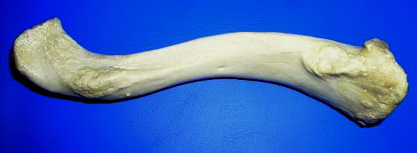
The clavicle, or collarbone, is a slender, S-shaped long bone approximately 6 inches (15 cm) long that serves as a strut between the shoulder blade and the sternum (breastbone). There are two clavicles, one on the left and one on the right. The clavicle is the only long bone in the body that lies horizontally. Together with the shoulder blade, it makes up the shoulder girdle. It is a palpable bone and, in people who have less fat in this region, the location of the bone is clearly visible. It receives its name from the Latin ''clavicula'' ("little key"), because the bone rotates along its axis like a key when the shoulder is abducted. The clavicle is the most commonly fractured bone. It can easily be fractured by impacts to the shoulder from the force of falling on outstretched arms or by a direct hit. Structure The collarbone is a thin doubly curved long bone that connects the arm to the trunk of the body. Located directly above the first rib, it acts as a strut to k ... [...More Info...] [...Related Items...] OR: [Wikipedia] [Google] [Baidu] |
Acromion
In human anatomy, the acromion (from Greek: ''akros'', "highest", ''ōmos'', "shoulder", plural: acromia) is a bony process on the scapula (shoulder blade). Together with the coracoid process it extends laterally over the shoulder joint. The acromion is a continuation of the scapular spine, and hooks over anteriorly. It articulates with the clavicle (collar bone) to form the acromioclavicular joint. Structure The acromion forms the summit of the shoulder, and is a large, somewhat triangular or oblong process, flattened from behind forward, projecting at first lateralward, and then curving forward and upward, so as to overhang the glenoid fossa.''Gray's Anatomy'' 1918, see infobox It starts from the base of acromion which marks its projecting point emerging from the spine of scapula. Surfaces Its superior surface, directed upward, backward, and lateralward, is convex, rough, and gives attachment to some fibers of the deltoideus, and in the rest of its extent is subcutaneous. ... [...More Info...] [...Related Items...] OR: [Wikipedia] [Google] [Baidu] |
Spine Of The Scapula
The spine of the scapula or scapular spine is a prominent plate of bone, which crosses obliquely the medial four-fifths of the scapula at its upper part, and separates the supra- from the infraspinatous fossa. Structure It begins at the vertical ertebral or medial borderborder by a smooth, triangular area over which the tendon of insertion of the lower part of the Trapezius glides, and, gradually becoming more elevated, ends in the acromion, which overhangs the shoulder-joint. The spine is triangular, and flattened from above downward, its apex being directed toward the vertebral border. Root The ''root of the spine'' of the scapula is the most medial part of the scapular spine. It is termed "triangular area of the spine of scapula", based on its triangular shape giving it distinguishable visible shape on x-ray images. The root of the spine is on a level with the tip of the spinous process of the third thoracic vertebra.Gray's Anatomy (1918)p.1306/ref> File:Root of spine - l ... [...More Info...] [...Related Items...] OR: [Wikipedia] [Google] [Baidu] |
Biceps Brachii
The biceps or biceps brachii ( la, musculus biceps brachii, "two-headed muscle of the arm") is a large muscle that lies on the front of the upper arm between the shoulder and the elbow. Both heads of the muscle arise on the scapula and join to form a single muscle belly which is attached to the upper forearm. While the biceps crosses both the shoulder and elbow joints, its main function is at the elbow where it flexes the forearm and supinates the forearm. Both these movements are used when opening a bottle with a corkscrew: first biceps screws in the cork (supination), then it pulls the cork out (flexion). Structure The biceps is one of three muscles in the anterior compartment of the upper arm, along with the brachialis muscle and the coracobrachialis muscle, with which the biceps shares a nerve supply. The biceps muscle has two heads, the short head and the long head, distinguished according to their origin at the coracoid process and supraglenoid tubercle of the sca ... [...More Info...] [...Related Items...] OR: [Wikipedia] [Google] [Baidu] |
Triceps Brachii
The triceps, or triceps brachii (Latin for "three-headed muscle of the arm"), is a large muscle on the back of the upper limb of many vertebrates Vertebrates () comprise all animal taxa within the subphylum Vertebrata () ( chordates with backbones), including all mammals, birds, reptiles, amphibians, and fish. Vertebrates represent the overwhelming majority of the phylum Chordata, .... It consists of 3 parts: the medial, lateral, and long head. It is the muscle principally responsible for Extension (kinesiology), extension of the elbow joint (straightening of the arm). Structure The long head arises from the infraglenoid tubercle of the scapula. It extends distally anterior to the Teres minor muscle, teres minor and posterior to the Teres major muscle, teres major. The medial head arises proximally in the humerus, just inferior to the Radial sulcus, groove of the radial nerve; from the dorsal (back) surface of the humerus; from the Medial intermuscular septum of arm ... [...More Info...] [...Related Items...] OR: [Wikipedia] [Google] [Baidu] |
Epicondyles
An epicondyle () is a rounded eminence on a bone that lies upon a condyle ('' epi-'', "upon" + ''condyle'', from a root meaning "knuckle" or "rounded articular area"). There are various epicondyles in the human skeleton, each named by its anatomic Anatomy () is the branch of biology concerned with the study of the structure of organisms and their parts. Anatomy is a branch of natural science that deals with the structural organization of living things. It is an old science, having its ... site. They include the following: Skeletal system {{musculoskeletal-stub ... [...More Info...] [...Related Items...] OR: [Wikipedia] [Google] [Baidu] |
Humerus
The humerus (; ) is a long bone in the arm that runs from the shoulder to the elbow. It connects the scapula and the two bones of the lower arm, the radius and ulna, and consists of three sections. The humeral upper extremity consists of a rounded head, a narrow neck, and two short processes (tubercles, sometimes called tuberosities). The body is cylindrical in its upper portion, and more prismatic below. The lower extremity consists of 2 epicondyles, 2 processes (trochlea & capitulum), and 3 fossae (radial fossa, coronoid fossa, and olecranon fossa). As well as its true anatomical neck, the constriction below the greater and lesser tubercles of the humerus is referred to as its surgical neck due to its tendency to fracture, thus often becoming the focus of surgeons. Etymology The word "humerus" is derived from la, humerus, umerus meaning upper arm, shoulder, and is linguistically related to Gothic ''ams'' shoulder and Greek ''ōmos''. Structure Upper extremity The upper or pr ... [...More Info...] [...Related Items...] OR: [Wikipedia] [Google] [Baidu] |
Latissimus Dorsi
The latissimus dorsi () is a large, flat muscle on the back that stretches to the sides, behind the arm, and is partly covered by the trapezius on the back near the midline. The word latissimus dorsi (plural: ''latissimi dorsorum'') comes from Latin and means "broadest uscleof the back", from "latissimus" ( la, broadest)' and "dorsum" ( la, back). The pair of muscles are commonly known as "lats", especially among bodybuilders. The latissimus dorsi is the largest muscle in the upper body. The latissimus dorsi is responsible for extension, adduction, transverse extension also known as horizontal abduction (or horizontal extension), flexion from an extended position, and (medial) internal rotation of the shoulder joint. It also has a synergistic role in extension and lateral flexion of the lumbar spine. Due to bypassing the scapulothoracic joints and attaching directly to the spine, the actions the latissimi dorsi have on moving the arms can also influence the movement of the sca ... [...More Info...] [...Related Items...] OR: [Wikipedia] [Google] [Baidu] |
Medial Intermuscular Septum Of Arm
The fascial compartments of arm refers to the specific anatomical term of the compartments within the upper segment of the upper limb (the arm) of the body. The upper limb is divided into two segments, the arm and the forearm. Each of these segments is further divided into two compartments which are formed by deep fascia – tough connective tissue septa (walls). Each compartment encloses specific muscles and nerves. The compartments of the arm are the anterior compartment of the arm and the posterior compartment of the arm, divided by the lateral and the medial intermuscular septa. The compartments of the forearm are the anterior compartment of the forearm and posterior compartment of the forearm. Intermuscular septa The lateral intermuscular septum extends from the lower part of the crest of the greater tubercle of the humerus, along the lateral supracondylar ridge, to the lateral epicondyle; it is blended with the tendon of the deltoid muscle, gives attachment to the tricep ... [...More Info...] [...Related Items...] OR: [Wikipedia] [Google] [Baidu] |


