|
Atrial Septum
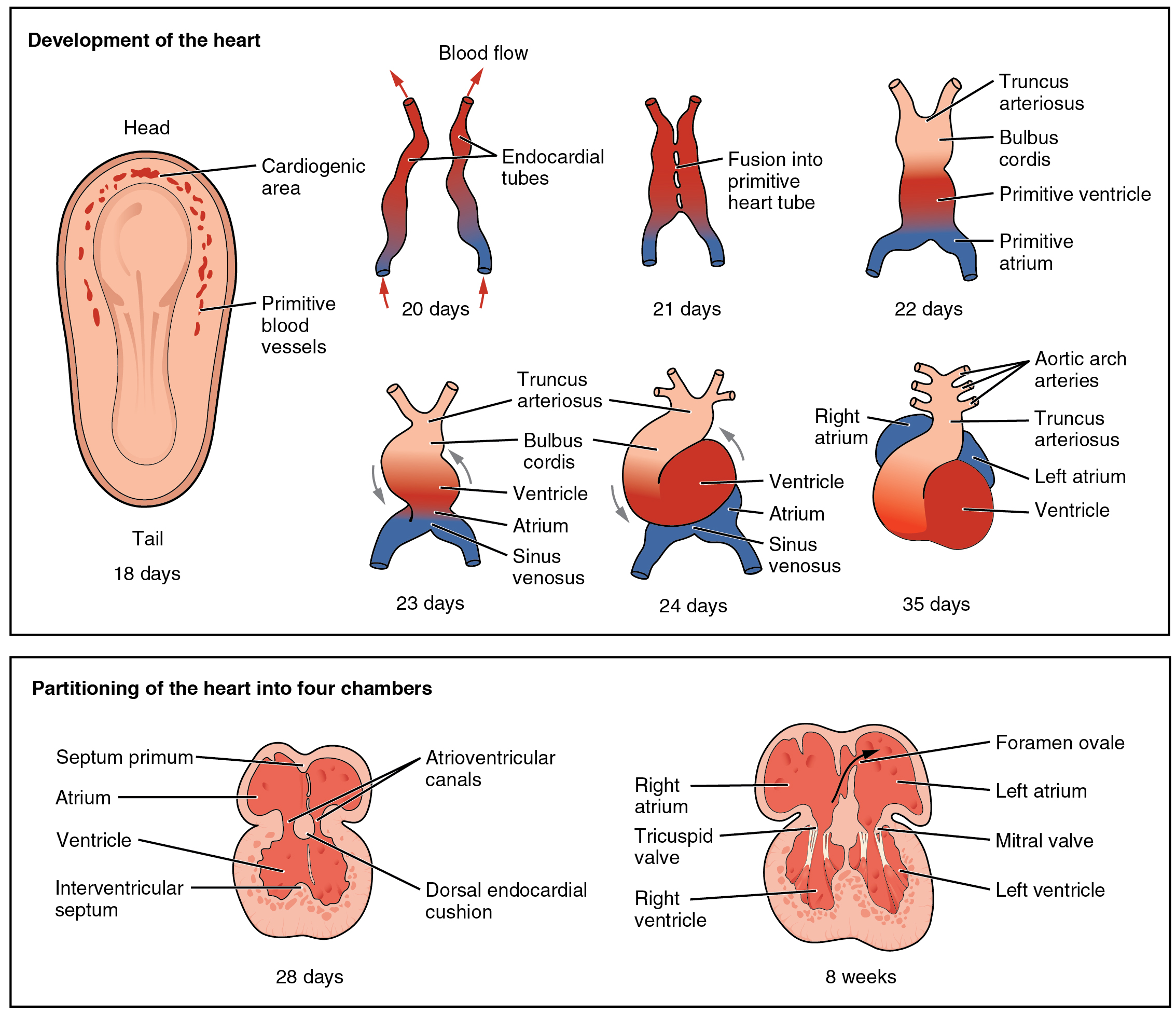
The interatrial septum is the wall of tissue that separates the right and left atria of the heart. Structure The interatrial septum is a that lies between the left atrium and right atrium of the human heart. The interatrial septum lies at angle of 65 degrees from right posterior to left anterior because right atrium is located at the right side of the body while left atrium is located at the left side of the body. Development The interatrial septum forms during the first and second months of fetal development. Formation of the septum occurs in several stages. The first is the development of the septum primum, a crescent-shaped piece of tissue forming the initial divider between the right and left atria. Because of its crescent shape, the septum primum does not fully occlude the space between the left and right atria; the opening that remains is called the ostium primum. During fetal development, this opening allows blood to be shunted from the right atrium to the left. As the ... [...More Info...] [...Related Items...] OR: [Wikipedia] [Google] [Baidu] |
Atrium (heart)
The atrium ( la, ātrium, , entry hall) is one of two upper chambers in the heart that receives blood from the circulatory system. The blood in the atria is pumped into the heart ventricles through the atrioventricular valves. There are two atria in the human heart – the left atrium receives blood from the pulmonary circulation, and the right atrium receives blood from the venae cavae of the systemic circulation. During the cardiac cycle the atria receive blood while relaxed in diastole, then contract in systole to move blood to the ventricles. Each atrium is roughly cube-shaped except for an ear-shaped projection called an atrial appendage, sometimes known as an auricle. All animals with a closed circulatory system have at least one atrium. The atrium was formerly called the 'auricle'. That term is still used to describe this chamber in some other animals, such as the ''Mollusca''. They have thicker muscular walls than the atria do. Structure Humans have a four-chambered ... [...More Info...] [...Related Items...] OR: [Wikipedia] [Google] [Baidu] |
Pulmonary Circulation
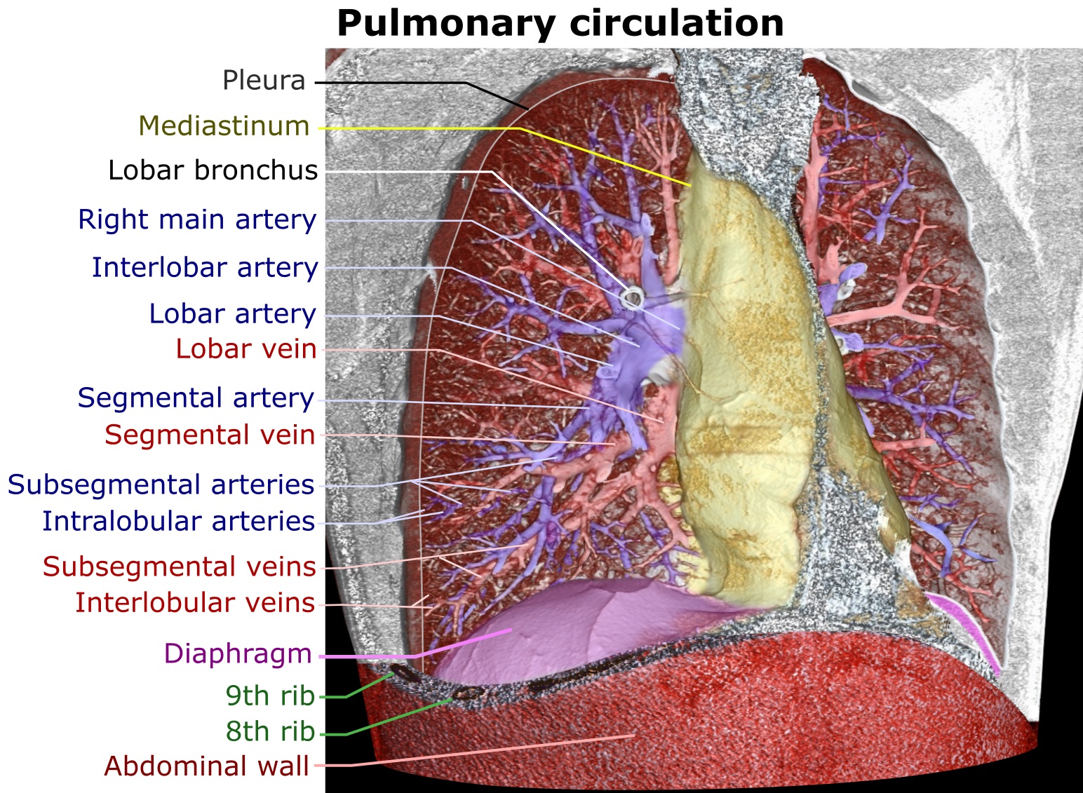
The pulmonary circulation is a division of the circulatory system in all vertebrates. The circuit begins with deoxygenated blood returned from the body to the right atrium of the heart where it is pumped out from the right ventricle to the lungs. In the lungs the blood is oxygenated and returned to the left atrium to complete the circuit. The other division of the circulatory system is the systemic circulation that begins with receiving the oxygenated blood from the pulmonary circulation into the left atrium. From the atrium the oxygenated blood enters the left ventricle where it is pumped out to the rest of the body, returning as deoxygenated blood back to the pulmonary circulation. The blood vessels of the pulmonary circulation are the pulmonary arteries and the pulmonary veins. A separate circulatory circuit known as the bronchial circulation supplies oxygenated blood to the tissue of the larger airways of the lung. Structure De-oxygenated blood leaves the heart, goe ... [...More Info...] [...Related Items...] OR: [Wikipedia] [Google] [Baidu] |
Interventricular Septum
The interventricular septum (IVS, or ventricular septum, or during development septum inferius) is the stout wall separating the ventricles, the lower chambers of the heart, from one another. The ventricular septum is directed obliquely backward to the right and curved with the convexity toward the right ventricle; its margins correspond with the anterior and posterior interventricular sulci. The lower part of the septum, which is the major part, is thick and muscular, and its much smaller upper part is thin and membraneous. During each cardiac cycle the interventricular septum contracts by shortening longitudinally and becoming thicker. Structure The interventricular septum is the stout wall separating the ventricles, the lower chambers of the heart, from one another. The ventricular septum is directed obliquely backward to the right and curved with the convexity toward the right ventricle; its margins correspond with the anterior and posterior longitudinal sulci. The gr ... [...More Info...] [...Related Items...] OR: [Wikipedia] [Google] [Baidu] |
Eisenmenger Syndrome
Eisenmenger syndrome or Eisenmenger's syndrome is defined as the process in which a long-standing left-to-right cardiac shunt caused by a congenital heart defect (typically by a ventricular septal defect, atrial septal defect, or less commonly, patent ductus arteriosus) causes pulmonary hypertension and eventual reversal of the shunt into a cyanotic right-to-left shunt. Because of the advent of fetal screening with echocardiography early in life, the incidence of heart defects progressing to Eisenmenger syndrome has decreased. Eisenmenger syndrome in a pregnant mother can cause serious complications, though successful delivery has been reported. Maternal mortality ranges from 30% to 60%, and may be attributed to fainting spells, blood clots forming in the veins and traveling to distant sites, hypovolemia, coughing up blood or preeclampsia. Most deaths occur either during or within the first weeks after delivery.Curr Cardiol Rev. 2010 November; 6(4): 363–372.The Adult Patient ... [...More Info...] [...Related Items...] OR: [Wikipedia] [Google] [Baidu] |
Hypertrophy
Hypertrophy is the increase in the volume of an organ or tissue due to the enlargement of its component cells. It is distinguished from hyperplasia, in which the cells remain approximately the same size but increase in number.Updated by Linda J. Vorvick. 8/14/1Hyperplasia/ref> Although hypertrophy and hyperplasia are two distinct processes, they frequently occur together, such as in the case of the hormonally-induced proliferation and enlargement of the cells of the uterus during pregnancy. Eccentric hypertrophy is a type of hypertrophy where the walls and chamber of a hollow organ undergo growth in which the overall size and volume are enlarged. It is applied especially to the left ventricle of heart. Sarcomeres are added in series, as for example in dilated cardiomyopathy (in contrast to hypertrophic cardiomyopathy, a type of concentric hypertrophy, where sarcomeres are added in parallel). Gallery File:*+ * Photographic documentation on sexual education - Hypertrophy of bre ... [...More Info...] [...Related Items...] OR: [Wikipedia] [Google] [Baidu] |
Down Syndrome
Down syndrome or Down's syndrome, also known as trisomy 21, is a genetic disorder caused by the presence of all or part of a third copy of chromosome 21. It is usually associated with physical growth delays, mild to moderate intellectual disability, and characteristic facial features. The average IQ of a young adult with Down syndrome is 50, equivalent to the mental ability of an eight- or nine-year-old child, but this can vary widely. The parents of the affected individual are usually genetically normal. The probability increases from less than 0.1% in 20-year-old mothers to 3% in those of age 45. The extra chromosome is believed to occur by chance, with no known behavioral activity or environmental factor that changes the probability. Down syndrome can be identified during pregnancy by prenatal screening followed by diagnostic testing or after birth by direct observation and genetic testing. Since the introduction of screening, Down syndrome pregnancies are often abor ... [...More Info...] [...Related Items...] OR: [Wikipedia] [Google] [Baidu] |
Atrial Septal Defect
Atrial septal defect (ASD) is a congenital heart defect in which blood flows between the atria (upper chambers) of the heart. Some flow is a normal condition both pre-birth and immediately post-birth via the foramen ovale; however, when this does not naturally close after birth it is referred to as a patent (open) foramen ovale (PFO). It is common in patients with a congenital atrial septal aneurysm (ASA). After PFO closure the atria normally are separated by a dividing wall, the interatrial septum. If this septum is defective or absent, then oxygen-rich blood can flow directly from the left side of the heart to mix with the oxygen-poor blood in the right side of the heart; or the opposite, depending on whether the left or right atrium has the higher blood pressure. In the absence of other heart defects, the left atrium has the higher pressure. This can lead to lower-than-normal oxygen levels in the arterial blood that supplies the brain, organs, and tissues. However, an ASD m ... [...More Info...] [...Related Items...] OR: [Wikipedia] [Google] [Baidu] |
Patent Foramen Ovale
Atrial septal defect (ASD) is a congenital heart defect in which blood flows between the atria (upper chambers) of the heart. Some flow is a normal condition both pre-birth and immediately post-birth via the foramen ovale; however, when this does not naturally close after birth it is referred to as a patent (open) foramen ovale (PFO). It is common in patients with a congenital atrial septal aneurysm (ASA). After PFO closure the atria normally are separated by a dividing wall, the interatrial septum. If this septum is defective or absent, then oxygen-rich blood can flow directly from the left side of the heart to mix with the oxygen-poor blood in the right side of the heart; or the opposite, depending on whether the left or right atrium has the higher blood pressure. In the absence of other heart defects, the left atrium has the higher pressure. This can lead to lower-than-normal oxygen levels in the arterial blood that supplies the brain, organs, and tissues. However, an ASD m ... [...More Info...] [...Related Items...] OR: [Wikipedia] [Google] [Baidu] |
Systemic Circulation
The blood circulatory system is a system of organs that includes the heart, blood vessels, and blood which is circulated throughout the entire body of a human or other vertebrate. It includes the cardiovascular system, or vascular system, that consists of the heart and blood vessels (from Greek ''kardia'' meaning ''heart'', and from Latin ''vascula'' meaning ''vessels''). The circulatory system has two divisions, a systemic circulation or circuit, and a pulmonary circulation or circuit. Some sources use the terms ''cardiovascular system'' and ''vascular system'' interchangeably with the ''circulatory system''. The network of blood vessels are the great vessels of the heart including large elastic arteries, and large veins; other arteries, smaller arterioles, capillaries that join with venules (small veins), and other veins. The circulatory system is closed in vertebrates, which means that the blood never leaves the network of blood vessels. Some invertebrates such as arthro ... [...More Info...] [...Related Items...] OR: [Wikipedia] [Google] [Baidu] |
Lung
The lungs are the primary organs of the respiratory system in humans and most other animals, including some snails and a small number of fish. In mammals and most other vertebrates, two lungs are located near the backbone on either side of the heart. Their function in the respiratory system is to extract oxygen from the air and transfer it into the bloodstream, and to release carbon dioxide from the bloodstream into the atmosphere, in a process of gas exchange. Respiration is driven by different muscular systems in different species. Mammals, reptiles and birds use their different muscles to support and foster breathing. In earlier tetrapods, air was driven into the lungs by the pharyngeal muscles via buccal pumping, a mechanism still seen in amphibians. In humans, the main muscle of respiration that drives breathing is the diaphragm. The lungs also provide airflow that makes vocal sounds including human speech possible. Humans have two lungs, one on the left and on ... [...More Info...] [...Related Items...] OR: [Wikipedia] [Google] [Baidu] |


